[ad_1]
In a recent study published in The New England Journal of Medicine, researchers investigated whether micro- and nano-plastics (MNPs) are detectable in atherosclerotic plaques.
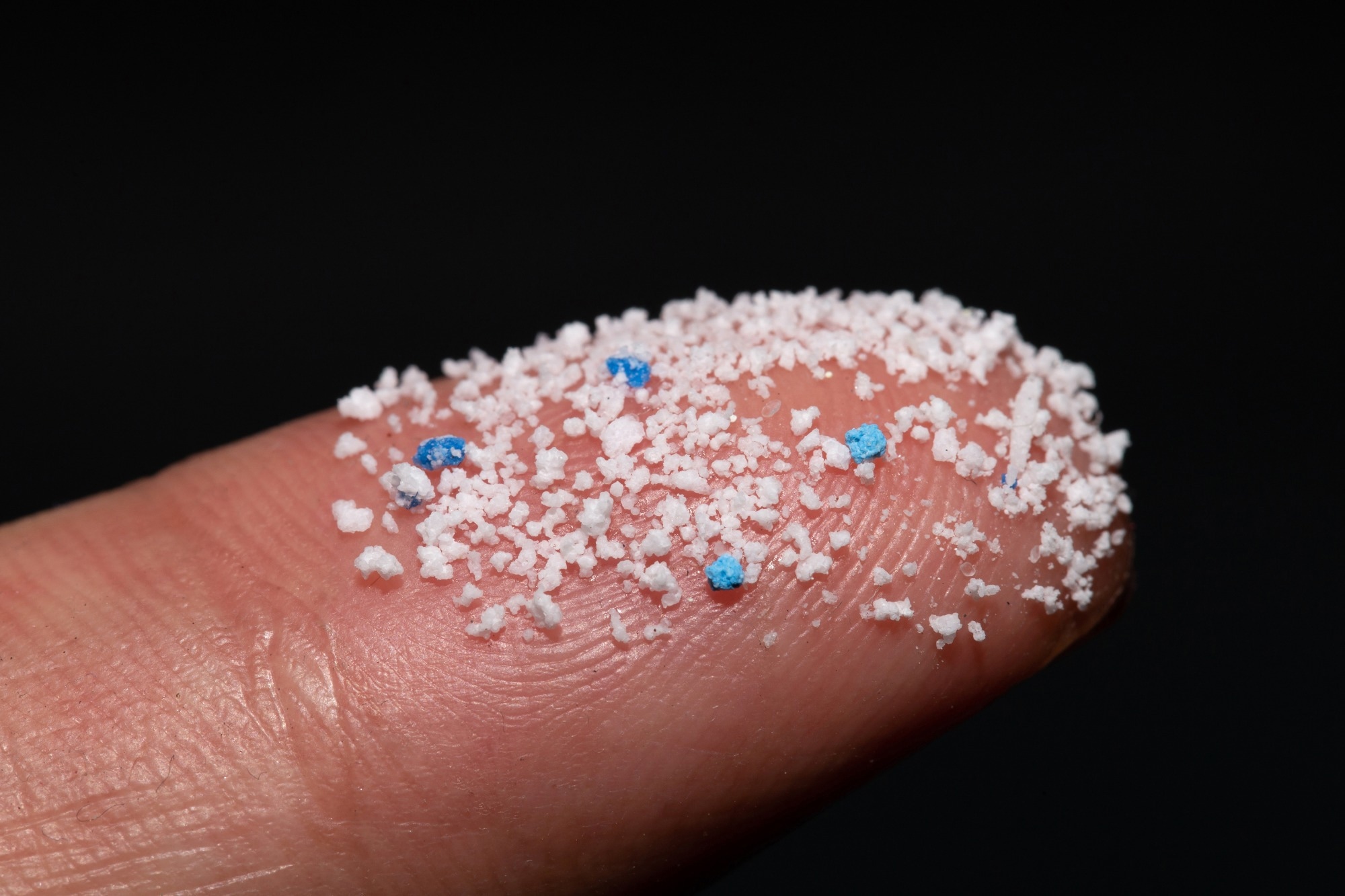 Study: Microplastics and Nanoplastics in Atheromas and Cardiovascular Events. Image Credit: chayanuphol/Shutterstock.com
Background
Plastic production has been constantly increasing and is likely to continue until 2050. Plastics can degrade and form MNPs, inducing toxic effects.
Studies have demonstrated the entry of MNPs into the body through skin exposure, inhalation, and ingestion, as well as their interactions with tissues/organs. Further, MNPs have been detected in the placenta, liver, lungs, urine, blood, and breast milk. Recent preclinical reports implicate MNPs as a cardiovascular risk factor.
In vitro findings indicate that some MNPs promote inflammation, oxidative stress, and apoptosis in endothelial cells. Moreover, animal studies support the role of MNPs in myocardial fibrosis, endothelial dysfunction, and cardiac function impairment.
However, their clinical relevance remains unknown. There is no evidence to suggest the infiltration of MNPs in human vascular lesions or associations between MNP burden and cardiovascular disease.
About the study
In the present study, researchers investigated the presence of MNPs in atherosclerotic plaques and the associations between MNP burden and cardiovascular disease.
Consecutive patients aged 18–75 with asymptomatic carotid artery stenosis indicated for carotid endarterectomy were screened. Patients with valvular defects, secondary causes of hypertension, malignant neoplasms, or heart failure were excluded.
Besides, patients who had complications in the postoperative period were also excluded. Baseline clinical examinations were performed, and health records were accessed for clinical, demographic, and intervention data.
Fasting blood specimens were collected for biochemical analyses. Participants were followed up after carotid endarterectomy.
Surgically excised atheromatous plaque specimens were obtained at atherectomy. MNP abundance was measured using pyrolysis–gas chromatography–mass spectrometry, and results were validated using electron microscopy (EM) and isotope analysis.
The primary endpoint was a composite of non-fatal stroke, non-fatal myocardial infarction, or death. Patients were grouped based on the presence/absence of MNPs in plaques.
Cox regression was performed to assess associations between the presence of MNPs in plaques and composite endpoint incidence.
Analyses were adjusted for sex, age, body mass index (BMI), creatinine, low- and high-density lipoprotein cholesterol, total cholesterol, triglycerides, hypertension, diabetes, and prior cardiovascular events.
Findings
The team screened 312 patients; of these, 47 were lost to follow-up or had missing data, and eight had a stroke or died before discharge.
Overall, 257 subjects were followed up for an average of 33.7 months. Polyethylene was detectable in the excised carotid plaque of 150 patients; thirty-one of these also had measurable levels of polyvinyl chloride in the plaque.
The average levels of polyethylene and polyvinyl chloride in plaques were 21.7 μg/mg and 5.2 μg/mg, respectively.
Patients with these MNPs were younger, male, smokers, had dyslipidemia, cardiovascular disease, diabetes, and higher levels of creatinine, and were less likely to have hypertension compared to those without MNPs.
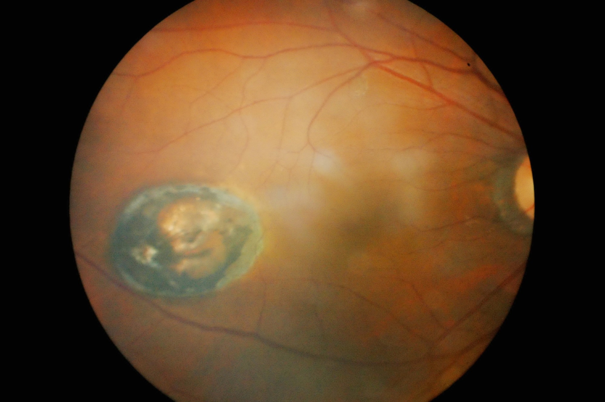
Ten random plaque samples with both polyvinyl chloride and polyethylene were analyzed using EM. Transmission EM (TEM) revealed particles (foreign origin) smaller than one μm with jagged edges within foamy macrophages.
Besides, the same slices were observed with scanning EM (SEM), and spectral X-ray maps were generated from particles resembling those observed with TEM.
The maps indicated decreased carbon and oxygen in plaque samples and increased chlorine. Given the probable non-biologic nature of chlorine, this might confirm polyvinyl chloride deposits.
The researchers performed the isotope analysis on 26 random plaque samples as petroleum-derived plastics exhibit lower δ13C values, i.e., the ratio between carbon-13 and carbon-12, than human tissues.
This analysis revealed two distinct patient clusters. One cluster included patients with higher δ13C values; the other cluster showed lower values, perhaps due to MNP contamination. Lower values were more evident in plaques with MNPs.
The primary endpoint event occurred in 30 and eight patients with and without evidence of MNPs, respectively. Patients with MNPs in plaques had a higher risk of the primary endpoint events than those without MNPs.
Conclusions
In patients with high-grade asymptomatic carotid stenosis indicated for carotid endarterectomy, those with MNPs in plaques had a higher incidence of the composite endpoint than those without MNPs.
Notably, the results do not prove causality; the association between MNPs in plaques and the primary endpoint might also entail risks from exposure to unmeasured, residual, or other confounding variables.
[ad_2]
Source link