Tag: bacteria

  • Genetically engineering skin bacteria to fight cancer

    Genetically engineering skin bacteria to fight cancer

    [ad_1]

    While studying a type of bacteria that lives on the healthy skin of every human being, researchers from Stanford Medicine and a colleague may have stumbled on a powerful new way to fight cancer.

    After genetically engineering the bacteria, called Staphylococcus epidermidis, to produce a tumor antigen (a protein unique to the tumor that’s capable of stimulating the immune system), they applied the live bacteria onto the fur of mice with cancer. The resulting immune response was strong enough to kill even an aggressive type of metastatic skin cancer, without causing inflammation.

    It seemed almost like magic. These mice had very aggressive tumors growing on their flank, and we gave them a gentle treatment where we simply took a swab of bacteria and rubbed it on the fur of their heads.”


    Michael Fischbach, PhD, associate professor of bioengineering

    Their research was published online April 13 in Science. Fischbach is the senior author, and Yiyin Erin Chen, MD, PhD, a former postdoctoral scholar at Stanford Medicine, now an assistant professor of biology at the Massachusetts Institute of Technology, is the lead author.

    Skin colonizers

    Millions of bacteria, fungi and viruses live on the surface of healthy skin. These friendly colonists play a crucial role in maintaining the skin barrier and preventing infection, but there are many unknowns about how the skin microbiota interacts with the host immune system. For instance, unique among colonizing bacteria, staph epidermidis triggers the production of potent immune cells called CD8 T cells — the “killer” cells responsible for battling severe infections or cancer.

    The researchers showed that by inserting a tumor antigen into staph epidermidis, they could trick the mouse’s immune system into producing CD8 T cells targeting the chosen antigen. These cells traveled throughout the mice and rapidly proliferated when they encountered a matching tumor, drastically slowing tumor growth or extinguishing the tumors altogether.

    “Watching those tumors disappear — especially at a site distant from where we applied the bacteria — was shocking,” Fischbach said. “It took us a while to believe it was happening.”

    The mystery of the T cells that do nothing

    Fischbach and his team didn’t start out trying to fight cancer. They wanted to answer a much more basic question: Why would a host organism waste energy making T cells designed to attack helpful colonizing bacteria? Especially as these T cells are “antigen-specific,” meaning each T cell has a homing receptor that matches a single fragment of the bacterium that activated it.

    Even stranger, the CD8 T cells induced by naturally occurring staph epidermidis don’t cause inflammation; in fact, they appear to do nothing at all. Most scientists thought colonist-induced T cells must be fundamentally different from regular T cells, Fischbach said, because instead of traveling throughout the body to hunt for their target, they seemed to stay right below the skin surface, somehow programmed to keep the peace between bacteria and host.

    To test whether these colonist-induced CD8 T cells could behave like regular killer T cells, the researchers engineered a strain of staph epidermidis to produce a different antigen — one that would generate T cells specific for a commonly studied tumor model in mice.

    They genetically grafted a small fragment of DNA encoding part of a protein called ovalbumin onto the surface of staph epidermidis. They chose ovalbumin because it’s been engineered into many commonly studied mouse tumor lines, including a type of aggressive melanoma, and therefore can act as a tumor antigen in multiple types of cancer. 

    The power of tumor-specific T cells

    Next, the scientists applied the genetically engineered bacteria to healthy mice. Because staph epidermidis is an efficient skin colonizer, they didn’t need to clean or shave the animals’ fur, but simply rubbed the bacteria on their heads. As expected, colonization didn’t cause any inflammation or infection.

    Source:

    Journal reference:

    Chen, Y. E., et al. (2024) Engineered skin bacteria induce antitumor T cell responses against melanoma. Science. doi.org/10.1126/science.abp9563.

    [ad_2]

    Source link

  • Antibiotics ineffective for cough treatment in lower respiratory tract infections

    Antibiotics ineffective for cough treatment in lower respiratory tract infections

    [ad_1]

    Use of antibiotics provided no measurable impact on the severity or duration of coughs even if a bacterial infection was present, finds a large, prospective study of people who sought treatment in U.S. primary or urgent care settings for lower-respiratory tract infections.

    The study by researchers at Georgetown University Medical Center and colleagues appeared April 15, 2024, in the Journal of General Internal Medicine.

    Upper respiratory tract infections usually include the common cold, sore throat, sinus infections and ear infections and have well established ways to determine if antibiotics should be given. Lower respiratory tract infections tend to have the potential to be more dangerous, since about 3% to 5% of these patients have pneumonia. But not everyone has easy access at an initial visit to an X-ray, which may be the reason clinicians still give antibiotics without any other evidence of a bacterial infection. Plus, patients have come to expect antibiotics for a cough, even if it doesn’t help. Basic symptom-relieving medications plus time brings a resolution to most people’s infections.”


    Dan Merenstein, MD, professor of family medicine at Georgetown University School of Medicine

    The antibiotics prescribed in this study for lower tract infections were all appropriate, commonly used antibiotics to treat bacterial infections. But the researchers’ analysis showed that of the 29% of people given an antibiotic during their initial medical visit, there was no effect on the duration or overall severity of cough compared to those who didn’t receive an antibiotic.

    “Physicians know, but probably overestimate, the percentage of lower tract infections that are bacterial; they also likely overestimate their ability to distinguish viral from bacterial infections,” says Mark H. Ebell, MD, MS, a study author and professor in the College of Public Health at the University of Georgia. “In our analysis, 29% of people were prescribed an antibiotic while only 7% were given an antiviral. But most patients do not need antivirals as there exist only two respiratory viruses where we have medications to treat them: influenza and SARS-COV-2. There are none for all of the other viruses.”

    To determine if there was an actual bacterial or viral infection present, beyond the self-reported symptoms of a cough, the investigators confirmed the presence of pathogens with advanced lab tests to look for microbiologic results classified as only bacteria, only viruses, both virus and bacteria, or no organism detected. Very importantly, for those with a confirmed bacterial infection, the length of time until illness resolution was the same for those receiving an antibiotic versus those not receiving one – about 17 days.

    Overuse of antibiotics can result in dizziness, nausea, diarrhea, and rash along with about a 4% chance of serious adverse effects including anaphylaxis, which is a severe, life-threatening allergic reaction; Stevens-Johnson syndrome, a rare, serious disorder of the skin and mucous membranes; and Clostridioides difficile-associated diarrhea. Another significant concern of the overuse of antibiotics is resistance. The World Health Organization released a statement on April 4, 2024, stating: “Uncontrolled antimicrobial resistance [due to the overuse of antibiotics] is expected to lower life expectancy and lead to unprecedented health expenditure and economic losses.”

    “We know that cough can be an indicator of a serious problem. It is the most common illness-related reason for an ambulatory care visit, accounting for nearly 3 million outpatient visits and more than 4 million emergency department visits annually,” says Merenstein. “Serious cough symptoms and how to treat them properly needs to be studied more, perhaps in a randomized clinical trial as this study was observational and there haven’t been any randomized trials looking at this issue since about 2012.”

    In addition to Merenstein and Ebell, the other co-author is Bruce Barrett MD, PhD at the University of Wisconsin, Madison,

    This work was supported by an AHRQ grant R01HS025584.

    Source:

    Journal reference:

    Merenstein, D.J., et al. (2024) Antibiotics Not Associated with Shorter Duration or Reduced Severity of Acute Lower Respiratory Tract Infection. Journal of General Internal Medicine. doi.org/10.1007/s11606-024-08758-y.

    [ad_2]

    Source link

  • Nigeria first to rollout new Men5CV vaccine against meningitis

    Nigeria first to rollout new Men5CV vaccine against meningitis

    [ad_1]

    In a historic move, Nigeria has become the first country in the world to roll out a new vaccine (called Men5CV) recommended by the World Health Organization (WHO), which protects people against five strains of the meningococcus bacteria. The vaccine and emergency vaccination activities are funded by Gavi, the Vaccine Alliance, which funds the global meningitis vaccine stockpile, and supports lower-income countries with routine vaccination against meningitis. 

    Nigeria is one of the 26 meningitis hyper-endemic countries of Africa, situated in the area known as the African Meningitis Belt. Last year, there was a 50% jump in annual meningitis cases reported across Africa.

    In Nigeria, an outbreak of Neisseria meningitidis (meningococcus) serogroup C outbreak led to 1742 suspected meningitis cases, including 101 confirmed cases and 153 deaths in seven of 36 Nigerian states (Adamawa, Bauchi, Gombe, Jigawa, Katsina, Yobe, Zamfara) between 1 October 2023 and 11 March 2024. To quell the deadly outbreak, a vaccination campaign has been undertaken on 25–28 March 2024 to initially reach more than one million people aged 1-29 years.

    Meningitis is a serious infection that leads to the inflammation of the membranes (meninges) that surround and protect the brain and spinal cord. There are multiple causes of meningitis, including viral, bacterial, fungal and parasitic pathogens. Symptoms often include headache, fever and stiff neck. Bacterial meningitis is the most serious, can also result in septicaemia (blood poisoning), and can seriously disable or kill within 24 hours those that contract it.

    Meningitis is an old and deadly foe, but this new vaccine holds the potential to change the trajectory of the disease, preventing future outbreaks and saving many lives. Nigeria’s rollout brings us one step closer to our goal to eliminate meningitis by 2030.”


     Dr Tedros Adhanom Ghebreyesus, WHO Director-General

    The revolutionary new vaccine offers a powerful shield against the five major strains of the meningococcal bacteria (A, C, W, Y and X) in a single shot. All five strains cause meningitis and blood poisoning. This provides broader protection than the current vaccine used in much of Africa, which is only effective against the A strain.

    The new vaccine has the potential to significantly reduce meningitis cases and advance progress in defeating meningitis. This is especially important for countries like Nigeria where multiple serogroups are prevalent. The new vaccine uses the same technology as the meningitis A conjugate vaccine (MenAfriVac®), which wiped out meningococcal A epidemics in Nigeria.

    “Northern Nigeria, particularly the states of Jigawa, Bauchi and Yobe were badly hit by the deadly outbreak of meningitis, and this vaccine provides health workers with a new tool to both stop this outbreak but also put the country on a path to elimination,” said Prof. Muhammad Ali Pate of the Nigerian Ministry of Health and Social Welfare. “We’ve done a lot of work preparing health workers and the health system for the rollout of this new vaccine. We got an invaluable support from our populations despite this fasting period and from our community leaders especially the Emir of Gumel in Jigawa state who personally launched the vaccination campaign in the state. We’ll be monitoring progress closely and hopefully expanding the immunization in the coming months and years to accelerate progress.”

    This new multivalent conjugate vaccine was 13 years in the making and was based on a partnership between PATH and the Serum Institute of India. Financing from the UK government’s Foreign, Commonwealth and Development Office was critical to its development.

    In July 2023, WHO prequalified the new Men5CV vaccine (which has brand name MenFive®) and in October 2023 issued an official recommendation to countries to introduce the new vaccine. Gavi allocated resources for the Men5CV rollout in December 2023, which is currently available for outbreak response through the emergency stockpile managed by the International Coordinating Group (ICG) on Vaccine Provision, while roll-out through mass preventive campaigns is expected to start in 2025 across countries of the Meningitis Belt.

    “The rollout of one million vaccines in northern Nigeria will help save lives, prevent long-term illness and boost our goal of defeating meningitis globally by 2030,” said Andrew Mitchell, UK Minister for Development and Africa. “This is exactly the kind of scientific innovation, supported by the UK, which I hope is replicated in years to come to help us drive further breakthroughs, including wiping out other diseases.”

    WHO has been supporting the Nigeria Centre for Disease Control and Prevention (NCDC) in responding to the meningitis outbreak in the country. This includes disease surveillance, active case finding, sample testing, and case management. WHO and partners have also played a vital role in supporting Nigeria to prepare for the rollout of the new vaccine and training health workers.

    “Year after year, meningococcal meningitis has tormented countries across Africa,” said Dr Nanthalile Mugala, PATH’s Chief of Africa Region. “The introduction of MenFive® in Nigeria heralds a transformative era in the fight against meningococcal meningitis in Africa. Building on the legacy of previous vaccination efforts, this milestone reflects over a decade of unwavering, innovative partnerships. The promise of MenFive® lies not just in its immediate impact but in the countless lives it stands to protect in the years to come, moving us closer to a future free from the threat of this disease.”

    In 2019, WHO and partners launched theglobal roadmap to defeating meningitis by 2030. The roadmap sets a comprehensive vision towards a world free of meningitis, and has three goals:

    – elimination of bacterial meningitis epidemics;
    – reduction of cases of vaccine-preventable bacterial meningitis by 50% and deaths by 70%; and

    – reduction of disability and improvement of quality of life after meningitis due to any cause.

    “With outbreaks of infectious diseases on the rise worldwide, new innovations such as MenFive® are critical in helping us fight back,” said Aurélia Nguyen, Chief Programme Officer at Gavi, the Vaccine Alliance, which funds the global stockpile as well as vaccine rollout in lower-income countries. “This first shipment signals the start of Gavi support for a multivalent meningococcal conjugate vaccine (MMCV) program, which, with the required donor funding for our next five years of work, will see pentavalent meningococcal conjugate vaccines rolled out in high-risk countries. Thanks to vaccines, we have eliminated large and disruptive outbreaks of meningitis A in Africa: now we have a tool to respond to other serogroups that still cause large outbreaks resulting in long-term disability and deaths.” 

    Following Nigeria’s meningitis vaccine campaign, a major milestone on the road to defeat meningitis is the international summit on meningitis taking place in Paris in April 2024 where leaders will come together to celebrate progress, identify challenges and assess next steps. It is also an opportunity for country leaders and key partners to commit politically and financially to accelerate progress towards eliminating meningitis as a public health problem by 2030. 

    [ad_2]

    Source link

  • Study aims to explore the underlying causes of excessive alcohol production in overweight people

    Study aims to explore the underlying causes of excessive alcohol production in overweight people

    [ad_1]

    The microorganisms in the intestines of many overweight people produce alcohol to an increased extent, as Max Nieuwdorp, professor of Internal Medicine at Amsterdam UMC discovered a few years ago. Breaking down that excessive alcohol leads to fatty liver, which in turn increases the risk of serious diseases such as diabetes and cardiovascular disease. Nieuwdorp has now received an ERC Advanced grant of 2.5 million euros for a major study into the underlying causes of excessive alcohol production. Ultimately, he hopes to find a way to prevent excess alcohol produced in the intestines, and thus the related diseases. In 2022, Nieuwdorp and his team published a study in Nature Medicine on alcohol production in the intestines of overweight patients. “Our findings showed that the turnover of sugars in the intestines of these patients releases far too much alcohol, equivalent to almost half a litre of whisky of alcohol. This is because the composition of the microbiome in their small and large intestines is disrupted. It seems that a change in acidity plays a role in this,” Nieuwdorp explains. 

    Liver has to work hard

    For patients, large quantities of alcohol in the intestines can have major consequences. “The liver, as with alcohol from liquor, has to work hard to breakdown the alcohol, and that is done by storing it as fat. This causes people to develop a fatty liver disease that can eventually become inflamed and lead to serious conditions such as cirrhosis of the liver and cardiovascular disease,” says Nieuwdorp.  

    Almost 1 in 5 adults in the Netherlands are overweight and more than 80% of them have fatty liver. Nieuwdorp suspects that the high quantities of sugar in our modern diet can lead to increased alcohol production in the intestines. With the European money from the ERC Advanced grant, he will investigate this further, for example by analyzing the medical data and eating patterns of participants in the long-term HELIUS study.  

    Bacteria in the gut  

    Nieuwdorp hopes that the discovery of the increased alcohol production due to the disrupted microbiome in the intestines will create a new path in the search for a way to treat fatty liver disease and liver inflammation. For example, he wants to see if it is possible to control alcohol production in the intestines by equipping bacteria in the intestine with properties that allow them to breakdown more alcohol.

    But whether and how that actually works is still unknown. That’s what we’re going to investigate in this FATGAP-project.”

    Max Nieuwdorp, Professor of Internal Medicine at Amsterdam UMC

    [ad_2]

    Source link

  • Hospital sinks fuel antibiotic-resistant bacteria spread

    Hospital sinks fuel antibiotic-resistant bacteria spread

    [ad_1]

    A new study published today in the American Journal of Infection Control (AJIC) reports the infection prevention steps taken to control a months-long multispecies outbreak of carbapenemase-producing Enterobacterales that occurred in a pediatric ward at the Toho University Omori Medical Center in Tokyo in 2017. This study highlights the particular vulnerability for contamination through sinks and other water sources; indeed, even replacing all sinks in the ward did not stop this outbreak.

    Carbapenemase-producing Enterobacterales (CPE) are a major public health threat because of their resistance to widely used antibiotics. The biological mechanism that confers multidrug resistance can be passed from one bacterial species to another, contributing to the growing epidemic of antimicrobial resistance. An outbreak of one CPE species in a hospital has the potential to turn into an outbreak involving many species, making it that much more difficult to stop. Many studies have shown that there is a high risk of CPE contamination in and around hospital sinks.

    This new report from an academic medical center in Tokyo details the detection of CPE in a single patient in June 2016, which appears to have triggered an outbreak starting in March 2017 and ending in October 2017. The outbreak involved a total of 19 pediatric patients. The infection prevention team sampled microbes from patients and the environment of the pediatric ward to better understand how the outbreak was spreading. This sampling identified nine sinks contaminated with CPE, including six in hospital rooms and three more in a nurse center, a waste room, and an ice machine. The CPE-positive sinks were all found in rooms where CPE-positive patients had been treated. In rooms with CPE-negative patients, no sink contamination was detected.

    As part of the outbreak control process, genome analysis was performed to identify the specific resistance mechanisms found in the bacterial strains, which included Klebsiella variicola, Klebsiella quasipneumoniae, and Escherichia coli, among others. Identical DNA sequences from all samples but one support the idea that the resistance mechanism could have been passed from one bacterial species to another within the hospital.

    To help rein in the outbreak, all sinks in the pediatric ward were replaced with new ones in June 2017, and the new sinks were thoroughly disinfected with hydrogen peroxide. However, CPE contamination continued even after that step. The discovery of the same bacterial species in sinks in adjoining rooms indicates that pathogen transmission may be possible from one sink to another via the drains and connected plumbing.

    Other measures implemented by the infection prevention team -; composed of doctors, nurses, pharmacists, and microbiologists -; included recommending hand disinfection after using sinks, introducing disposable tools for cleaning sinks, prohibiting mouth-washing with sink water, enacting disinfection and drying procedures to any items exposed to sink water, and more. Finally, after October 2017, no further CPE contamination was identified in patient samples or environmental surveillance.

    After months of intense infection control protocols, we were at last able to declare an end to this outbreak. Our experience highlights the importance of focusing on sinks and other water-related areas in hospital wards, as these are critical for CPE transmission and therefore major fronts in the fight against antibiotic resistance.”

    Sadako Yoshizawa, MD, PhD, Associate Professor in the Department of Microbiology and Infectious Diseases at Toho University School of Medicine, Deputy Director of Clinical Laboratory at Toho University Omori Medical Center, and corresponding author of the study

    Additional details from the study include:

    • Toho University Omori Medical Center is a 916-bed academic medical center with 55 pediatric inpatient beds.
    • The first patient to be detected with CPE in June 2016 was a one-year-old boy hospitalized with cardiac disease.
    • The resistance mechanism detected in this outbreak was a plasmid enabling the production of carbapenemase, which can make bacteria resistant to the carbapenem class of antibiotics. In this outbreak, all CPE strains except one harbored blaIMP-1, with identical blaIMP-1-carrying IncM1 plasmids.

    The tremendous effort that went into controlling this outbreak is representative of the comprehensive and holistic approach to infection prevention that is required in these situations. Even a measure as definitive as replacing contaminated sinks may not be enough to stop the spread of antibiotic-resistant organisms. This outbreak ended because the infection prevention team implemented a bundle of core processes related to hand hygiene, and the use and disinfection of hospital sinks.”


    Tania Bubb, PhD, RN, CIC, FAPIC, 2024 APIC president

    Source:

    Journal reference:

    Tsukada, M., et al. (2024) The outbreak of multispecies carbapenemase-producing Enterobacterales associated with pediatric ward sinks: IncM1 plasmids act as vehicles for cross-species transmission. American Journal of Infection Control. doi.org/10.1016/j.ajic.2024.02.013.

    [ad_2]

    Source link

  • Blocking polyphosphates could help treat chronic infections

    Blocking polyphosphates could help treat chronic infections

    [ad_1]

    Most disease-causing bacteria are known for their speed: In mere minutes, they can double their population, quickly making a person sick. But just as dangerous as this rapid growth can be a bacterium’s resting state, which helps the pathogen evade antibiotics and contributes to severe chronic infections in the lungs and blood, within wounds, and on the surfaces of medical devices.

    Now, Scripps Research scientists have discovered how long chains of molecules called polyphosphates (polyP) are needed for bacteria to slow down movements within cells and let them enter this resting state. The findings, published in Proceedings of the National Academy of Sciences on April 02, 2024, could eventually lead to new ways of treating chronic infections in which typical antibiotics aren’t effective.

    Many current antibiotics block bacterial growth, but bacteria spend a lot of their time not growing. We really need new and creative strategies for targeting bacteria’s slow-growing and non-growing phases.”


    Lisa Racki, assistant professor in the Department of Integrative Structural and Computational Biology at Scripps Research and senior author of the new paper

    Researchers have long known that bacteria can survive for especially long periods of time when they stop growing, entering a dormant and energy-saving state. They also knew that when bacteria enter this resting state, they use valuable energy to produce polyP strands, which form large clumps inside their cells. But scientists had been historically unsure about the purpose of polyP.

    To study polyP, Racki and her collaborators turned to Pseudomonas aeruginosa, bacteria that can cause pneumonia and blood infections in people who are hospitalized or have weakened immune systems. One of the reasons P. aeruginosa can be so hard to treat is that it forms biofilms-;tightly joined, slimy communities of bacteria, many of which are in a resting state and can evade typical antibiotics.

    When P. aeruginosa is starved of nitrogen-;one of the key nutrients it needs for growth-;it produces lots of polyP. In the new work, Racki and her collaborators at EPFL and Caltech discovered that a mutant unable to make polyP cannot enter its resting state. To better understand why this happens and the consequences, the researchers genetically engineered P. aeruginosa to make small, labeled particles that let them track how molecules within the bacteria were moving around.

    “What we found is that when you get rid of polyP, everything in the cell moves too much,” says Racki. “The cells are partying when they should be taking a break.”

    When starved of most nutrients, P. aeruginosa slows the movement of materials within its interior and stops dividing. But without nitrogen and polyP, the bacteria keep moving materials around at top-speed, become bigger, loosen their genetic material and continue dividing.

    Racki’s team concluded that polyP is usually responsible for helping P. aeruginosa-;and likely other bacterial species-;slow down. It also leads them to hypothesize that preventing cells from producing polyP could keep them active and make them more susceptible to some antibiotics.

    “This not only helps point in possible directions for treating pathogenic bacteria, but also reveals answers for fundamental questions about how things diffuse throughout a bacterial cell,” says Racki.

    Racki and her lab are now planning more experiments to better probe exactly why cells cannot slow their interior movements without polyP, and whether blocking the bacterial production of polyP could be an effective tactic to treat some infections.

    Source:

    Journal reference:

    Magkiriadou, S., et al. (2024). Polyphosphate affects cytoplasmic and chromosomal dynamics in nitrogen-starved Pseudomonas aeruginosaProceedings of the National Academy of Sciences. doi.org/10.1073/pnas.2313004121.

    [ad_2]

    Source link

  • Rising antibiotic resistance prompts shift to ecological research strategies in infection control

    Rising antibiotic resistance prompts shift to ecological research strategies in infection control

    [ad_1]

    In a recent article published in The Lancet Microbe, researchers discussed the need to enhance research on the interactions between bacterial pathogens and commensals within human microbiomes and host biology to develop innovative infection prevention and treatment strategies.

    Study: Integrating research on bacterial pathogens and commensals to fight infections—an ecological perspective. Image Credit: TopMicrobialStock/Shutterstock.comStudy: Integrating research on bacterial pathogens and commensals to fight infections—an ecological perspective. Image Credit: TopMicrobialStock/Shutterstock.com

    Background

    The growing appreciation of the human microbiome’s role in health drives innovative disease prevention and treatment approaches.

    This includes using microbiome signatures as biomarkers for diagnosing various diseases and exploring interventions like fecal microbiota transplantation.

    Historically, research focused more on pathogens, but now, understanding the dynamic roles of pathogens and commensals is essential. Interdisciplinary collaboration offers new insights into the ecological factors driving bacterial behavior and the development of preventive and therapeutic strategies for infections.

    This convergence of microbiome science and innovative approaches holds great potential for promoting human health.

    Future research on commensal bacteria inspired by that on pathogens

    Research on commensal bacteria is gaining momentum, inspired by the extensive study of major bacterial pathogens like Shigella flexneri, Staphylococcus aureus, and Streptococcus pneumoniae.

    While pathogens have been extensively studied, commensals, particularly those from genera like Bacteroides, Clostridium, and Cutibacterium, have received less attention despite their potential clinical significance.

    Evidence suggests that certain commensals play crucial roles in human health, producing compounds that inhibit pathogens and those that directly benefit the host. Understanding the host-specificity and biology of these beneficial commensals could offer insights into combating bacterial infections.

    Additionally, exploring the mechanisms behind commensal-host interactions, such as adhesion to epithelial surfaces and modulation of mucosal immunity, could provide further avenues for research.

    Expanding our understanding of fitness mechanisms of facultative human pathogens

    Research on facultative human pathogens, which typically exist as commensals but can cause disease under certain conditions, is gaining importance, considering the global rise in antimicrobial resistance.

    While much attention has been given to understanding the virulence mechanisms of major pathogens like S. aureus and S. pneumoniae, the factors influencing the fitness of these organisms during their commensal phase have been largely overlooked.

    This oversight is particularly concerning given the increasing prevalence of antibiotic-resistant pathogens, including those grouped as ESKAPE pathogens (Enterococcus faecium, Staphylococcus aureus, Klebsiella pneumoniae, Acinetobacter baumannii, Pseudomonas aeruginosa, and Enterobacter spp).

    Understanding why certain antibiotic-resistant clones spread more effectively than others is crucial for developing effective control strategies.

    Applying ecological concepts to studying pathogenic bacteria could shed light on their success in competing with other microbiome members. Factors such as nutrient utilization, resistance to antimicrobial molecules, and adhesion to host epithelial surfaces play important roles in determining a bacterial species’ long-term success within a host and its ability to spread to new hosts.

    Investigating the mechanisms underlying vertical and lateral transmission of bacteria is also crucial, as it can provide insights into the dynamics of microbiome colonization and infection spread.

    The severity and type of infection caused by pathogens can influence their spread among hosts. For example, pathogens like S. flexneri, causing diarrheal diseases, may spread through contaminated sewage, while those like S. aureus, causing skin infections, may spread through direct contact between individuals.

    Accidental pathogens, which typically lack aggressive toxins but possess immune-evasion mechanisms, may persist in human microbiomes more readily than professional pathogens.

    However, professional pathogens can also cause opportunistic infections, particularly in immunocompromised individuals.

    Integrative understanding of fitness of bacterial commensals and pathogens can help with infection prevention

    Understanding commensal and pathogenic bacteria within their natural habitats can provide valuable insights for controlling infections and addressing microbiome-related disorders.

    Commensal bacteria exhibit complex effects on infectious diseases, employing various defense strategies such as antimicrobial production and metabolic interference to inhibit pathogen colonization.

    Microbiome restoration through fecal microbiota transplantation has shown promise in treating infections, but standardized approaches are needed. Utilizing defined commensal communities for microbiome editing could offer more consistent results and personalized treatment options.

    However, antibiotic treatment, primarily with broad-spectrum antibiotics, can disrupt the microbiome and fuel antibiotic resistance.

    Commensals play a crucial role in resistance gene transfer to pathogens, highlighting the need to monitor key commensal species alongside major pathogens.

    Ecological approaches to infection research can help identify vulnerabilities in pathogens, leading to the development of targeted anti-fitness drugs and anti-virulence compounds. Understanding the interplay between pathogens and mucosal immunity is essential for developing mucosal vaccines against potentially pathogenic microbiome members.

    Conclusion

    In conclusion, understanding the interaction between commensal and pathogenic bacteria within their ecological contexts offers promising avenues for infection prevention and treatment.

    By leveraging the protective mechanisms of commensals and targeting pathogen virulence, tailored interventions can enhance host defenses and suppress pathogen proliferation.

    Moreover, the development of mucosal vaccines and microbiome-based therapies offers new strategies for infection control. Integrating ecological principles into infection research provides a holistic framework for combating infectious diseases and addressing antimicrobial resistance, ultimately optimizing human health.

    [ad_2]

    Source link

  • Study reveals key gut microbiome differences in prediabetic patients

    Study reveals key gut microbiome differences in prediabetic patients

    [ad_1]

    A recent Nutrients study compares the composition of the gut microbiome in prediabetic patients and healthy individuals.

    Study: Gut Microbiota in Patients with Prediabetes. Image Credit: Sebastian Kaulitzki / Shutterstock.com

    What is prediabetes?

    Prediabetes is defined as glycated hemoglobin levels between 5.7-6.5% and fasting blood glucose levels between 100-126 mg/dL. Prediabetes significantly increases the mortality risks and burden on the healthcare system; therefore, several interventions have been designed to maintain healthy glucose levels.

    For example, lifestyle changes, bariatric surgery, and medications are commonly used to prevent the progression of prediabetes to diabetes. Despite these interventions, a recent American Diabetes Association report indicated that most prediabetic patients eventually develop diabetes. Therefore, it is imperative to develop more effective interventions to prevent or reverse the prediabetic condition.

    The gut microbiome and diabetes

    The gut microbiome plays a vital role in regulating lipid and glucose metabolism, as gut microbial dysbiosis leads to the development of many diseases. For example, intestinal bacterial composition and abundance changes influence intestinal permeability, which induces insulin resistance and the introduction of bacterial lipopolysaccharides into the bloodstream.

    Gut microbial dysbiosis is directly associated with increased gut permeability, which promotes low-grade systemic inflammation. This condition is a key contributor to metabolic syndrome and various chronic diseases, such as type 2 diabetes.

    Considering these observations, it is important to understand the mechanism by which intestinal bacteria alterations influence the development of diabetes. This information could ultimately support the development of novel approaches to prevent the progression of prediabetes to diabetes.

    Diet, drugs, and age play an important role in altering the gut microbiota. Although several studies have established differential intestinal bacteria between diabetic and healthy individuals, few studies have assessed intestinal bacteria composition in prediabetic patients and its impact on the physiological mechanisms of this health condition.

    About the study

    The current study compared gut microbial composition between prediabetic patients and healthy individuals. The impact of diet on the gut microbiome of prediabetic patients was also assessed in an effort to identify nutrition-based interventions that could potentially prevent the progression of prediabetes to diabetes.

    A total of 57 study participants were recruited from Taipei Tzu-Chi Hospital in Taiwan. Gut microbiota data of 60 healthy individuals between 18 and 65 years were obtained from biobanks and used as the reference group.

    Study participants were advised to keep a three-day food record and collect fecal samples on the third day. The nutrient composition of the participants’ diet was measured using Nutritionist Professional software 2.0.

    Study findings

    The composition, diversity, and abundance of the gut microbiota were significantly reduced in prediabetic patients compared to healthy individuals. This finding was in line with previous studies that indicated differential microbial composition in patients with diabetes. Prediabetic patients also exhibited a higher body mass index (BMI) than controls.

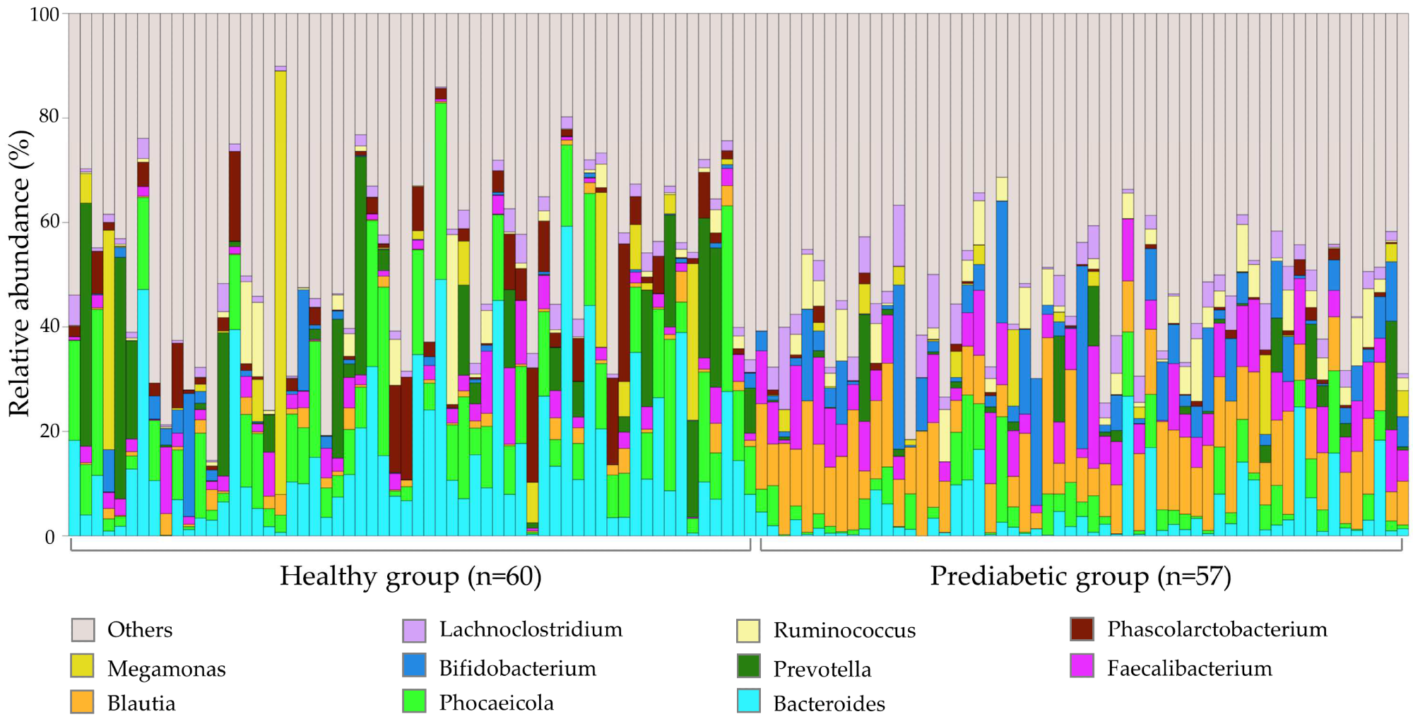
    Gut microbiome profile of 117 fecal samples at the genus level. The remaining bacterial genera are summed as ‘Others’.Gut microbiome profile of 117 fecal samples at the genus level. The remaining bacterial genera are summed as ‘Others’.

    Consistent with previous studies on type 2 diabetes, the current study reported higher levels of MediterraneibacterBifidobacteriumBlautiaAnaerostipes, Clostridium, and Butyricicoccus in the fecal samples of healthy individuals than prediabetic patients. 

    Previous studies have shown that butyrate maintains the integrity of the intestinal mucosa. This metabolite is synthesized by gut bacteria, namely, Anaerostipes and Faecalibacterium.

    Maintaining the integrity of intestinal mucosa can prevent the invasion of pathogenic bacteria in the blood and the destruction of pancreatic β-cells. This finding indicates the indirect role of Anaerostipes and Faecalibacterium in regulating blood glucose levels.

    In contrast to prediabetic samples, healthy fecal samples exhibited high levels of Eggerthella and Streptococcus. However, a higher abundance of PhascolarctobacteriumBacteroidesParaprevotella, and Parabacteroides was observed in prediabetic fecal samples.

    Prediabetic patients exhibited multiple altered physiological metabolic pathways, which affect insulin transmembrane signaling and overexpression of retinoic acid-inducible gene I (RIG-I). This metabolic dysfunction triggers immune cells to attack β cells, affecting blood glucose levels. Previous studies have also indicated that abnormal sphingolipid metabolism leads to insulin resistance and neuronal apoptosis.

    Diet plays a vital role in maintaining gut bacterial diversity and abundance. Therefore, prediabetic patients are advised to consume a low carbohydrate (LC) diet with a higher dietary fiber intake. This combination could improve intestinal barrier integrity, thereby preventing the progression of prediabetes to diabetes.

    Conclusions

    Differential gut microbial composition and abundance were observed in prediabetic patients compared to healthy controls. This difference is also associated with altered metabolic and physiological responses. These findings suggest that improving the gut microbiome could prevent the onset of diabetes by maintaining normal physiological metabolism.

    Journal reference:

    • Chang, W., Chen, Y., Tseng, H., et al. (2024). Gut Microbiota in Patients with Prediabetes. Nutrients 16(8); 1105. doi:10.3390/nu16081105

    [ad_2]

    Source link

  • Gut bacteria play a pivotal role in obesity’s impact on body fat metabolism

    Gut bacteria play a pivotal role in obesity’s impact on body fat metabolism

    [ad_1]

    In a recent review published in the journal Nutrients, researchers explored how the dysregulation of gut microbiota in obesity impacts adipose tissue (AT) metabolism through direct and indirect effects on the mitochondria within white (WAT) and brown adipose tissue (BAT).

    Study: The Crosstalk between Gut Microbiota and White Adipose Tissue Mitochondria in Obesity. Image Credit: KateStudio / ShutterstockStudy: The Crosstalk between Gut Microbiota and White Adipose Tissue Mitochondria in Obesity. Image Credit: KateStudio / Shutterstock

    Background 

    Obesity, affecting 13% of the global population as of 2016, has reached epidemic levels, challenging both developed and developing nations. By 2039, it is projected that more than 30% of adults in Europe and even more in the United States of America (USA) will be obese. This condition arises from a complex interplay of genetic, lifestyle, and environmental factors, leading to excessive energy storage in AT. This storage exceeds the tissue’s capacity for oxygenation, causing inflammation, insulin resistance, and increased cardiometabolic and cancer risks. Despite extensive study, the role of cellular and mitochondrial metabolism in obesity, especially the influence of gut microbiota on AT, needs clearer understanding. Identifying how gut microbiota affects AT mitochondria could lay the groundwork for novel obesity treatments, highlighting the need for further research.

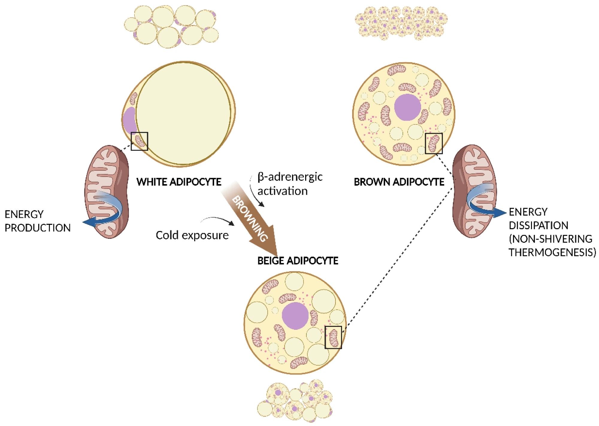
    WAT, BAT, and WAT browning. White adipocyte has one large droplet in the centre of the cell that compresses nucleus and mitochondria at one pole. Brown adipocyte has multiple small lipid droplets and more mitochondria, spread out between the droplets. Beige adipocyte has intermediate characteristics. Cold exposure and β-adrenergic activation determine the browning of WAT. Both brown and beige mitochondria are involved in non-shivering thermogenesis.  Abbreviations: BAT, brown adipose tissue; WAT, white adipose tissue.WAT, BAT, and WAT browning. White adipocyte has one large droplet in the centre of the cell that compresses nucleus and mitochondria at one pole. Brown adipocyte has multiple small lipid droplets and more mitochondria, spread out between the droplets. Beige adipocyte has intermediate characteristics. Cold exposure and β-adrenergic activation determine the browning of WAT. Both brown and beige mitochondria are involved in non-shivering thermogenesis.  Abbreviations: BAT, brown adipose tissue; WAT, white adipose tissue.

    AT: An active endocrine organ

    AT has transcended its traditional view as a mere energy reservoir and insulator and is now recognized as an active endocrine organ instrumental in metabolic regulation. This shift is attributed to its secretion of hormones like leptin and adiponectin and a variety of cytokines termed adipokines, marking its profound impact on metabolism. Within this tissue, adipocytes and other cell types, such as pre-adipocytes and immune cells, form a complex cellular environment underlying its multifaceted functions.

    Diverse functions of WAT and BAT

    AT, classified into WAT for energy storage and BAT for thermogenic energy expenditure, plays vital roles in metabolic health. WAT’s large cells store fat, contributing to mechanical protection and metabolic regulation, while BAT’s smaller, lipid-rich cells generate heat through non-shivering thermogenesis, offering potential in treating metabolic disorders.

    The metabolic and endocrine role of AT

    Ectopic fat deposition and the activation of ATs highlight the complexity of their roles in health and disease. While ectopic fats are associated with metabolic complications, the process of “browning” in WAT, wherein cells adopt BAT-like characteristics, offers therapeutic prospects for metabolic diseases. The endocrine functions of WAT further elucidate its role in energy and metabolic homeostasis, with adipokines like leptin and adiponectin playing critical roles. BAT’s contribution to energy expenditure through non-shivering thermogenesis represents a fundamental aspect of metabolic health, distinguishing the unique contributions of WAT and BAT to the body’s energy balance and metabolic regulation.

    Mitochondrial function in AT

    Mitochondria play a critical role in energy metabolism within both WAT and BAT, driving adenosine triphosphate (ATP) production through nutrient oxidation and regulating lipid metabolism. In WAT, they support lipid synthesis and breakdown, influencing adipocyte differentiation and metabolic health. Dysfunctional mitochondria in WAT are linked to metabolic diseases due to impaired regulation of adipokines and fatty acid oxidation. Conversely, BAT mitochondria facilitate non-shivering thermogenesis via uncoupling protein-1 (UCP-1), showcasing their essential role in energy expenditure. This distinct mitochondrial function in BAT versus WAT underscores their significance in metabolic regulation and the potential for therapeutic targets in obesity and related conditions.

    Mitochondrial dysfunction and obesity: A close connection

    Obesity’s impact on mitochondrial metabolism in AT, particularly in WAT and BAT, underscores a critical aspect of its pathophysiology. Mitochondrial dysfunction, characterized by altered bioenergetics and impaired lipid and glucose metabolism, plays an important role in intensifying obesity-related metabolic complications. Studies have revealed significant mitochondrial alterations in obesity, including diminished expression of mitochondrial proteins, reduced mitochondrial deoxyribonucleic acid (mtDNA) copy numbers, and decreased activity of oxidative phosphorylation complexes. These changes not only contribute to inefficient energy utilization and storage but also foster a shift towards adipocyte hypertrophy, further promoting inflammation and insulin resistance. The compromised mitochondrial function in WAT affects fatty acid oxidation and adipocyte differentiation, while in BAT, it impairs thermogenic efficiency, potentially shifting its metabolism towards a WAT-like phenotype. This connection between mitochondrial dysfunction and obesity highlights the urgent need for therapeutic strategies aimed at restoring mitochondrial health, offering a promising avenue for obesity management and the reduction of its associated metabolic disorders.

    The role of gut microbiota in obesity and AT mitochondrial function

    The interplay between gut microbiota and AT mitochondria significantly influences obesity management and metabolic health. The diverse gut ecosystem plays a fundamental role in metabolic processes, impacting lipid and glucose metabolism through the production of microbial metabolites like short-chain fatty acids (SCFAs). These metabolites affect mitochondrial function in ATs, modifying fatty acid oxidation and adipocyte differentiation, which are critical in obesity progression. Dysbiosis, characterized by an imbalance in gut microbial composition, has been linked to obesity-related metabolic impairments, suggesting that modulating gut microbiota could offer therapeutic avenues for improving mitochondrial function and combating obesity. This highlights the necessity for continued exploration into the gut microbiota- AT mitochondria axis to develop targeted interventions for obesity and its associated metabolic disorders.

    Journal reference:

    • Colangeli L, Escobar Marcillo DI, Simonelli V, Iorio E, Rinaldi T, Sbraccia P, Fortini P, Guglielmi V. The Crosstalk between Gut Microbiota and White Adipose Tissue Mitochondria in Obesity. Nutrients. (2023). DOI – 10.3390/nu15071723, https://www.mdpi.com/2072-6643/15/7/1723 

    [ad_2]

    Source link

  • Tumor microbiomes offer new insights for enhancing cancer therapies

    Tumor microbiomes offer new insights for enhancing cancer therapies

    [ad_1]

    In a recent study published in the journal Cell, researchers used metagenomics, genomics, and transcriptomics to examine microbiome genomes in over 4,000 metastatic tumor tissues. They analyzed the tumor microbiome and tumor microenvironment (TME), offering biological information and influencing the development of bacteria-focused techniques to supplement and improve cancer treatments.

    Microbial communities play a crucial role in the human body, influencing the immune system and anticancer therapies. They are present in primary tumors and interact with the commensal microbiota. The gut microbiota can modulate immune checkpoint blockers (ICB) and conventional chemotherapies. Fecal microbial transplants may improve clinical responsiveness to ICB agents. Understanding how tumor-resident bacteria shape tumor biology, immune infiltration, and treatment responsiveness is essential for understanding tumor response to ICB.

    Study: A pan-cancer analysis of the microbiome in metastatic cancerStudy: A pan-cancer analysis of the microbiome in metastatic cancer

    About the study

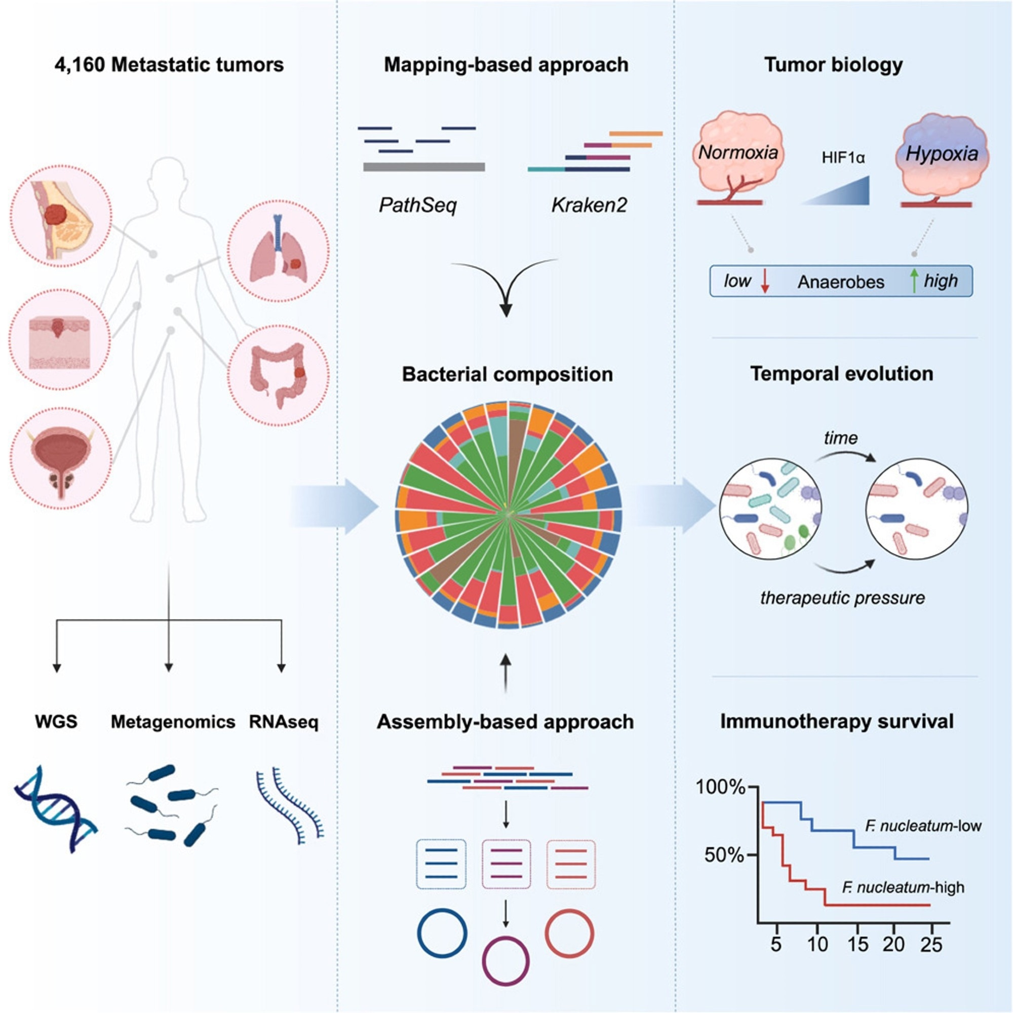
    In the present study, researchers used bioinformatics to investigate the microbiota in metastatic malignancies, evaluating 4,160 specimens from diverse cancer types.

    The researchers used mapping and assembly-based metagenomics, genomes, transcriptomics, and clinical data to develop a pan-cancer repository that might help advance treatment techniques. They used two distinct computational approaches, PathSeq and Kraken2, to define tumor-resident microbiome communities at the genus level and a metagenomic assembly-based approach at the species level. The team then shaped the metastatic tumor microbiome by identifying the elements that influence its makeup and evaluating cancer-type-specific microbial communities. They used the characteristic hypoxia gene profile to assess the degree of hypoxia in metastatic cancers and then performed gene set enrichment analysis (GSEA). They also investigated whether microbial communities may affect host immunity and the TME.

    The researchers investigated the relationship between gram-negative bacteria in metastases and Toll-like receptor (TLR) expression and whether lipopolysaccharide (LPS), obtained from dead or active bacteria, plays a primary role in TLR4 signaling in metastases. They additionally examined the relationship between bacterial makeup and tumor gene expression and the relationships between particular bacteria and immune cells.

    To further understand the impact of metastatic heterogeneity and the durability of tumor-resident microorganisms over time, the team examined 185 pairs of 370 repeated tumor specimens obtained from 173 different individuals. They examined bacterial enrichment changes before and after tumor treatment with immunotherapy, targeted therapy, or hormone therapy. They also investigated bacterial count reductions following immunotherapy in responsive patients and whether these germs were more prevalent in non-responsive individuals before treatment. Lastly, they examined pre-treatment bacterial communities associated with a lack of response to immunosuppressive medication in an ICB-monotherapy cohort of NSCLC patients.

    Results

    The researchers detected tumor-resident bacteria deoxyribonucleic acid (DNA) in a pan-cancer metastasis cohort, and assembling tumor-derived bacterial DNA provided species-level genomic characterization. Bacterial diversity correlated with cellular and molecular tumor immunity characteristics. In an NSCLC cohort, high levels of fusobacterium DNA imply a poor immunotherapy response. Researchers found organ-specific microbe tropisms, anaerobic bacteria enrichments in hypoxic tumors, links between microbial diversity and tumor-infiltrating neutrophils, and Fusobacterium’s relationship with resistance to ICB therapy in lung cancer.

    Using mapping-based techniques and screening genera to eliminate technical contamination and seldom-seen genera, the team cataloged 165 microbial genera from 3,526 specimens, with 68% facultative/anaerobic and 49% gram-negative anaerobes. They built 514 metagenomic-assembled genomes (MAGs) of medium- to almost high-quality using tumor-derived microbial sequences. The most common tumor types were colorectal, breast, prostate, lung, and melanoma, with the lymph node, liver, and lung being the most common metastatic locations for tumor samples.

    The quantity of bacterial-derived reads expressed as a human-mapped genetic read proportion varied by cancer type, with higher fractions in renal and uterine malignancies and lower burdens in tumors originating from the brain and spinal cord. Renal and colorectal metastases were the most diverse, but head and neck metastatic tumors showed more dominant microbial genera.

    Tumor-resident microbial communities were associated with tumor biology, with a strong correlation between LPS load and TLR4 signaling but not gram-positive lipoteichoic acid (LTA) load.  Multivariate Cox proportional-hazards modeling showed lower overall survival (OS) and progression-free survival (PFS) rates significantly correlated with continuous Fusobacterium abundance, considering the genome-wide mutational load. Using the pan-cancer dataset, the researchers classified all tumors as Fuso-high or Fuso-low based on an upper quartile relative abundance cutoff similar to previously established criteria. Fuso-high tumors showed considerably decreased cytotoxic, interferon-gamma (IFN-γ), and major histocompatibility complex (MHC) class II gene expression profiles.

    The study provides the first large-scale pan-cancer map of intratumor microbiomes in metastatic malignancies, examining diversity across anatomical regions, initial tumor type, and treatment responses, including immunotherapy. The study showed that the metastatic microbiome partially comprises anaerobic bacteria that may get altered during treatment. The study also discovered links between intra-tumoral microorganisms and the activation of innate immune sensing pathways, indicating that the tumor microenvironment alters via direct identification of bacterial ligands.

    [ad_2]

    Source link