Tag: Biochemistry

  • Scientists Unveil Groundbreaking Single-Molecule Detection Technique

    Scientists Unveil Groundbreaking Single-Molecule Detection Technique

    [ad_1]

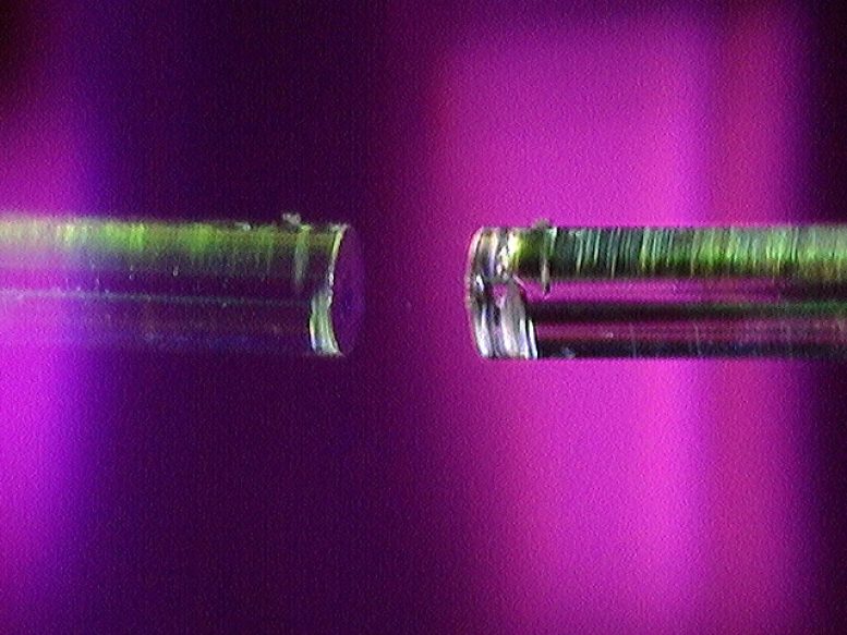
    Microcavity With Two Concave Mirrors

    The heart of this study is a fiber microcavity. Here, one can see a small concave depression in the surface of an optical fiber. The researchers used a microcavity with two concave mirrors, but this image of a single concave microcavity makes it easier to see the fiber mirror setup. Credit: Photo by Carlos Saavedra / UW–Madison

    University of Wisconsin–Madison scientists have developed a new, highly sensitive method to detect and analyze single molecules without using fluorescent labels, potentially transforming research in drug discovery and materials science.

    Researchers at the University of Wisconsin–Madison have developed the most sensitive method yet for detecting and profiling a single molecule — unlocking a new tool that holds potential for better understanding how the building blocks of matter interact with each other. The new method could have implications for pursuits as varied as drug discovery and the development of advanced materials.

    The technical achievement, detailed in a paper published in the journal Nature, marks a significant advance in the burgeoning field of observing individual molecules without the aid of fluorescent labels. While these labels are useful in many applications, they alter molecules in ways that can obscure how they naturally interact with one another. The new label-free method makes the molecules so easy to detect, it is almost as if they had labels.

    “We’re very excited about this,” says Randall Goldsmith, a UW–Madison professor of chemistry who led the work. “Capturing behaviors at the level of single molecules is an amazingly informative way of understanding complex systems, and if you can build new tools that grant better access to that perspective, those tools can be really powerful.”

    While researchers can glean useful information from studying materials and biological systems at larger scales, Goldsmith says that observing the behavior of and interactions between individual molecules is important for contextualizing that information, sometimes leading to new insights.

    “When you see how nations interact with each other, it all comes down to interactions between individuals,” says Goldsmith. “You wouldn’t even think of understanding how groups of people interact with each other while ignoring how individuals interact with each other.”

    The Importance of Single Molecule Observation

    Goldsmith has been chasing the allure of single molecules since he was a postdoctoral researcher at Stanford University more than a decade ago. There, he worked under the chemist W.E. Moerner, who received the Nobel Prize in chemistry in 2014 for developing the first method of using light to observe a single molecule.

    Since Moerner’s initial success, researchers around the world have devised and refined new ways to observe these tiny bits of matter.

    The method that the UW–Madison team developed relies on a device called an optical microresonator, or microcavity. As its name suggests, the microcavity is an extremely tiny space where light can be trapped in both space and time — at least for a few nanoseconds — where it can interact with a molecule. Microcavities are more commonly found in physics or electrical engineering laboratories, not chemistry labs. Goldsmith’s history of combining concepts from disparate scientific fields was recognized in 2022 with a Polymath award from Schmidt Futures.

    Microcavities are built from incredibly small mirrors fashioned right on top of a fiber optic cable. These fiber optic mirrors bounce the light back and forth many times very quickly within the microcavity.

    Potential Applications and Future Developments

    The researchers let molecules tumble into the cavity, let the light pass through it, and can not only detect the molecule’s presence, but also learn information about it, such as how fast it moves through water. This information can be used to determine the molecule’s shape, or conformation.

    “Conformation at the molecular level is incredibly important, particularly for thinking about how biomolecules interact with each other,” says Goldsmith. “Let’s say you have a protein and you have some small-molecule drug. You want to see if the protein’s druggable, which is to say, ‘Does the drug have some kind of major interaction with the protein?’ One way you might be able to see that is if it introduces a conformational change.”

    There are other ways to do that, but they require large amounts of sample material and time-consuming analyses. With the newly developed microcavity technique, Goldsmith says, “We can potentially build a black-box tool to give us the answer in tens of seconds.”

    The team, which included Lisa-Maria Needham, a former postdoctoral researcher who is now a laboratory director at the University of Cambridge, has filed a patent for the device. Goldsmith says the device and methods will now be refined over the next couple of years. In the meantime, he says he and his collaborators are already thinking about the many ways it could be useful.

    “We’re excited about many other applications in spectroscopy,” he says. “We hope we can use this as a stepping stone to other ways to learn about molecules.”

    Reference: “Label-free detection and profiling of individual solution-phase molecules” by Lisa-Maria Needham, Carlos Saavedra, Julia K. Rasch, Daniel Sole-Barber, Beau S. Schweitzer, Alex J. Fairhall, Cecilia H. Vollbrecht, Sushu Wan, Yulia Podorova, Anders J. Bergsten, Brandon Mehlenbacher, Zhao Zhang, Lukas Tenbrake, Jovanna Saimi, Lucy C. Kneely, Jackson S. Kirkwood, Hannes Pfeifer, Edwin R. Chapman and Randall H. Goldsmith, 8 May 2024, Nature.
    DOI: 10.1038/s41586-024-07370-8

    This research was primarily funded by the National Institutes of Health (R01GM136981), with resonator construction supported by the Q-NEXT Quantum Center, a U.S. Department of Energy, Office of Science, National Quantum Information Science Research Center, under award number DE-FOA-0002253.


    [ad_2]

    Source link

  • Boronic Acid-Powered Enzyme Yields Groundbreaking Catalysis

    Boronic Acid-Powered Enzyme Yields Groundbreaking Catalysis

    [ad_1]

    Abstract Molecules Chemistry

    Scientists have created an enzyme that incorporates boronic acid, which enhances its catalytic power and selectivity through directed evolution, offering a more sustainable alternative to traditional chemical reactions. This innovation not only speeds up catalysis but also achieves high enantioselectivity, crucial for producing chiral molecules needed in pharmaceuticals.

    Boronic acid has been utilized in organic chemistry for decades, despite not being found in any living organism.

    ‘It gives rise to different chemical reactions than those we find in nature,’ explains Gerard Roelfes, Professor of Biomolecular Chemistry & Catalysis at the University of Groningen. His group created an enzyme with boronic acid at its reactive center and then used directed evolution to make it more selective and to improve its catalytic power.

    Geard Roelfes

    This is Gerard Roelfes, Professor of Biomolecular Chemistry & Catalysis at the University of Groningen and lead author of the Nature paper. Credit: University of Groningen

    Furthermore, enzymatic reactions are more sustainable than classical chemical reactions, as they take place at low temperatures and without toxic solvents. The study was presented online in the journal Nature on 8 May.

    The application of boron in organic chemistry dates back some seventy years and was awarded a Nobel Prize for Chemistry in 1979. In recent years, the interest in boron as a catalyst has grown, but as yet, its use in the chemical industry is limited. Roelfes: ‘So far, boron catalysis is too slow and it is not very suitable for enantioselective reactions.’

    These types of reactions are used to create chiral molecules, which can exist in two versions that are mirror images of each other, like a left and a right hand. In many drugs, both ‘hands’ can have a different effect. It is, therefore, important to selectively produce the proper ‘hand’, especially for the pharmaceutical industry.

    Expanded genetic code

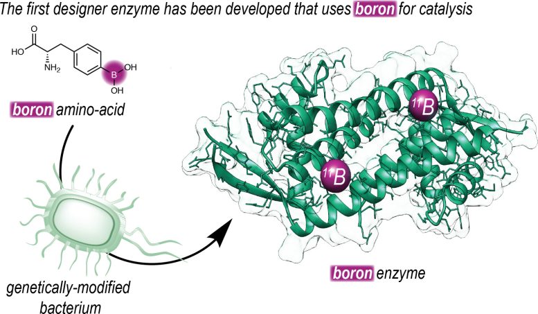
    ‘To make this possible, we set out to introduce boron into an enzyme. Our group has a long history of designing enzymes that don’t exist in nature.’ The Roelfes group used an expanded genetic code to introduce a non-natural amino acid that contains a reactive boronic acid group into an enzyme. ‘Using this technique, we can determine at the DNA level where we place the amino acid in a protein.’

    Insertion of Boron Into an Enzyme

    This is a schematic representation of how University of Groningen chemists introduced an artificial amino acid containing boron into an enzyme. Credit: Roefels Lab / University of Groningen

    Once they made an enzyme with boronic acid at its reactive center, they could use directed evolution to increase its efficiency, resulting in faster catalysis. ‘Furthermore, by placing the boronic acid in the chiral context of an enzyme, we were able to achieve highly enantioselective catalysis.’ The reaction that is described in the journal Nature is a ‘proof of principle’ and shows the way to harnessing the catalytic power of boron in enzymes.

    Biocatalysis

    Using enzymes to create organic compounds is important for the pharmaceutical industry.

    ‘In their push towards greener and more sustainable ways of producing drugs, they are looking at biocatalysis to replace conventional chemical reactions.’ At the University of Groningen, concerted efforts are being made towards this goal. ‘We have a number of research groups at the Faculty of Science and Engineering engaged in this kind of work, using different approaches to create biocatalytic solutions for the chemical industry.’ In this context, Roelfes and his team will continue to develop their boronic acid enzymes and create other such new-to-nature enzymes.

    Reference: “Boron catalysis in a designer enzyme” by Lars Longwitz, Reuben B. Leveson-Gower, Henriëtte J. Rozeboom, Andy-Mark W. H. Thunnissen and Gerard Roelfes, 8 May 2024, Nature.
    DOI: 10.1038/s41586-024-07391-3


    [ad_2]

    Source link

  • Designing Molecular Shapes for Medical Miracles

    Designing Molecular Shapes for Medical Miracles

    [ad_1]

    Alpha Helices

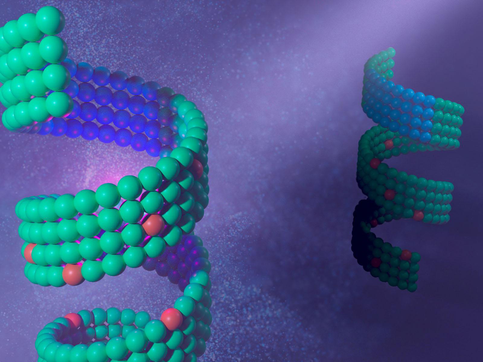
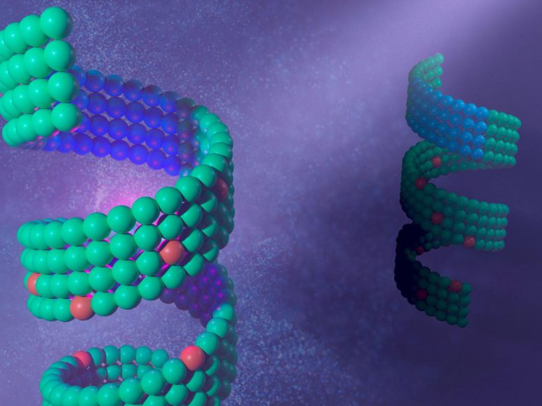
    Scientists have developed methods to control the precise shapes and “handedness” of peptoids, protein-like molecules inspired by natural molecular structures, which are vital for applications like drug delivery and disease therapy. Their research highlights their innovative techniques in creating functional peptoid-based materials for various technological and medical uses. Credit: Illustration by Nathan Johnson | Pacific Northwest National Laboratory

    Researchers have learned to control the “handedness” of peptoids, a crucial factor in precision drug delivery and diagnostics.

    Nature is filled with extraordinarily precise molecular shapes that fit together like a hand in glove. Proteins, for example, can assemble into a wide variety of well-defined shapes that grant them their function.

    “Depending on their shape, proteins can fit together with other proteins to perform functions or malfunction by clumping together, as observed in Alzheimer’s disease,” said materials scientist Chun-Long Chen. “Understanding how they assemble, and the origins of their particular shape, could be significant for various applications such as drug delivery, diagnostics, and therapeutics.”

    In studies published in Nature Communications and Angewandte Chemie, Chen and his PNNL colleagues investigated how to control these shapes by creating peptoid-based materials inspired by nature. He uses these sophisticated protein-like molecules to design substances for energy applications, such as harvesting light or breaking down woody lignin. In the last decade, Chen and his team at Pacific Northwest National Laboratory have developed a platform for creation of designer peptoid-based functional materials and characterization of their behavior.

    “Peptoids have the potential to be used in a variety of applications,” said Chen. “Based on their assembled shapes and other properties, it’s possible to design peptoids as drug delivery agents or artificial enzymes.”

    Like a hand in a glove

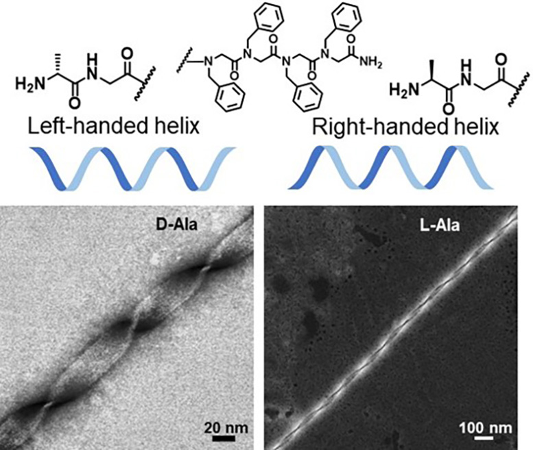
    Chen and his colleagues teamed with the University of Washington, the University of Chicago, and the Georgia Institute of Technology to design peptoids assemblies with precise shapes. Their experiment involves directing the “handedness” of the helix. Helices can be “left-handed” or “right-handed” depending on the direction in which they spiral. Their results were published in Nature Communications.

    “Handedness is extremely important when designing specialized molecules, like medications,” said Chen. “Understanding and controlling this handedness can provide insights into processes like protein assembly and could be valuable to finding cures to protein folding-related diseases such as Alzheimer’s disease.”

    Chiral Helices

    Chun-Long Chen and his team developed a way to control the shape of the peptoid helix. Credit: Chun-Long Chen | Pacific Northwest National Laboratory

    For this experiment, Chen and his team chose to pursue corkscrew-like helical structures because of their biological importance. In fact, most proteins contain these basic helical structures.

    Previous peptoid synthesis methods would yield a mix of left- and right-handed helices. In nature, proteins need to be in a specific conformation to perform their functions—most being left-handed.

    “Other groups before us were able to synthesize peptoid nanohelices, but precisely controlling their shapes and handedness remained a challenge,” said Chen. “Being able to control their shapes would not only open the door for designing future materials, it would also provide insights into biological processes involving these structures.”

    Using a combination of experimental and computational techniques, Chen and his team discovered a way to control the handedness of a peptoid helix. Similar to proteins, peptoids are created from amino acid-like building blocks. Every building block has the same “backbone” atoms that form peptoid bonds, however, each individual link in the chain can vary tremendously. Chen’s group found that they could control the shape of the helix by manipulating the sequence of the peptoid side chains.

    Adding another dimension to peptoid research

    To further investigate how peptoids can assemble, Chen collaborated with colleagues from the University of Washington, Harvard University, Binghamton University, and Zhejiang Sci-Tech University. Expanding on their previous two-dimensional studies of peptoid structures, the team was able to successfully develop a three-dimensional helical nanostructure.

    They observed that the inclusion of special “functional groups” of atoms in their peptoid sequences allowed them to create structures with special functions—similar to protein assemblies. Their work was published in Angewandte Chemie.

    “While this is a fundamental study, this research gives us additional insights into how we can create better, more precise materials—like those found in nature—for specific applications,” said Chen. “Peptoids have the potential to be used in a variety of applications. Based on their structure and other properties, it’s possible to design peptoids as drug delivery agents or artificial light-harvesting systems.”

    In the future, Chen and his team hope to create a wide range of peptoid-based nanomaterials for applications. Controlling peptoid shape, as outlined in their research papers, is just the first step.

    References: “Assembly of short amphiphilic peptoids into nanohelices with controllable supramolecular chirality” by Renyu Zheng, Mingfei Zhao, Jingshan S. Du, Tarunya Rao Sudarshan, Yicheng Zhou, Anant K. Paravastu, James J. De Yoreo, Andrew L. Ferguson and Chun-Long Chen, 16 April 2024, Nature Communications.
    DOI: 10.1038/s41467-024-46839-y

    “Hierarchical Self-Assembly of Multidimensional Functional Materials from Sequence-Defined Peptoids” by Li Shao, Dehong Hu, Shao-Liang Zheng, Thi Kim Hoang Trinh, Wenhao Zhou, Haoyu Wang, Yanxu Zong, Changning Li and Chun-Long Chen, 24 April 2024, Angewandte Chemie International Edition.
    DOI: 10.1002/anie.202403263

    Both studies were primarily supported by the Department of Energy, Office of Science, Basic Energy Sciences program as part of the Energy Frontier Research Centers program: CSSAS – The Center for the Science of Synthesis Across Scales.


    [ad_2]

    Source link

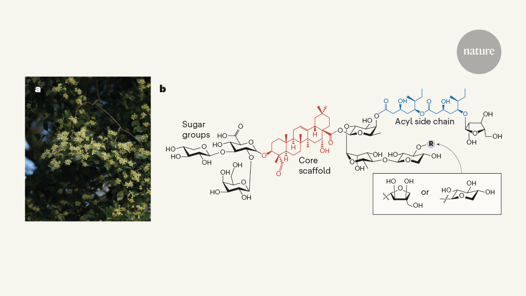
  • De Luca, V., Salim, V., Atsumi, S. M. & Yu, F. Science 336, 1658–1661 (2012).

    Article 
    PubMed 

    Google Scholar
     

  • Jacobowitz, J. R. & Weng, J.-K. Annu. Rev. Plant Biol. 71, 631–658 (2020).

    Article 
    PubMed 

    Google Scholar
     

  • Liu, Y. et al. Nature https://doi.org/10.1038/s41586-024-07345-9 (2024).

    Article 

    Google Scholar
     

  • Pifferi, C., Fuentes, R. & Fernández-Tejada, A. Nature Rev. Chem. 5, 197–216 (2021).

    Article 
    PubMed 

    Google Scholar
     

  • Kensil, C. R., Patel, U., Lennick, M. & Marciani, D. J. Immunol. 146, 431–437 (1991).

    Article 
    PubMed 

    Google Scholar
     

  • San Martín, R. & Briones, R. Econ. Bot. 53, 302–311 (1999).

    Article 

    Google Scholar
     

  • Perrot, T. et al. Curr. Opin. Biotechnol. 87, 103098 (2024).

    Article 
    PubMed 

    Google Scholar
     

  • Reed, J. et al. Science 379, 1252–1264 (2023).

    Article 
    PubMed 

    Google Scholar
     

  • Martin, L. B. B. et al. Nature Chem. Biol. 20, 493–502 (2024).

    Article 
    PubMed 

    Google Scholar
     

  • Srinivasan, P. & Smolke, C. D. Nature 585, 614–619 (2020).

    Article 
    PubMed 

    Google Scholar
     

  • Galanie, S., Thodey, K., Trenchard, I. J., Interrante, M. F. & Smolke, C. D. Science 349, 1095–1100 (2015).

    Article 
    PubMed 

    Google Scholar
     

  • Luo, X. et al. Nature 567, 123–126 (2019).

    Article 
    PubMed 

    Google Scholar
     

[ad_2]

Source link

  • First glowing animals lit up the oceans half a billion years ago

    First glowing animals lit up the oceans half a billion years ago

    [ad_1]

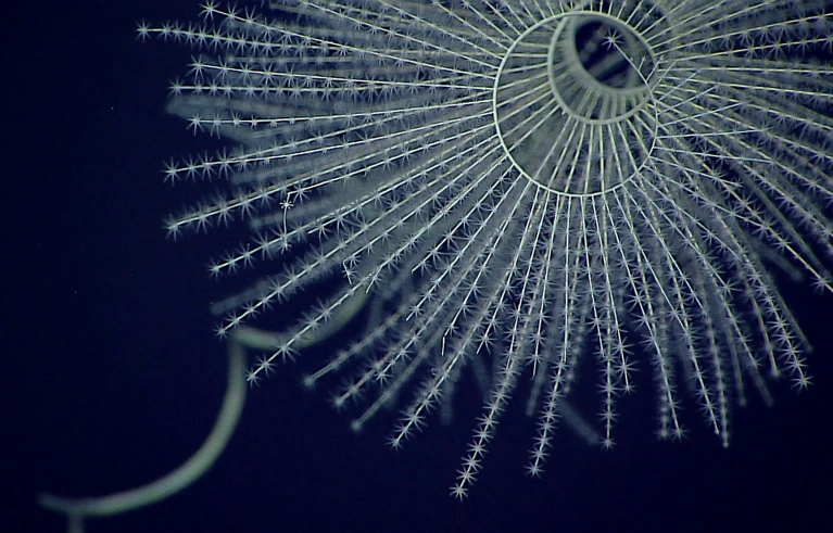
    A magnificent coral Iridogorgia magnispiralis, a deep-sea octocorals that are known to be bioluminescent.

    A bioluminescent octocoral, Iridogorgia magnispiralis.Credit: NOAA Office of Ocean Exploration and Research, Deepwater Wonders of Wake

    Some 540 million years ago, an ancient group of corals developed the ability to make its own light1.

    Scientists have previously found that bioluminescence is an ancient trait — with one group of tiny crustaceans first making their own light an estimated 267 million years ago. But this new finding pushes back the origins of bioluminescence even further by around 270 million years.

    “We had no idea it was going to be this old,” says Danielle DeLeo, an evolutionary marine biologist at Florida International University in Miami, who led the study, which was published on 24 April in Proceedings of the Royal Society B. “The fact that this trait has been retained for hundreds of millions of years really tells us that it is conferring some type of fitness advantage.”

    Bioluminescence has evolved independently at least 100 times in animals and other organisms. Some glowing species, such as fireflies, use their light to communicate in the darkness. Other animals, including anglerfish, use it as a lure to attract prey, or to scare away predators.

    However, it’s not always clear why bioluminescence evolved. Take octocorals. These soft-bodied organisms are found in both shallow water and the deep ocean, and produce an enzyme called luciferase to break down a chemical to make light. But whether glowing octocorals use their light to attract zooplankton as prey or for some other purpose is unclear.

    First light

    Searching for answers, DeLeo and her colleagues analysed a large data set of genetic sequences and the sparse octocoral fossil record to reconstruct the animals’ evolutionary history. They then used a computer model to determine how likely it was that ancestral species were bioluminescent.

    The model revealed that the common ancestor of all octocorals — which lived around 540 million years ago — was probably bioluminescent. The finding suggests that luciferase-based biofluorescence evolved early and was lost by non-bioluminescent descendants of ancient glowing octocorals.

    The study shows that bioluminescence has been around since at least the Cambrian period (around 540 million to 485 million years ago), when the first animal species developed eyes. That’s surprising, says evolutionary biologist Todd Oakley, at the University of California, Santa Barbara, because bioluminescence is a trait that “tends to blink on and off” across evolutionary time.

    Luciferase is just one way animals make light. Other organisms use different chemistry to get their telltale glow. In the case of octocorals, the luciferase system could have evolved for the production of an antioxidant, says DeLeo. Later, the light-generating aspect of the reaction would have become useful for communication.

    In any case, the deep origin of bioluminescence suggests that it could be one of the oldest forms of communication on Earth, she says. “If you’re producing light — whether or not it’s intentional — you are signalling other animals,” she says. “Like, ‘Hey! I’m over here!”

    [ad_2]

    Source link

  • Study reveals how DNA gyrase resolves DNA entanglements

    Study reveals how DNA gyrase resolves DNA entanglements

    [ad_1]

    Picture in your mind a traditional “landline” telephone with a coiled cord connecting the handset to the phone. The coiled telephone cord and the DNA double helix that stores the genetic material in every cell in the body have one thing in common; they both supercoil, or coil about themselves, and tangle in ways that can be difficult to undo. In the case of DNA, if this overwinding is not dealt with, essential processes such as copying DNA and cell division grind to a halt. Fortunately, cells have an ingenious solution to carefully regulate DNA supercoiling.

    In this study published in the journal Science, researchers at Baylor College of Medicine, Université de Strasbourg, Université Paris Cité and collaborating institutions reveal how DNA gyrase resolves DNA entanglements. The findings not only provide novel insights into this fundamental biological mechanism but also have potential practical applications. Gyrases are biomedical targets for the treatment of bacterial infections and the similar human versions of the enzymes are targets for many anti-cancer drugs. Better understanding of how gyrases work at the molecular level can potentially improve clinical treatments.

    Some DNA supercoiling is essential to make DNA accessible to allow the cell to read and make copies of the genetic information, but either too little or too much supercoiling is detrimental. For example, the act of copying and reading DNA overwinds it ahead of the enzymes that read and copy the genetic code, interrupting the process. It’s long been known that DNA gyrase plays a role in untangling the overwinding, but the details were not clear.

    DNA minicircles and advanced imaging techniques reveal first step to untangle DNA

    We typically picture DNA as the straight double helix structure, but inside cells, DNA exists in supercoiled loops. Understanding the molecular interactions between the supercoils and the enzymes that participate in DNA functions has been technically challenging, so we typically use linear DNA molecules instead of coiled DNA to study the interactions. One goal of our laboratory has been to study these interactions using a DNA structure that more closely mimics the actual supercoiled and looped DNA form present in living cells.”


    Dr. Lynn Zechiedrich, study author, Kyle and Josephine Morrow Chair in Molecular Virology and Microbiology and professor of the Verna and Marrs McLean Department of Biochemistry and Molecular Pharmacology at Baylor College of Medicine

    After years of work, the Zechiedrich lab has created small loops of supercoiled DNA. In essence, they took the familiar straight linear DNA double helix and twisted it in either direction once, twice, three times or more and connected the ends together to form a loop. Their previous study looking at the 3-D structures of the resulting supercoiled minicircles revealed that these loops form a variety of shapes that they hypothesized enzymes such as gyrase would recognize.

    In the current study, their hypothesis was proven correct. The team of researchers combined their expertise to study the interactions of DNA gyrase with DNA minicircles using recent technology advances in electron cryomicroscopy, an imaging technique that produces high-resolution 3-D views of large molecules, and other technologies.

    “My lab has long been interested in understanding how molecular nanomachines operate in the cell. We have been studying DNA gyrases, very large enzymes that regulate DNA supercoiling,” said co-corresponding author Dr. Valérie Lamour, associate professor at the Institut de Génétique et de Biologie Moléculaire et Cellulaire, Université de Strasbourg. “Among other functions, supercoiling is the cell’s way of confining about 2 meters (6.6 feet) of linear DNA into the microscopic nucleus of the cell.”

    As the DNA supercoils inside the nucleus, it twists and folds in different forms. Imagine twisting that telephone cord mentioned at the beginning, several times on itself. It will overwind and form a loop by crossing over DNA chains, tightening the structure.

    “We found, just as we had hypothesized, that gyrase is attracted to the supercoiled minicircle and places itself in the inside of this supercoiled loop,” said co-author, Dr. Jonathan Fogg, senior staff scientist of molecular virology and microbiology, and biochemistry and molecular pharmacology in the Zechiedrich lab.

    “This is the first step of the mechanism that prompts the enzyme for resolving DNA entanglements,” Lamour said.

    “DNA gyrase, now surrounded by a tightly supercoiled loop, will cut one DNA helix in the loop, pass the other DNA helix through the cut in the other, and reseal the break, which relaxes the overwinding and eases the tangles, regulating DNA supercoiling to control DNA activity,” Zechiedrich said. “Imagine watching the rodeo. Like roping cattle with a lasso, supercoiled looped DNA captures gyrase in the first step. Gyrase then cuts one double-helix of the DNA lasso and passes the other helix through the break to get free.”

    Co-corresponding author, Dr. Marc Nadal, professor at the École Normale in Paris confirmed the observation of the path of the DNA wrapped in the loop around gyrase using magnetic tweezers, a biophysical technique that allows to measure the deformation and fluctuations in the length of a single molecule of DNA. Observing a single molecule provides information that is often obscured when looking at thousands of molecules in traditional so-called “ensemble” experiments in a test tube.

    Interestingly, the “DNA strand inversion model” for gyrase activity was proposed in 1979 by Drs. Patrick O. Brown and the late Nicholas R. Cozzarelli, also in a Science paper, well before researchers had access to supercoiled minicircles or the 3-D molecular structure of the enzyme. “It’s especially meaningful to me that 45 years later, we finally provide experimental evidence supporting their hypothesis because Nick was my postdoctoral mentor,” Zechiedrich said.

    “This work opens a myriad of perspectives to study the mechanism of this conserved class of enzymes, which are of great clinical value,” Lamour said.

    “This work supports new ideas on how DNA activities are regulated. We propose that DNA is not a passive biomolecule acted upon by enzymes, but an active one that uses supercoiling, looping and 3-D shapes to direct accessibility of enzymes such as gyrase to specific DNA sequences in a variety of situations, which will likely impact cellular responses to antibiotics or other treatments,” Fogg said.

    Contributors to this work also include Marlène Vayssières (lead author), Nils Marechal, Long Yun, Brian Lopez Duran and Naveen Kumar Murugasamy. The authors are affiliated with one or more of the following institutions: Baylor College of Medicine, Université de Strasbourg, Institut de Génétique et de Biologie Moléculaire et Cellulaire, INSERM, Université Paris and Hôpitaux Universitaires de Strasbourg.

    Source:

    Journal reference:

    Vayssières, M., et al. (2024) Structural basis of DNA crossover capture by Escherichia coli DNA gyrase. Science. doi.org/10.1126/science.adl5899.

    [ad_2]

    Source link

  • Study sheds light on how androgens shape sex differences at the cellular and molecular levels

    Study sheds light on how androgens shape sex differences at the cellular and molecular levels

    [ad_1]

    Sex differences are widespread across human development, physiological processes, and diseases, making it important to characterize the impact of sex differences in these areas. Understanding the regulatory mechanisms associated with these differences, including the role of androgens, is also vital for clinical translation-;especially for diseases more prevalent in one sex.

    To answer these questions, a team led by Prof. GAO Dong and Prof. CHEN Luonan from the Center for Excellence in Molecular Cell Science, Shanghai Institute of Biochemistry and Cell Biology of the Chinese Academy of Sciences, Prof. BAI Fan from Peking University, and Prof. YU Chen from the Shenzhen Bay Laboratory, deeply explored the role of androgens in shaping sex differences at the molecular and cellular levels. Their study was published in Nature. 

    The researchers developed a detailed single-cell transcriptomic map from 17 different tissues of the mouse (Mus musculus). Using this dataset, they analyzed sex differences in depth and investigated how androgens influence these differences through specific molecular pathways and cell types. They also explored the implications of their findings on sex-biased diseases. 

    They then pinpointed the genes (i.e., AASB-DEGs) among these various tissues and cell types whose expression is sex-biased and directly influenced by androgens. These genes, including Egfr, Fos, and Il33, were highlighted as potential targets for precision medicine by modulating the androgen pathway. 

    The researchers also detailed how androgens affect the prevalence of certain cell types across sexes in various tissues, notably within immune cell populations. A key finding was the identification of group 2 innate lymphoid cells (ILC2s), which play a role in inflammation and enhancing PD-1 blockade therapy. Interestingly, ILC2s exhibited the highest androgen receptor (Ar) expression levels among the major immune cell types. The presence of these cells was notably affected by androgen levels, suggesting a mechanism by which androgens influence immune responses and disease susceptibility. 

    By integrating their findings with data from the UK Biobank, the researchers discovered that the most common risk genes for multiple sex-biased diseases were major histocompatibility complex (MHC) genes, some of which showed sex differences or were androgen-responsive. Cross-species analyses based on this atlas also identified associations between cell types and sex-biased diseases. 

    Overall, this study sheds light on the intricate ways in which androgens contribute to sex differences at the cellular and molecular levels and provides the foundation for developing targeted therapies for sex-biased diseases by modulating the androgen pathway. 

    Source:

    Journal reference:

    Li, F., et al. (2024). Sex differences orchestrated by androgens at single-cell resolution. Nature. doi.org/10.1038/s41586-024-07291-6.

    [ad_2]

    Source link

  • Scientists Solve Early Earth RNA Puzzle

    Scientists Solve Early Earth RNA Puzzle

    [ad_1]

    Abstract Biochemistry Origin of life Concept

    A groundbreaking study reveals how life’s complexity could originate from simple RNA molecules on early Earth. Through experiments demonstrating RNA’s recycling and replication abilities under specific conditions, such as low salinity and high pH, the research suggests life could emerge from minimal molecular sets in environments akin to volcanic islands. This finding challenges prior assumptions about RNA’s evolution and underscores the potential for life’s emergence in simple, cold prebiotic conditions.

    Recent research illustrates how RNA molecules’ chemical characteristics might have played a crucial role in the development of complex life forms.

    How did complex life manage to evolve on the early, inhospitable Earth? Initially, ribonucleic acid (RNA) must have existed to carry the first genetic information. For these biomolecules to build-up complexity in their sequences, they needed to release water. However, on the early Earth, which was predominantly covered in seawater, this process was challenging.

    In a paper recently published in the Journal of the American Chemical Society (JACS), researchers from the team of LMU professor Dieter Braun have shown that in RNA’s struggle with the surrounding water, its natural recycling capabilities, and the right ambient conditions could have been decisive.

    “The building blocks of RNA release a water molecule for every bond they form in a growing RNA chain,” explains Braun, spokesperson for the Collaborative Research Centre (CRC) Molecular Evolution in Prebiotic Environments and coordinator at the ORIGINS Excellence Cluster. “When, conversely, water is added to an RNA molecule, the RNA building blocks are fed back into the prebiotic pool.”

    This turnover of water works particularly well under low saline conditions with high pH levels. “Our experiments indicate that life could emerge from a very small set of molecules, under conditions such as those prevailing on volcanic islands on the early Earth,” says Adriana Serrão, lead author of the study.

    A New Understanding of RNA Evolution

    Under these conditions, RNA has the ability to split without adding a water molecule. The end of the RNA strand remains water-free and can spontaneously re-form new RNA bonds. Braun’s laboratory demonstrated that the rebinding of this split RNA works efficiently and with remarkable precision when copying the sequence information. This process only takes place when the RNA building blocks are bound to a template RNA molecule with precisely matching base pairs in a double-stranded configuration. This produces a copy of the existing RNA strand before it disintegrates through the addition of water.

    It had previously been assumed that RNA can only copy itself by ‘randomly’ constructing sequences of around 200 nucleotides in length – so-called ribozymes. However, ribozymes can operate only in saline, and thus RNA-hostile, environments. As a result of this new research, these complex ribozyme sequences in the early stages of RNA evolution are not necessary. “The precision is comparable to the copying of RNA achieved by ribozymes,” says Sreekar Wunnava, also lead author of the study. “This means that an RNA world could arise without the prior necessity for long complex sequences.”

    Early life thus consisted of a very simple metabolic process whereby RNA sequences were copied by means of continuous replacement with recycled molecules. All that is needed for this to happen is an alkaline freshwater environment such as still exist today on volcanic islands like the Hawaiian archipelago or Iceland. “And so life could have emerged from a simple, cold prebiotic primordial soup of RNA building blocks,” explains Braun. Although the reactions take place very slowly under these conditions and require several days to complete, there was no shortage of time at the start of evolution and the cold freshwater refuges on primeval volcanic islands allowed RNA to survive on the otherwise inhospitable early Earth.

    Reference: “High-Fidelity RNA Copying via 2′,3′-Cyclic Phosphate Ligation” by Adriana Calaça Serrão, Sreekar Wunnava, Avinash V. Dass, Lennard Ufer, Philipp Schwintek, Christof B. Mast and Dieter Braun, 19 March 2024, Journal of the American Chemical Society.
    DOI: 10.1021/jacs.3c10813


    [ad_2]

    Source link

  • Unraveling Nature’s Ancient Molecular Kitchen

    Unraveling Nature’s Ancient Molecular Kitchen

    [ad_1]

    Origin Emergence of Life Concept

    Research reveals that ancient heat flows could have organized prebiotic molecules into life’s building blocks, providing a natural mechanism for the complex molecular interactions essential for life’s origin.

    Life is complicated. What is true for our everyday existence also holds for the many complex processes that take place inside cells. Proteins are continuously synthesized, cell walls are constructed, and DNA is replicated This can only work when reaction partners converge at the right time in sufficiently high concentrations while suffering little disruption from other substances. Over the course of billions of years, evolution has perfected these mechanisms and ensured that such vital processes occur with high efficiency at the correct place.

    Circumstances were probably a lot more chaotic four billion years ago, when prebiotic reactions created the conditions for the emergence of the first lifeforms. For these reactions, too, it was necessary for the ‘right’ substances to be brought together at the ‘right’ time in one place, so that more complex biomolecules like RNA and amino acid chains could form.

    While such reactions are possible to recreate in the laboratory thanks to manual intermediate steps, it is highly challenging for them to come about in a simple ‘primordial soup’ – that is to say, a very dilute mixture of prebiotic building blocks. So how could nature create suitable conditions for the origin of life?

    A collaboration of LMU biophysicists Dr. Christof Mast and Professor Dieter Braun, coordinator at the ORIGINS Excellence Cluster, and geoscience Professor Bettina Scheu have found a possible answer to this question and recently published their results in the journal Nature. “Our investigations show how simple heat flows could have yielded order amid the chemical chaos of primordial times and fostered the first prebiotic reactions,” explains Mast.

    Heat is produced by a wide range of geological and chemical processes, so presumably heat flows occurred almost everywhere in those distant times. If this heat flows through thin, water-filled cracks, such as arise through the rapid cooling of molten rock, it leads to water convection and to a directed movement of the dissolved molecules in the water along the heat flow. Combined, these two effects – convection and thermophoresis – drive accumulation and selective up-concentration of the various solutes in different places.

    A pinch of this and a sprinkle of that: ingredients for life

    Mast’s group has now experimentally demonstrated this selective accumulation for over 60 different prebiotic building blocks, such as nucleobases and amino acids. They found that these substances can differ strongly in their thermophoresis and therefore enrich differently in the rock fissures. “In a system of interconnected cracks and fissures in the rock, this effect is reinforced and produces mixtures with different compositions of prebiotic substances in each fissure,” explains Thomas Matreux, lead author of the paper. “Although the initial solution was uniformly dilute, and therefore unreactive, simple heat flows can generate an astonishing variety of possible starting conditions for prebiotic chemistry in this manner,” adds Paula Aikkila, the other lead author of the study.

    Without the aid of modern lab technology or the advanced reaction mechanisms of life today, nature could thus have created a ‘molecular kitchen’ in large geological network systems, in which all the ingredients of life were sorted and ready. As part of the Collaborative Research Centre “Molecular Evolution in Prebiotic Environments” (CRC 392), the researchers now plan to investigate how many ‘dishes’ of life can be ‘prepared’ in this system.

    Reference: “Heat flows enrich prebiotic building blocks and enhance their reactivity” by Thomas Matreux, Paula Aikkila, Bettina Scheu, Dieter Braun and Christof B. Mast, 3 April 2024, Nature.
    DOI: 10.1038/s41586-024-07193-7


    [ad_2]

    Source link

  • Newly discovered molecule may be a potential therapeutic target for triple-negative breast cancer

    Newly discovered molecule may be a potential therapeutic target for triple-negative breast cancer

    [ad_1]

    A team of researchers from Hiroshima University has discovered a molecule that promotes the production of cancer cells. This molecule may prove to be a potential therapeutic target in the treatment of triple-negative breast cancer, an aggressive form of breast cancer.

    Their work was published in the journal Molecular Cancer Research on January 18, 2024.

    Breast cancer is the most common type of cancer, ranking fifth among all cancers in cancer-related deaths. In 2020, there were 2.3 million new cases of breast cancers reported around the globe. In that year, breast cancer caused 685,000 deaths.

    Several studies have reported that a molecule called AIbZIP (androgen-induced basic leucine zipper) promotes malignant behavior in different cancer types. So the research team examined the potential role of AIbZIP in malignant tumors. Their computer simulation analysis revealed that AIbZIP was highly expressed in the luminal androgen receptor subtype of triple negative breast cancer, playing a significant role in cell cycle regulation. They identified a novel mechanism by which AIbZIP regulates cancer cell proliferation in this type of breast cancer.

    We found that AIbZIP is highly upregulated in triple negative breast cancer. AIbZIP plays a crucial role for hyper proliferation of triple negative breast cancer cells by promoting the degradation of p27, a negative regulator for cell proliferation. Our study indicates that AIbZIP may be potential therapeutic target of triple negative breast cancer” said Atsushi Saito, an associate professor and Kazunori Imaizumi, a professor in the Department of Biochemistry, Graduate School of Biomedical and Health Sciences, Hiroshima University.

    Breast cancer is divided into three types. These types, defined mainly by receptor and protein expression, are luminal breast cancer, HER2-positive breast cancer, and triple-negative breast cancer. Triple-negative breast cancer does not have the estrogen receptor, progesterone receptor, or HER2 (human epidermal growth factor receptor 2) found in the other two types of breast cancer. Without these receptors, or targets, this cancer is challenging to treat. “Among breast cancer, triple negative breast cancer has no known therapeutic targets and a poor prognosis. Therefore, new therapeutic targets and strategies against TNBC are required,” said Taichi Ito, a student in the Department of Biochemistry, Graduate School of Biomedical and Health Sciences, Hiroshima University.

    Triple negative breast cancer has been further divided into subtypes, based on gene expression. The luminal androgen receptor is a subtype of triple negative breast cancer. This subtype represents 15 to 20 percent of all triple negative breast cancer cases.

    Recent treatments for triple-negative breast cancer include chemotherapy, immunotherapy, and targeted therapies. These treatments hold promise, but they are not ideal solutions. Researchers continue to explore new therapeutic targets and strategies against triple-negative breast cancer.

    The luminal androgen receptor subtype of triple negative breast cancer has a high expression of the androgen receptor and genes associated with androgenic hormonally-regulated pathways. As a result, researchers have made many attempts to develop new strategies to treat the luminal androgen receptor subtype of triple negative breast cancer with drugs that inhibit the androgen receptor activity. However, their therapeutic value is limited, and effective treatment for luminal androgen receptor subtype of triple negative breast cancer has not yet been achieved. So scientists have worked to identify new targets that can inhibit proliferation, invasion, and metastasis of the luminal androgen receptor subtype of triple negative breast cancer.

    Further studies are required to verify whether this regulation of cell cycle progression occurs in other cell types. The team knows that AIbZIP is highly upregulated in many cancers, including luminal and HER2-positive breast cancer. The novel pathway they have discovered may contribute to cancer treatments in other cancer types besides triple negative breast cancer.

    Looking ahead, the team sees more work to be done. They want to check the expression level of AIbZIP in tumors derived from triple negative breast cancer patients to confirm the robust link between AIbZIP and the development of triple negative breast cancer. “If we can downregulate the AIbZIP activity, it may lead to develop the novel therapeutic strategy against triple negative breast cancer,” said Imaizumi.

    The research team includes Taichi Ito, Atsushi Saito, Yasunao Kamikawa, Nayuta Nakazawa, and Kazunori Imaizumi from the Department of Biochemistry, Graduate School of Biomedical and Health Sciences, Hiroshima University, Japan.

    The research is funded by JSPS KAKENHI; Takeda Science Foundation; MSD Life Science Foundation; The UBE Foundation; the Program of the Network-type Joint Usage/Research Center for Radiation Disaster Medical Science, Analysis Center of Life Science, the Natural Science Center for Basic Research and Development, Hiroshima University.

    Source:

    Journal reference:

    Ito, T., et al. (2024). AIbZIP/CREB3L4 Promotes Cell Proliferation via the SKP2-p27 Axis in Luminal Androgen Receptor Subtype Triple Negative Breast Cancer. Molecular Cancer Research. doi.org/10.1158/1541-7786.mcr-23-0629.

    [ad_2]

    Source link