Tag: Biomarker

  • Research reveals potential target for enfortumab vedotin therapy in urothelial carcinoma

    Research reveals potential target for enfortumab vedotin therapy in urothelial carcinoma

    [ad_1]

    Under the leadership of PD Dr. Niklas Klümper, Assistant Physician at the Department of Urology at the University Hospital Bonn (UKB) and Clinician Scientist of the BMBF-funded ACCENT program and PD Dr. Markus Eckstein, senior physician at the Institute of Pathology at the Uniklinikum Erlangen of the Friedrich-Alexander-University Erlangen-Nürnberg (FAU), an interdisciplinary research team has published new findings that indicate which patients with advanced urothelial carcinoma could benefit in particular from the new targeted therapy with the antibody-drug conjugate enfortumab vedotin. The study, published yesterday in the prestigious Journal of Clinical Oncology, identifies NECTIN4 amplification as a promising genomic biomarker for predicting treatment response to enfortumab vedotin. These findings could represent a significant advance in the improved treatment of this cancer.

    As an alternative to chemotherapies used to treat aggressive advanced and metastatic urothelial carcinoma, a new class of drugs, known as antibody-drug conjugates, has recently become available. Enfortumab vedotin (EV) is the first drug in this new class to be approved by the EMA at for the treatment of patients and patients with metastatic urothelial carcinoma. Given the extremely promising results of the EV-302 trial, which showed a near doubling of survival with the combination therapy of EV and pembrolizumab, an immune checkpoint inhibitor, compared to conventional platinum-based chemotherapy in untreated patients with metastatic urothelial carcinoma, it is expected that the use of EV will increase significantly in the future.

    Modern targeted oncology therapy

    Antibody-drug conjugates consist of an antibody directed against a target structure on tumor cells and combined with a highly toxic chemotherapeutic agent. This combines the selectivity of targeted antibody therapy with the cytotoxic potential of conventional chemotherapy, which represents an innovative and new oncological therapeutic approach.

    Research for more targeted therapy: precision oncology

    Long-term efficacy of the new drug EV has so far been shown in an uncharacterized group of patients with metastatic urothelial carcinoma. The research team led by PD Dr. Niklas Klümper (UKB) and PD Dr. Markus Eckstein (Uniklinikum Erlangen) therefore wanted to analyze in more detail which patients benefit effectively from EV therapy in order to be able to use it in a more targeted manner – and conversely to identify patients who benefit less or not from EV, as they could possibly be treated more effectively with other therapies.

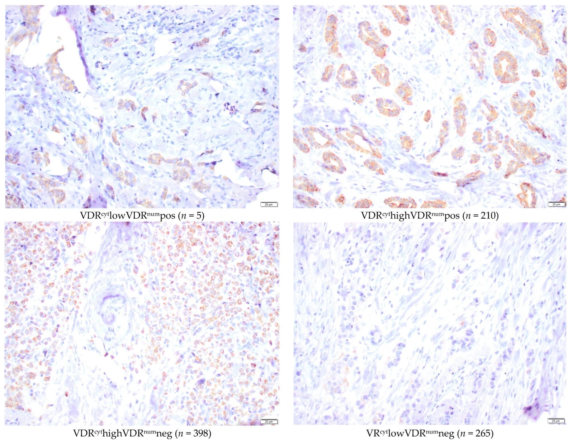
    Nectin-4, the target structure of EV, is localized on chromosome 1q23.3. This gene segment is increased in about 20-25 percent of urothelial carcinomas, which is referred to as amplification. The new study aimed to investigate NECTIN4 amplifications as a potential genomic biomarker to predict treatment response to the drug EV in patients with advanced urothelial carcinoma.

    We have successfully developed and applied a simple FISH test (fluorescence in situ hybridization) that is specific for NECTIN4. This test proved to be a reliable method for identifying NECTIN4 amplification. Our studies showed that the presence of NECTIN4 amplification is a robust biomarker for response to EV therapy. In fact, over 90 percent of patients with NECTIN4 amplification showed tumor response to EV therapy, compared to about 30 percent of patients without this amplification”, says PD Dr. Markus Eckstein. These new findings can help to better select patients for this promising therapy in the future. “NECTIN4 amplification is a promising biomarker for predicting treatment response to EV. Excitingly, NECTIN4 amplification is also common in other solid tumors besides urothelial carcinoma, e.g. lung and breast cancer. The consideration of NECTIN4 amplification could therefore also be an exciting option for other tumor types in order to select patients for anti-NECTIN4-directed therapy in a more targeted manner. Further studies on this topic are needed, but our work could be the starting signal for the establishment of new targeted treatment strategies “, says PD Dr. Niklas Klümper.

    Both study PIs also agree that without the great support of all the colleagues involved from the numerous oncology centers in Germany, Austria, the Netherlands and the USA, and of course without the patients’ consent to participate in the study, it could never have been carried out successfully.

    The study was funded and initiated by the German Research Foundation (DFG) as part of the DFG Young Investigator Academy UroAgeCare of the German Society of Urology (DGU). “This underlines the high relevance of funding clinician scientist programs for medical progress,” says Prof. Michael Hölzel, mentor of PD Dr. Klümper within the program.

    Source:

    Journal reference:

    Klümper, N., et al. (2024) NECTIN4 Amplification Is Frequent in Solid Tumors and Predicts Enfortumab Vedotin Response in Metastatic Urothelial Cancer. Journal of Clinical Oncology. doi.org/10.1200/JCO.23.01983.

    [ad_2]

    Source link

  • Study reveals human gut plasmid with biomarker potential

    Study reveals human gut plasmid with biomarker potential

    [ad_1]

    A component of the human intestinal flora that has been little studied to date is the focus of a new study. Plasmids are small extrachromosomal genetic elements that frequently occur in bacterial cells and can influence microbial lifestyles – yet their diversity in natural habitats is poorly understood. An international team led by Prof. Dr. A. Murat Eren from the Helmholtz Institute for Functional Marine Biodiversity at the University of Oldenburg (HIFMB) reports in the science journal Cell, a mysterious plasmid, is one of the most numerous genetic elements in the human gut that could potentially serve as a powerful biomarker for identifying health hazards such as fecal contamination of water or human disorders such as Inflammatory Bowel Disease. According to the team’s analyses, this plasmid is present in the intestines of more than 90 percent of individuals in industrialized countries.

    Plasmids are extrachromosomal DNA sequences which are common to cells from all domains of life. Eren describes them as “typically small genetic entities that carry additional genes”. They can be exchanged between different bacterial cells and even between different types of bacteria. The replication of plasmids is dependent on their host cells: but they make up for it by providing their hosts with in some cases extremely important fitness determinants. For instance, some plasmids contain genes that encode antibiotic resistance, which help their bacterial hosts to survive antibiotics, contributing one of the most pressing public health concerns around the globe.

    There are also other plasmids which, according to the research to date, do not contain genes encoding obvious beneficial functions for their host. “These so-called ‘cryptic plasmids’ are often referred to as genetic parasites. They remain a mystery in microbial ecology because from an evolutionary perspective they should not exist at all,” explains Eren, a computer scientist and Professor of Ecosystem Data Science at the University of Oldenburg.

    Identifying plasmids has been a difficult undertaking so far. For some time now, scientists have been able to extract genetic material directly from environmental samples and, for example, analyze the microbial community in the human gut in its entirety, without having to cultivate individual bacterial organisms. However, the ability to confidently distinguish what is a plasmid among this conglomeration of genetic material, referred to as the metagenome, poses a considerable challenge.

    To solve this problem, Eren and his colleagues developed a new machine learning approach. As the team reported in an article recently published in the science journal Nature Microbiology, using this approach they identified over 68,000 plasmids in human intestinal flora, and also discovered that a certain cryptic plasmid called pBI143 was particularly abundant in their dataset.

    One of the most numerous genetic entities in the human gut

    In the study published in Cell, the team of researchers took a closer look at this plasmid, which consists of only two genes that rather surprisingly only serves for its own replication and mobilization across bacterial cells with no other clear benefit. To better understand the ecology of pBI143, the team analyzed 60,000 human and 40,000 environmental metagenomes generated from various habitats.

    “We found that pBI143 has a list of very interesting features,” Eren explains. The team discovered that more than 90 percent of people in industrialized countries carry the plasmid and that on average it is one of the most numerous genetic entities in the human gut. “On average it was more than ten times as numerous as a viral genome which was previously thought to be the most abundant genetic extrachromosomal element in the human gut,” says the researcher.

    Further analyses revealed that the plasmid occurred almost exclusively in the human gut but was virtually absent in datasets from other environments such as the oceans, soils, plants and the digestive organs of animals and their feces. The only other samples in which the researchers were able to detect the characteristic gene sequence for these plasmids was in samples from environments that are influenced by humans, such as waste water, hospital surfaces and laboratory rats.

    Due to its sheer numbers, prevalence across humans, and its conservancy across human populations, the team of researchers hypothesized that pBI143 could, for instance, be used as a biomarker in testing for fecal contamination.

    In fact, we were able to show that this plasmid is a more sensitive marker for detecting fecal contamination in drinking water compared to state-of-the-art biomarkers based on specific gene sequences of human intestinal bacteria.”


    Dr. A. Murat Eren, Professor of Ecosystem Data Science at the University of Oldenburg

    Non-invasive method to quantify progress of IBD

    The team also identified another potential application of this prevalent genetic entity in the context of human disorders such as Inflammatory Bowel Disease (IBD), a medical condition that affects 3 million people in Europe alone. They were able to demonstrate that the relative copy number of this cryptic plasmid increased almost four times in the intestines of people suffering from IBD compared as in the intestines of healthy individuals, suggesting that the changes of the copy number of the plasmid can serve as a non-invasive method to quantify the disease progress or severity.

    At the HIFMB, Eren’s team is developing new tools at the intersection of computer science and microbiology to identify and characterize naturally occurring plasmids and other mobile genetic elements in bacteria that live in the ocean. They strive to gain a better understanding of the ecology and evolution of microbes, and strategies that enable to them to respond to their everchanging environments for new biotechnological applications that can ameliorate crises we face.

    Source:

    Journal reference:

    Fogarty, E. C., et al. (2024). A cryptic plasmid is among the most numerous genetic elements in the human gut. Cell. doi.org/10.1016/j.cell.2024.01.039.

    [ad_2]

    Source link

  • Heavy alcohol use linked to increased risk of Type 2 diabetes in middle-aged adults

    Heavy alcohol use linked to increased risk of Type 2 diabetes in middle-aged adults

    [ad_1]

    Heavy alcohol use may increase middle-aged adults’ risk of developing Type 2 diabetes, according to research to be presented this week at the American Physiology Summit in Long Beach, California. The Summit is the flagship annual meeting of the American Physiological Society (APS).

    Heavy alcohol use is defined by the National Institute on Alcohol Abuse and Alcoholism (NIAAA) as five or more drinks per day or 15 or more drinks per week for men and, for women, more than four drinks per day or eight or more drinks per week.

    Heavy alcohol use can diminish liver and pancreas function. Reduced function of these organs in turn can affect control of glucose (blood sugar) in the body. In regard to heavy drinking, research suggests young adults do not typically experience severe impairment of fasting glucose levels and insulin resistance. However, as the U.S. population ages, the number of people that develop Type 2 diabetes is increasing. The association between alcohol use and diabetes in older adults is not clear.

    Researchers from University of Texas at Arlington’s Cardiovascular Laboratory of Health studied two groups of middle-aged adults between the ages of 50 and 64. One group of volunteers (“heavy drinkers”) was at risk for developing alcohol use disorder, as defined by the U.S. Alcohol Use Disorders Identification Test-;a questionnaire that asks about drinking frequency and volume-;and a dried blood spot phosphatidylethanol (PEth) test. A PEth test measures levels of a biomarker that forms in the blood after consuming alcohol. The PEth biomarker can be detected in the blood up to four weeks after drinking alcohol. A PEth score of 20 nanograms per milliliter (ng/mL) or higher indicates alcohol consumption that exceeds NIAAA recommendations. The second group of volunteers was labeled “nonheavy drinkers,” with PEth scores under 20 ng/mL.

    The research team analyzed additional blood samples from both volunteer groups. The heavy drinkers had higher fasting glucose levels, which “suggest[s] that heavy alcohol use may have negative effects on glucose regulation in aged populations,” the researchers wrote.

    Although there were no significant differences in kidney or liver function between the two groups, “alcohol drinking that exceeds the recommended limits causes organ damage throughout the body and increases not only the risk of potentially developing Type 2 diabetes, but also other diseases. Our team recommends not drinking every day. If [people] do drink alcohol, make sure for men, [it’s] no more than four drinks on any single day … and for women, no more than three drinks on any single day.”


    Chueh-Lung “Laura” Hwang, PhD, PT, senior author of the study

    This study was funded by NIAAA AA028537.

    [ad_2]

    Source link

  • Blood-based DNA test spares bladder cancer patients from unnecessary treatments

    Blood-based DNA test spares bladder cancer patients from unnecessary treatments

    [ad_1]

    Testing for tumor DNA in the blood can successfully identify advanced bladder cancer patients who will not relapse following surgery, new research shows.

    This could allow doctors to target treatments more effectively to those who need it, and spare those patients for whom further treatment is unnecessary, researchers say.

    The findings from the screening phase of the IMvigor011 Phase III trial are presented today [Friday 5 April] at the European Association of Urology Congress in Paris.

    They show that just over 90% of muscle invasive bladder cancer (MIBC) patients with a negative circulating DNA (ctDNA) test following surgery, which remained negative on follow up, did not relapse. The findings mean that use of a ctDNA test could allow some patients to be spared further treatment with minimal risk.

    MIBC is an advanced form of bladder cancer, where the tumor has spread into the bladder wall. The disease is usually treated by surgery to remove the bladder. Around half of patients see cancer return, often in the lungs and usually within two-to-three years. All patients are currently offered follow-up treatment such as chemotherapy or immunotherapy to prevent recurrence, for which the side effects can be serious and lifechanging.

    Other Phase III trial results, also presented at the EAU Congress today, show that patients given immunotherapy, nivolumab, as a follow up to surgery have an average survival of nearly six years, compared to four for patients on placebo.

    The CheckMate 274 trial has already shown that nivolumab can reduce recurrence of disease, but these interim results are the first to show the potential benefit in overall survival for MIBC patients.

    Joost Boormans, Professor of Urology at Erasmus University Medical Centre in Rotterdam, and member of the EAU Scientific Congress Office, is chairing the session where both trials will present their findings. He said:

    “Although we already knew that nivolumab improved disease-free survival in MIUC patients who received radical surgery, overall survival is what really matters following local treatment, such as radical surgery. These interim findings, which show that overall survival also improves, are very encouraging, particularly as this hasn’t been the case in other recent immunotherapy trials.

    “The question for regulators and healthcare authorities is whether the improvement in overall survival is enough to justify licensing or prescribing the drug for all patients, in the knowledge that some of these patients would have been cured of their cancer by surgery alone. This is where the findings from the IMvigor011 trial could really make a difference, by allowing us to select patients at highest risk who will benefit the most from treatment while sparing others for whom it isn’t needed.

    “At a time when healthcare resources are under pressure, this kind of innovation is really needed.”

    IMvigor011

    IMvigor011 is a global, double-blind, randomized Phase III trial looking at the efficacy of the immunotherapy atezolizumab vs placebo in patients with high-risk MIBC.

    The trial is recruiting MIBC patients post-surgery and testing their blood for circulating tumour DNA. Those with a positive ctDNA result are randomized to receive either atezolizumab or placebo. Those with a negative result are given no further treatment, but were followed up with scans and further ctDNA tests for up to two years. For the analysis presented at the EAU Congress today, 171 patients with a negative ctDNA test were included, with follow up continuing on a further 115.

    Just 17 patients of the 171 patients (9.9%) saw their cancer return within two years. These outcomes were irrespective of the stage their tumor was at or whether it showed elevated levels of PD-L1, a protein biomarker that plays a role in cancer.

    Professor Thomas Powles of Barts Cancer Institute leads the IMvigor011 trial.

    These results are even better than we were hoping. The risk of relapse in this ctDNA group of patients is just 1 in 10. It appears this test can effectively filter patients into two groups: those who are likely to relapse and those at much lower risk. Focusing treatment on those at risk and sparing the very low risk group potentially life-altering treatment-related side effects is attractive. Hopefully these data will allow patients to remain treatment free with the reassurance they need, that they’re unlikely to see their cancer return.”


    Professor Thomas Powles of Barts Cancer Institute

    The study is sponsored by F. Hoffmann-La Roche Ltd.

    CheckMate 274

    CheckMate 274 is a global, Phase III, randomized, double-blind trial of nivolumab vs placebo in high-risk MIBC after surgery.

    The trial recruited just over 700 patients, with half given nivolumab and the other half given a placebo every two weeks for 12 months following an operation to remove the bladder. Patients were also tested to see if their cancer had elevated levels of the biomarker PD-L1, which nivolumab specifically targets.

    The trial has already reported positive results in preventing recurrence, particularly for PD-L1 patients. Across all patients, those on nivolumab had an average of 22 months before recurrence, compared to 10 months for those on placebo. However, of the PD-L1 group, those on nivolumab had an average of over four years without recurrence, compared to just over eight months for those on placebo.

    The latest results, although still early stage, show a similar benefit in overall survival. For all patients, those on nivolumab survive on average for nearly six years (69.5 months) compared to just over four years (50.1 months) for those on placebo. The researchers do not yet have enough follow-up data to separate out the PD-L1 patients, but the analysis so far shows that overall survival is likely to also be even better for this group when treated with nivolumab versus placebo.

    Professor Matthew Galsky from the Tisch Cancer Institute, Icahn School of Medicine at Mount Sinai in New York, leads the CheckMate 274 trial. He said: “We know that patients with high-risk urothelial cancer are at highest risk for recurrence within the first three years after surgery. We’ve now followed a substantial subset of patients for longer than that on this study without recurrence. It looks as if the improvement in disease free survival is ultimately going to translate into improvement in overall survival. And that’s for all patients, but particularly patients with the PD-L1 biomarker. Our hope is that this improvement will then translate into an increased likelihood of curing cancer in these patients.”

    The trial is funded by Bristol Myers Squibb and Ono Pharmaceutical. Dr. Matthew D. Galsky is a paid consultant to Bristol Myers Squibb.

    [ad_2]

    Source link

  • Metabolic markers tied to increased risk of depression and anxiety, study finds

    Metabolic markers tied to increased risk of depression and anxiety, study finds

    [ad_1]

    In a recent study published in the JAMA Network Open, a group of researchers determined if carbohydrate, lipid, and apolipoprotein (Apo) biomarkers are linked to the future development of depression, anxiety, and stress-related disorders in a large Swedish cohort.

    Study: Metabolic Profile and Long-Term Risk of Depression, Anxiety, and Stress-Related Disorders. Image Credit: hikrcn / ShutterstockStudy: Metabolic Profile and Long-Term Risk of Depression, Anxiety, and Stress-Related Disorders. Image Credit: hikrcn / Shutterstock

    Background 

    About one-third of people experience depression, anxiety, and stress-related disorders in their lifetime, with growing evidence suggesting a link between these psychiatric conditions and metabolic dysregulation, such as lipid and glucose abnormalities that trigger inflammation. This inflammation may increase the risk of psychiatric disorders by affecting brain health. However, existing research on metabolic biomarkers and their association with psychiatric disorders has been inconsistent, often limited by methodological issues like short follow-up periods and reliance on self-reported depression measures, mainly in older adults. Furthermore, there is a notable gap in research on the connection between these biomarkers and anxiety or stress-related disorders, particularly the long-term effects of Apo’s. Further research is crucial to clarify these associations and explore potential preventative and therapeutic strategies.

    About the study 

    The Swedish Apolipoprotein-Related Mortality Risk (AMORIS) cohort, spanning from 1985 to 1996 and predominantly in the Stockholm region, includes 812,073 participants (49% men, 51% women) who underwent routine health screenings or were referred for laboratory testing due to health conditions. For this study, 211,200 participants over 16, free from mental disorders at baseline and with at least one biomarker measurement, were selected. Mental disorder histories were confirmed using the Swedish Patient Register, employing various International Classification of Diseases (ICD) revisions for accuracy.

    The study included a broad spectrum of stress-related conditions, with both primary and secondary diagnoses considered. Biomarkers of interest include glucose, cholesterol types, triglycerides, apolipoproteins, and their ratios, analyzed by consistent laboratory methods. Covariates like sex, age, fasting status, socioeconomic status, and birth country were also recorded, offering a detailed background for each participant.

    Statistical analysis involved Cox proportional hazards regression models to explore the relationship between initial biomarker levels and the risk of psychiatric disorders, adjusting for relevant covariates and employing both categorical and continuous variable analyses. Sensitivity analyses further refined these findings by focusing on employed individuals, outpatient referrals and excluding those with missing socioeconomic data. A case-control study embedded within the larger cohort provided a longitudinal perspective, examining biomarker trends up to 30 years before diagnosis, with controls matched to cases by sex, age, and enrollment year. 

    Study results 

    In the substantial cohort of 211,200 participants, consisting of 58% males and 42% females, with the vast majority born in Sweden (89.4%), the study observed notable trends over a mean (SD) follow-up period of 21 years. The participants had an average age of 42.1 years at their first blood sampling, with diagnoses of depression, anxiety, or stress-related disorders emerging at a mean age of 60.5 years. The incidence rates for these disorders varied, with depression, anxiety, and stress-related disorders recorded at rates of 21.5, 16.6, and 10.5 per 10,000 person-years, respectively. Notably, a segment of the cohort was diagnosed with multiple disorders, yet only 0.4% received diagnoses across all three categories.

    The analysis revealed a correlation between metabolic biomarker levels and psychiatric health risks. Elevated glucose and triglyceride (TG) levels significantly increased the risk of psychiatric disorders, whereas higher levels of high-density lipoprotein cholesterol (HDL-C) offered a protective effect. The delineation of risk did not change markedly between low and normal glucose levels, suggesting a particular risk threshold. This pattern persisted across gender lines and when evaluating each psychiatric condition individually, reinforcing the robustness of these findings.

    Further analysis, specifically among employed individuals, yielded consistent results with the primary analysis, underscoring the relationship between metabolic health and psychiatric conditions regardless of employment status. When examining the impact of outpatient care referrals on biomarker measurements, the associations remained similar for glucose and TGs, though the protective role of HDL-C lessened. Additionally, higher levels of low-density lipoprotein cholesterol (LDL-C), total cholesterol (TC), ApoB, and the ApoB/ApoA-I ratio were inversely associated with the risk of psychiatric disorders, indicating a complex interplay between various lipid biomarkers and mental health.

    Socioeconomic status also emerged as a significant factor, with lower incidence rates of psychiatric disorders observed among those with higher socioeconomic standing. This trend held true even after adjusting for potential confounders, including missing socioeconomic data, further emphasizing the socioeconomic gradient in psychiatric disorder risk.

    Longitudinal analysis, tracking biomarker levels up to 30 years prior to diagnosis, illustrated that patients eventually diagnosed with anxiety, depression, or stress-related disorders exhibited consistently higher levels of TGs, glucose, and TC two decades before diagnosis. Additionally, higher levels of ApoA-I and ApoB were noted in the decade leading up to diagnosis, indicating a prolonged period of metabolic dysregulation preceding psychiatric diagnoses. 

    [ad_2]

    Source link

  • Focusing on oncogenic driver mutations and resistance mechanisms in the treatment of NSCLC

    Focusing on oncogenic driver mutations and resistance mechanisms in the treatment of NSCLC

    [ad_1]

    Lung cancer is the leading cause of cancer-related death worldwide. Improved understanding of driver mutations of non-small cell lung cancer (NSCLC) has led to more biomarker-directed treatment for patients with advanced stages. The expanding number of drugs targeting these driver mutations offers more opportunity to improve patient’s survival benefit.

    To date, NSCLCs, especially those with non-squamous histology, are recommended for testing epidermal growth factor receptor (EGFR) mutations, anaplastic lymphoma kinase (ALK) gene rearrangements, ROS proto-oncogene receptor tyrosine kinase 1 (ROS-1) rearrangements, B-raf proto-oncogene (BRAF) mutations, rearranged during transfection (RET) fusions, Met proto-oncogene (MET) amplification and exon 14 skipping alterations, neurotrophic receptor tyrosine kinase (NTRK) gene fusions, and immunohistochemistry (IHC) testing for the programmed death receptor-ligand 1 (PD-L1) expression.

    EGFR-activating mutations are the most common driver mutations in NSCLC. Targeted therapies included first-generation epidermal growth factor inhibitor tyrosine kinase inhibitors (EGFR-TKIs), erlotinib, gefitinib, and icotinib; second-generation pan-human epidermal growth receptor (HER) family inhibitor, afatinib and dacotinib; and third-generation EGFR-TKI, osimertinib, that inhibits both EGFR-sensitive mutations and resistant mutation EGFR T790M. ALK inhibitors included first-generation, crizotinib; second generation, ceritinib, alectinib, and brigatinib; and third-generation, lorlatinib, with increasing capacity for ALK inhibition generation-by-generation. The resistance caused by secondary ALK mutations can be overcome by next-generation ALK-TKIs. Moreover, alectinib, brigatinib, and lorlatinib are all recommended as first-line treatment choice for ALK-rearranged NSCLC for their superior survival compared with crizontinb. Crizotinib, ceritinib, brigatinib, and lorlatinib can also inhibit ROS-1. BRAF inhibitor dabrafenib combined with MEK inhibitor trametinib is now the recommended treatment for BRAF V600E-mutated NSCLC attributing to improved outcomes compared with vemurafenib or dabrafenib monotherapy. Drugs targeting MET aberrations with different binding modes included type Ia, crizotinib; type Ib, tepotinib, capmatinib, and savolitinib; and type II, cabozantinib. Entrectinib and larotrectinib are the current standard treatment for NTRK fusion-positive NSCLC. Kirsten ratsarcoma viral oncogene homolog (KRAS) mutations occur in ~20%–30% of patients with NSCLC. Recently, several small molecules including sotorasib (AMG510), adagrasib (MRTX-849) have been developed to specifically target KRAS G12C.

    Although drugs targeting oncogenic driver mutations have significantly improved survival but their long-term responses are still uncommon for most patients, the emergence of acquired drug resistance is inevitable. Therefore, exploration and understanding of the resistance mechanism of target therapy are important to enhance the clinical outcomes and ultimately increase the treatment rate of NSCLC. Meanwhile, improved understanding of biological characteristics in various molecular subtypes of each driver mutation helps explore new classified treatment strategies. Until recently, multiomics analysis has ushered in a new era of precision targeted therapy in lung cancer and led to a deeper understanding of the underlying resistant mechanism. All these scientific and clinical progresses ultimately lead to improved survival in NSCLC and achieve more refined individualized treatment.

    The discovery of oncogenic driver alterations and target therapy have brought significant clinical benefits and established an individualized treatment approach. The management of advanced NSCLC has shifted from a histology based on a biomarker-driven process.

    The treatment landscape of oncogenic-addicted NSCLC has become complex. On the one hand, more individualized treatment based on fine stratification has become the focus of research nowadays. The choice of optimal treatment strategy should consider gene subtype, concomitant mutation, dynamic gene alternation, and metastasis site. On the other hand, understanding primary and acquired resistance to targeted therapy provides an insight into the molecular evolution of tumor development. The recognition of resistant mechanisms is the basis to design new drugs or combinatorial therapeutic strategies.

    Combination strategies require integration with immunotherapy, and the immunosuppressive microenvironment should be reversed to improve the sensitivity of ICIs by the drug combination. We should also be aware of the possible risk of combined toxicity and thereby explore the optimal timing and combined regimen of immunotherapy. Meanwhile, the interaction between driver mutations and immune-microenvironment is essential to uncover after drug resistance and to establish robust predictive biomarker for the NSCLC with driver mutations, in order to identify specific oncogenic driving patients with NSCLC who can benefit from immunotherapy.

    Source:

    Journal reference:

    Zhong, J., et al. (2023). Treatment of advanced non-small cell lung cancer with driver mutations: current applications and future directions. Frontiers of Medicine. doi.org/10.1007/s11684-022-0976-4.

    [ad_2]

    Source link

  • Skin biopsy for α-synuclein detection proves effective

    Skin biopsy for α-synuclein detection proves effective

    [ad_1]

    In a recent study published in JAMA, researchers evaluated the positivity rate of cutaneous phosphorylated alpha-synuclein protein (P-SYN) deposition among individuals with dementia with Lewy body presence (DLB), Parkinson’s disease (PD), pure autonomic failure (PAF), and multiple system atrophy (MSA).

    Study: Skin Biopsy Detection of Phosphorylated α-Synuclein in Patients With Synucleinopathies. Image Credit: BLACKDAY/Shutterstock.comStudy: Skin Biopsy Detection of Phosphorylated α-Synuclein in Patients With Synucleinopathies. Image Credit: BLACKDAY/Shutterstock.com

    Background

    Synucleinopathies are neurodegenerative illnesses that cause P-SYN accumulation in the peripheral and central nervous systems. They include PD, MSA, DLB, and PAF. These illnesses share clinical characteristics, including progressive impairment and neurodegeneration.

    Current pharmacology lacks disease-modifying medication for these illnesses, and many individuals diagnosed with synucleinopathies face diagnostic delays or misdiagnoses.

    A reliable biomarker for identifying synucleinopathies, such as immunohistochemistry of cutaneous phosphorylated α-synuclein, is urgently needed. This test might be sensitive and specific.

    About the study

    In the present prospective, multicenter, cross-sectional study, researchers investigated whether skin biopsy could detect P-SYN in PD, MSA, DLB, and PAF patients.

    The researchers enrolled clinically confirmed cases of DLB, PD, PAF, or MSA recruited from 19 community-based and 11 academic neurology practices between February 2021 and March 2023, aged between 40 and 99 years.

    Individuals without history or clinical features indicative of a synucleinopathy (such as constipation, hyposmia, dementia, rapid-eye movement [REM] sleep disorder, or mild cognitive impairments) or neurodegenerative disorders comprised the control group.

    The researchers excluded individuals with biopsy-associated risks and synucleinopathy-mimicking diseases. They also excluded those with missing data from questionnaires and clinical examinations. The study exposure was a cutaneous biopsy for P-SYN detection.

    The primary outcome was cutaneous P-SYN detection frequency among individuals with MSA, PD, PAF, or DLB and controls.

    The researchers obtained skin biopsy specimens from the posterior cervical area, 3.0 cm from the spinous process of C-7, and the distal aspects of the leg at a distance of 10 cm from the lateral malleolus, and the thigh 10 cm from the lateral knee.

    The team examined the participants using the Hoehn and Yahr scale, the Movement Disorders Society Unified PD Rating Scale (MDS-UPDRS), the Montreal Cognitive Assessment, and orthostatic blood pressure.

    The participants filled out questionnaires such as the 39-component Parkinson’s Disease Questionnaire, the European Quality of Life questionnaires (EQ-VAS and EQ-5D), the MSA Quality of Life assessment Questionnaire, the rapid eye movement (REM) sleep disorder questionnaire, and the Orthostatic Hypotension Questionnaire.

    The researchers obtained disease symptom and duration data from participant medical records. The referral physician, who evaluated the individual, provided a clinical diagnosis.

    The participant history, examination scores, medical records, and ancillary test results were transmitted to two disease specialists for central review to validate the diagnosis of the particular synucleinopathy based on specified consensus and eligibility criteria for diagnoses or controls.

    Results

    Out of 428 patients (277 with synucleinopathy and 151 controls), 343 were included in the primary analysis [mean age, 70 years; 175 (51%) men]; 223 fulfilled the consensus criteria for synucleinopathy, and 120 met the criterion as controls following expert panel evaluation.

    Among those with synucleinopathy, 96 (28%) were diagnosed with Parkinson’s disease, 50 (15%) with Lewy body dementia, 55 (16%) with multiple system atrophy, and 22 (6.4%) with complete autonomic failure.

    The proportion of participants with P-SYN in their skin was 93% (n = 89) with Parkinson’s disease, 98% (n=54) with multiple system atrophy, 96% (n=48) with Lewy body dementia, and 100% (n=22) with complete autonomic failure; 3.30% (n=4) of controls had cutaneous phosphorylated-SYN deposition.

    P-SYN detection in the subepidermal plexus varied by synucleinopathy subtype, with MSA (49%, n=27) having a higher prevalence than Parkinson’s disease (3.1%, n=3), DLB (10%, n=5), or PAF (9.1%, n=2). There were no infections or significant problems.

    The length-dependent small fiber neuropathy varies amongst synucleinopathy subtypes. Neuropathy was most prevalent in DLB patients (78%, n=39), followed by those with Parkinson’s disease (63%, n = 60), PAF (46%, n=10), and MSA (22%, n=12).

    The overall P-SYN for all research participants corresponded with their exam results and surveys. P-SYN deposition by the study participants was associated with the period since MSA, PAF, and PD diagnosis.

    Conclusions

    The study findings showed that skin biopsy may identify phosphorylated alpha-synuclein among individuals with DLB, PD, PAF, and MSA. The findings demonstrated that cutaneous P-SYN in >92% of participants, with skin biopsies, was tolerated well with minor side effects.

    However, there were 21% of misdiagnosed cases. Accurate diagnosis is critical for patient and family counseling, starting symptomatic treatment, and conducting clinical studies of possible disease-modifying medications.

    Cognitive neurology experts recommend skin biopsy as a novel method for movement problems. The findings might speed up medication development for synucleinopathies by enhancing patient homogeneity in clinical trials.

    More studies are needed to confirm the findings and better understand the potential relevance of skin biopsy detection in clinical treatment.

    Journal reference:

    • Christopher H. Gibbons, MD, MMSc, et al., (2024) Skin Biopsy Detection of Phosphorylated α-Synuclein in Patients With Synucleinopathies, JAMA 2024, doi: 10.1001/jama.2024.0792.

    [ad_2]

    Source link

  • Vitamin D receptor presence in breast cancer tumors linked to better survival outcomes

    Vitamin D receptor presence in breast cancer tumors linked to better survival outcomes

    [ad_1]

    In a recent study published in the journal Nutrients, researchers explored the role of vitamin D receptors (VDR) as a potential prognostic marker for breast cancer.

    Their results indicate significant associations between VDR, the mode of detection, and the size of invasive tumors, suggesting a promising avenue for further study.

    Study: The Vitamin D Receptor as a Prognostic Marker in Breast Cancer—A Cohort Study. Image Credit: Peddalanka Ramesh Babu / ShutterstockStudy: The Vitamin D Receptor as a Prognostic Marker in Breast Cancer—A Cohort Study. Image Credit: Peddalanka Ramesh Babu / Shutterstock

    Background

    People with breast cancer often show lower serum levels of vitamin D compared to healthy individuals; some studies have also found links between low vitamin D levels and the probability of an adverse prognosis, while others suggest that supplementation could reduce the risk of developing advanced cancer.

    Calcitriol, an active metabolite of vitamin D, binds to the VDR before it translocates into the nucleus, modifying regulatory genes that control cell signaling, apoptosis, and cell growth.

    This could be a mechanism that underlies the association between VDR and favorable breast cancer prognoses, but further research is needed to understand its potential role as a biomarker for the progression of tumors.

    About the study

    In this study, researchers attempted to validate prior work that found positive associations between breast cancer prognoses and nuclear VDR presence. They additionally explored if prognostic information can be refined by VDR subcellular localization.

    The dataset included tumor samples obtained from individuals who received a diagnosis of primary breast cancer between October 2002 and June 2012. The samples were utilized for tissue microarray (TMA) construction after excluding people who had received a prior diagnosis of any cancer in the previous 10 years.

    During preoperative visits, patients were asked to complete questionnaires that included information on reproductive factors, lifestyles, and medication and supplements consumed during the previous week. Vitamin D consumption was calculated from the product information for supplements.

    Nurses measured hip and waist circumference, height, weight, and breast volume. Pathology reports were used to obtain characteristics of tumors, including size, histologic type, grade, involvement of axillary lymph nodes, and status of progesterone receptors (PR) and estrogen receptors (ER).

    Pathological assessment of human epidermal growth factor receptor 2 (HER2) status and TMA were assessed jointly using microscopic methods after blinding. In multiple subcellular locations of invasive tumor cells, VDR staining was examined in the nuclear membrane (VDRnum), cytoplasm (VDRcyt), and nucleus (VDRnuc).

    During the final follow-up in 2019, medical records, pathology reports, and national registries for population and tumors were used to calculate the breast cancer-free interval (BCFI) and calculate endpoints (death or last follow-up) for survival analyses.

    Findings

    On average, patients were 61 years old, and VDR was obtained for 878 tumors; cytoplasmic intensity evaluations showed that 7% were VDR-negative, while VDRnum was positive in 25% of patients. Tumors included in the analysis were, on average, large, of a higher grade, and more likely to be lymph node-negative than those that were excluded.

    Microscopic representative images of immunohistochemical staining intensities of nuclear membrane and cytoplasmic VDR (40×) in the TMA. Bar represents 20 µm.Microscopic representative images of immunohistochemical staining intensities of nuclear membrane and cytoplasmic VDR (40×) in the TMA. Bar represents 20 µm.

    Patients with VDRnum-positive tumors had smaller waist circumferences and breast volumes on average and were more likely to have screening-detected tumors. VDRnum-positive tumors were linked with lower tumor grade, positive hormone receptors, and the absence of HER2 amplification.

    After surgery, more VDRnum-negative patients received chemotherapy and trastuzumab, while VDRnum-positive patients received further endocrine therapy. In terms of prognosis, patients with VDRnum-positive tumors had better outcomes in both BCFI and overall survival (OS) in univariable analysis. This effect persisted after adjusting for various factors.

    When VDR localization was considered, VDRnum-positive tumors showed the best prognosis regardless of VDRcyt. Patients with both ER-positive and VDRnum-positive tumors had the best prognosis, while those with ER-negative and VDRnum-negative tumors had the worst. Even among ER-positive tumors, VDRnum-positive tumors were associated with lower tumor grade and longer OS.

    Interaction analyses suggested that VDRnum interacts with the mode of detection and size of tumors in the case of BCFI. Larger VDRnum-positive tumors (>20 mm) were associated with significantly fewer breast cancer events than small ones. Clinically detected VDRnum-positive tumors showed better BCFI than screening-detected ones.

    Conclusions

    This study highlights significant correlations between positive VDR staining in the nuclear membranes of breast cancer cells and favorable characteristics of tumors, OS, and longer BCFI.

    Results indicate that evaluating nuclear membrane VDR levels may be a better prognosis predictor compared to cytoplasmic levels. VDRnum status could refine luminal breast cancer selection with favorable prognoses, especially when considering interactions with tumor size and detection mode.

    VDR expression inversely correlates with aggressiveness, particularly in triple-negative and HER2-amplified cancers, possibly due to mutations in the tumor protein p53. Further research is needed to standardize VDR evaluation methods and explore associations with vitamin D levels.

    The study suggests potential clinical relevance for VDR as a prognostic marker and underscores the need for understanding the interplay between vitamin D, VDR, and breast cancer outcomes. Since most participants in the study were of European descent, heterogeneous populations should be included in future studies to ensure the generalizability of findings.

    Journal reference:

    • The vitamin D receptor as a prognostic marker in breast cancer – a cohort study. Huss, L., Gulz-Haake, I., Nilsson, E., Tryggvadottir, H., Nilsson, L., Nodin, B., Jirström, K., Isaksson, K., Jernström, H. Nutrients (2024). 10.3390/nu16070931, https://www.mdpi.com/2072-6643/16/7/931

    [ad_2]

    Source link

  • Study reveals e-cigarette users with limited smoking history show similar DNA alterations as smokers

    Study reveals e-cigarette users with limited smoking history show similar DNA alterations as smokers

    [ad_1]

    A recent Cancer Research study assessed the effect of tobacco smoking and electronic cigarette (e-cigarettes) use on DNA methylation changes associated with carcinogenesis.

    Study: Cigarette smoking and e-cigarette use induce shared DNA methylation changes linked to carcinogenesis. Image Credit: Andrey_Popov/Shutterstock.com
    Study: Cigarette smoking and e-cigarette use induce shared DNA methylation changes linked to carcinogenesis. Image Credit: Andrey_Popov/Shutterstock.com

    Background

    In comparison to tobacco smoking, the use of e-cigarettes and smokeless, non-combustible tobacco has often been considered to be less harmful. However, recent studies have highlighted some of the potential adverse effects linked to e-cigarette use, including DNA damage and endothelial dysfunction. Therefore, it is imperative to understand the molecular changes and their long-term effects on health.

    E-cigarette use has been associated with similar biomarker changes to cigarette smoking. It is essential to identify biomarkers that indicate the risk of cancer. Some of the characteristic features that must be present in biomarkers are (a) modifiability through tobacco smoking and e-cigarette use, (b) lie in genes linked to carcinogenesis, (c) induce cancer progression in a premalignant lesion, and (d) reflective of long-term cancer risk in a surrogate tissue to aid non-invasive monitoring.

    The epigenome is a set of chemical modifications of DNA or proteins linked to DNA. Many studies have elucidated the role of epigenomics in carcinogenesis. This information has helped shed light on the long-term impacts of tobacco smoking and e-cigarette use.

    DNA methylation (DNAme) at the cytosine C-5 position is an epigenetic modification. Its alterations enriched in genes are associated with smoking-related diseases. Some epigenetic changes remain persistent even after smoking cessation. These biomarkers could be used as an indicator of lung cancer. 

    Epithelial cells that are exposed (e.g., oral mucosa and lungs) or not (e.g., cervix) to smoking or e-cigarette use are the key cells of origin for tobacco-related malignancies. Furthermore, smoking-related DNAme changes found in buccal samples are primarily of epithelial origin.

    About the study

    The current study addressed the aforementioned issues to understand better tissue- and cell-specific epigenetic impacts of e-cigarette or tobacco use on DNAme. It used more than 3,500 cervical, buccal/saliva, or blood samples from immune and epithelial cells at directly and indirectly exposed sites. Additionally, a control sample set was used for validation.

    This study is a part of the female cancer prediction using cervical omics to individualize screening and prevention (FORECEE) study. The participants came from five European countries, were between 18 and 86 years of age, and completed an epidemiological questionnaire. The effect of tobacco use on epithelial and immune cells was analyzed systematically using computational deconvolution and cell type-specific DNAme inference.

    The effect of direct (proximal) and indirect (distal) exposure to the use of smoking, smokeless tobacco (e.g., snuffs), or e-cigarettes on epithelial and immune cells was assessed. Furthermore, whether these uses also affected lung cancer tissue and prognosis were evaluated. The evaluation of the biomarkers at the cell-level is a key contribution of this study, as the majority of existing studies, including those predicting lung cancer, have used blood samples.

    Study findings

    The cell-specific alterations following cigarette and e-cigarette use that are associated with carcinogenesis have been uncovered in this study. Smoking was found to elicit changes in protected stem and submucosal gland cells. Cigarette smoking affected epithelial hypoM and this change was found in both proximal and distal exposure. Furthermore, DNAme alterations linked to specific proximal epithelial hyperM and distal epithelial hyperM were also identified.

    Smoking was seen to affect the myeloid more prominently than the lymphoid lineage. No significant genetic overlap linked with specific functions was observed in the samples obtained from five different sites. Mostly, epithelial hypoM sites were linked with detoxification responses, whereas proximal epithelial hyperM sites entailed DNA damage responses.

    The smoking-related DNAme loci (CpGs) identified here were clustered into four functional group based on anatomical site and cell type. Loci hypermethylated in cheek cells of smokers associated with the NOTCH1/RUNX3/growth factor receptor signaling showed a higher level of methylation in progressing lung carcinoma in situ lesions and cancer tissue. Alarmingly, the aforementioned CpGs were also noted to be hypermethylated in e-cigarette users who reported a limited smoking history.

    This study further highlighted a partial reversibility of smoking-induced epigenetic alterations in former tobacco smokers. This observation was based on the fact that epithelial hypoM could not be distinguished between ex-smokers and those who never smoked. This observation could imply that the hypermethylated cells disappeared due to cell death or the displacement of the methyl group in the living cell.

    Smokeless tobacco induces similar changes in DNAme in the epithelial hypoM and proximal epithelial hyperM sets, as cigarette smoking. It must be noted that only cigarette smokers exhibited changes in DNAme at immune hypoM sites. Proximal epithelial hypermethylation was robustly associated with lung cancer progression and cervical cancer.

    Conclusions

    In sum, the results presented here shed light on cell type-specific epigenetic changes following cigarette smoking. Some of these changes, which could also predict lung cancer, are similar to e-cigarette users.

    A key limitation of this study was the use of pathway analysis based on gene names, which limited the investigation to cis genes alone. In the future, scientists must perform multi-omics profiling to investigate the association between methylation changes and gene transcription function more comprehensively. 

    [ad_2]

    Source link

  • Study reveals breakthrough in non-invasive detection of endometrial cancer

    Study reveals breakthrough in non-invasive detection of endometrial cancer

    [ad_1]

    In a recent study published in eBioMedicine, researchers evaluated proteomic signatures in blood plasma and cervicovaginal fluid to detect endometrial cancer.

    Study: Detection of endometrial cancer in cervicovaginal fluid and blood plasma: leveraging proteomics and machine learning for biomarker discovery. Image Credit: crystal light / Shutterstock.comStudy: Detection of endometrial cancer in cervicovaginal fluid and blood plasma: leveraging proteomics and machine learning for biomarker discovery. Image Credit: crystal light / Shutterstock.com

    Diagnosing endometrial cancer

    The prevalence of endometrial cancer, which is the most common gynecological malignancy in high-income countries, continues to rise throughout the world. Endometrial cancer is amenable to curative hysterectomy when diagnosed early, with a five-year survival rate of over 90% following treatment. Comparatively, individuals with metastatic or advanced disease often have poor outcomes, with the five-year survival rate estimated at 15%.

    Over 90% of females with endometrial cancer present with postmenopausal bleeding, thus triggering urgent investigations through sequential transvaginal ultrasound, hysteroscopy, and endometrial biopsy, all of which could be anxiety-provoking and painful procedures. Therefore, developing simple, cost-effective, and non-invasive tests for early cancer diagnosis is crucial for both patients and clinicians.

    Cervicovaginal fluid, which is a mix of vaginal, uterine, and cervical secretions, has been investigated as a source of biomarkers for inflammatory conditions of the lower reproductive tract, pregnancy-related pathologies, and cervical neoplasia. In fact, one recent study found that cervicovaginal fluid can be used to detect endometrial cancer.

    About the study

    In the present study, researchers evaluate the performance of proteomic signatures from cervicovaginal fluid and plasma for endometrial cancer detection. Cases comprised females with histopathological evidence of endometrial cancer based on hysterectomy, whereas controls included symptomatic females without endometrial cancer or atypical hyperplasia. Individuals with a history of gynecological malignancy or hysterectomy were excluded.

    Cervicovaginal fluid and blood were collected, and mass spectrometry was performed. Digitized proteomic maps were derived using sequential window acquisition of all theoretical mass spectra.

    Spectral data were converted and searched against a human plasma library and a previously published library of 19,394 peptides and 2,425 proteins in the cervicovaginal fluid. Random forest (RF) modeling was used for feature selection. The most discriminatory proteins were ranked based on the mean decrease in accuracy.

    Nested logistic regression models were built by sequentially adding proteins based on their rank. The parsimonious model was identified, and its performance was evaluated by plotting the receiver operating characteristic curve and calculating the area under the curve (AUC). Likelihood ratio tests and Akaike information criteria (AIC) were used to compare the performance of nested models.

    Study findings

    Overall, 118 postmenopausal females with symptoms were included in the study, 53 of whom had confirmed endometrial cancer and 65 with no evidence of cancer. About 86% of the study cohort were White. Individuals with endometrial cancer were likely to be older and have a higher body mass index (BMI) than controls.

    Taken together, 597, 310, and 533 proteins were quantified in the cervicovaginal fluid supernatant, cell pellets, and plasma samples, respectively. Overall, 941 unique proteins were identified across sample types. There was evidence of separation between cancers and controls based on cervicovaginal fluid supernatant proteins.

    Classifiers were selected based on the mean decrease accuracy metric of the RF model. Principal component analyses (PCA) using the top discriminatory proteins revealed more substantial discrimination between cancers and controls.

    The model with the top five discriminatory proteins had the lowest AIC value and was selected as a parsimonious model. This model predicted endometrial cancer with AUC, sensitivity, and specificity of 0.95, 91%, and 86%, respectively.

    Feature selection analysis indicated that 38 proteins were important for discrimination between cancers and controls. Proteins in cervicovaginal fluid cell pellets were less promising as cancer biomarkers than supernatant-derived proteins.

    Fewer differentially expressed proteins were observed in plasma samples between cases and controls as compared to the cervicovaginal fluid, with little evidence of discrimination based on plasma proteins. PCA indicated a modest separation between cancers and controls. A three-plasma biomarker panel predicted endometrial cancer with AUC, sensitivity, and specificity of 0.87, 75%, and 84%, respectively.

    Feature selection analysis revealed six plasma proteins as important classifiers. Furthermore, three- and four-marker panels of cervicovaginal fluid and plasma proteins predicted early-stage endometrial cancer with AUCs of 0.92 and 0.88, respectively. Five- and six-marker panels of cervicovaginal fluid and plasma proteins predicted advanced-stage endometrial cancer with AUCs of 0.96 and 0.93, respectively.

    Conclusions

    Cervicovaginal fluid proteins were more accurate in detecting endometrial cancer than plasma proteins. The five-marker panel of cervicovaginal fluid proteins comprised the immunoglobulin heavy constant mu (IGHM), haptoglobin (HPT), fibrinogen alpha chain (FGA), lymphocyte antigen 6D (LY6D), and galectin-3-binding protein (LG3BP), whereas the three-marker panel of plasma proteins included HPT, proteasome 20S subunit alpha 7 (PSMA7), and apolipoprotein D (APOD).

    Further confirmatory studies using larger cohorts are needed to validate these findings.

    Journal reference:

    • Njoku, K., Pierce, A., Chiasserini, D., et al. (2024). Detection of endometrial cancer in cervicovaginal fluid and blood plasma: leveraging proteomics and machine learning for biomarker discovery. eBioMedicine. doi:10.1016/j.ebiom.2024.105064

    [ad_2]

    Source link