A new review article in npj Precision Oncology summarizes the current state of knowledge about the role of artificial intelligence (AI) in the diagnosis, treatment, and prognosis of brain tumors.
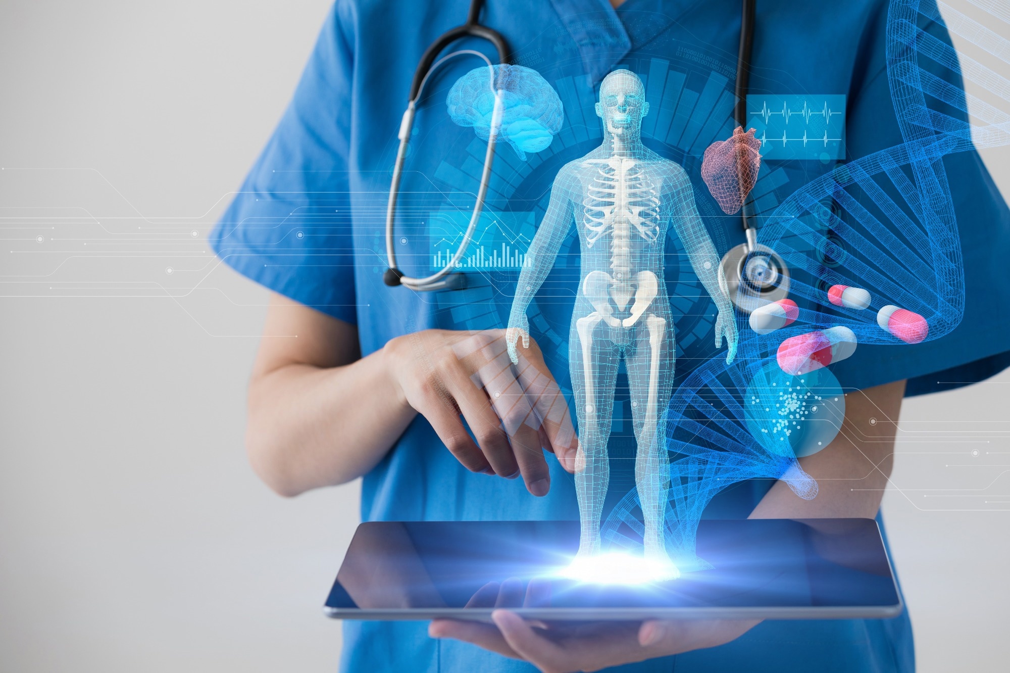 Study: Artificial intelligence in neuro-oncology: advances and challenges in brain tumor diagnosis, prognosis, and precision treatment. Image Credit: metamorworks/Shutterstock.com
Background
Brain tumors, although uncommon, pose a significant health challenge globally, with approximately 250,000 new cases each year. In the United States alone, over 96,000 brain tumor cases were reported in 2022, with around 26,600 of these being cancerous.
Glioblastoma is the most frequently diagnosed type of brain tumor and has a particularly poor prognosis, with only a 7% survival rate five years after diagnosis.
This highlights the urgent need for improved methods of diagnosing, treating, and forecasting the progression of brain tumors.
Challenges in managing brain tumors
Diffuse midline glioma (DMG) in children and glioblastoma in adults are among the toughest brain tumors to treat and are often considered incurable with current medical approaches.
Tailored treatments stand the best chance of providing a cure with the least possible harm. However, the challenge is that information on diagnosing and treating brain tumors is scattered and hard to come by.
Only a select number of medical centers have access to the latest treatment techniques. Moreover, much of the available data on these treatments is sourced from just one or a few institutions, limiting the breadth of knowledge and accessibility for many.
Management approaches and diagnostic criteria based on such data are open to a lack of demographic data and may not be generalizable globally.
Socioeconomic inequity also contributes to late diagnosis, therapeutic challenges, and reduced survival by restricting access to some key tests and reducing the odds of combination therapies. This includes 06-Methylguanine-DNA-methyltransferase (MGMT) testing for glioblastoma.
The need for precise diagnosis, staging, and treatment monitoring is difficult to meet in many cases.
Taking into account the contribution of tumor genotype to the prognosis, limited accessibility for imaging and biopsy, intratumor heterogeneity, and poorly reliable biomarkers to monitor the progress of therapy, there are significant obstacles to the optimal care of these patients.
The brain tumor paradigm
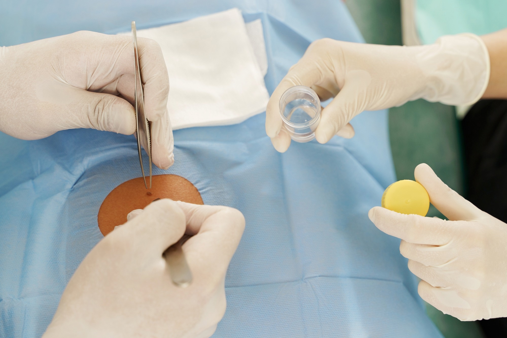
In most cases, a suspected brain tumor is diagnosed, beginning with a physical examination and neuroimaging. A biopsy follows this. If possible, the tumor and other biomarkers are removed and subjected to histologic and molecular analysis.
The choice of therapy depends on available and recommended care practices, clinical trials that are currently going on, the patient’s medical status, and toxicity risks. Magnetic resonance imaging (MRI) is the follow-up modality of choice, sometimes supplemented with cerebrospinal fluid (CSF) or blood tests.
“Decisions regarding brain tumor treatment often involve multidisciplinary meetings between neuro-oncologists, neurosurgeons, neuroradiologists, molecular pathologists, and neuropathologists, underscoring the complexity of these decisions.”
The advantages of AI
AI includes machine learning (ML) and deep learning (DL) techniques, computer vision (CV), and the integration of these as Computational Biology. ML excels at pattern recognition and DL in extracting detailed features. CV improves visual interpretation of imaging data to provide medical data.
Computational biology uses all these methods to parse biological data, helping to understand tumor genetics and molecular biology.
This study aims to uncover AI-assisted tumor radiology, pathology, and genomics advancements. AI contributes synergistically to all these domains to improve their role as a combined dataset in brain tumor management.
AI may help clinicians navigate tumor management decisions by improving MRI imaging accuracy and enhancing the speed at which results are available.
It offers increased sensitivity to anomalies picked up on imaging, detailed image analysis, optimized workflows, comprehensive data analysis from multiple sources, and detecting patterns that could be missed by the human observer.
AI algorithms help localize tumors more efficiently, avoiding human error. The nnU-Net algorithm excels at tumor segmentation, reducing radiation or surgical harm.
This enables AI to help diagnose and grade the tumor, determine the prognosis, and plan treatment while setting up a monitoring framework.
AI may become part of new clinical trials, exploring the feasibility of personalized therapy by leveraging its ability to handle large volumes of data.
AI uses various data types, including imaging data from MRI and computerized tomography (CT), radiomics, histopathologic data, genomics, molecular biomarkers from tumor cells, and clinical data.
Neuroimaging often uses pre- and post-contrast T1-weighted, T2-weighted, fluid-attenuated inversion recovery (FLAIR), diffusion-weighted (DWI), and susceptibility-weighted imaging (SWI), as well as, in specialized centers, MR spectroscopy and perfusion imaging.
Molecular biomarkers include IDH mutations for astrocytomas and oligodendrogliomas, TERT promoter mutations for glioblastomas, EGFR amplification for glioblastomas, gain of chromosome 7 and loss of chromosome 10 for glioblastomas, and MGMT promoter methylation for glioblastomas.
Non-invasive circulating tumor DNA (ctDNA) analysis is a newer method for diagnosing such tumors.
AI platforms
3D U-Net, DeepMedic, and V-Net are AI architectures that help preprocess tumor images, making the analysis more robust and precise. Methylome profiling is useful in classifying brain tumors using AI/MI and systems like DeepGlioma. This uses stimulated Raman histology (SRH) to offer results on GMB molecular diagnosis within 90 seconds.
Other systems to predict IDH and other mutations based on radiomics data from MRI perfusion scans or 18F-FET PET/CT scans are being explored, such as a deep learning imaging signature (DLIS) and Terahertz spectroscopy.
‘Sturgeon’ is another DL method to classify brain tumors intraoperatively using nanopore-sequenced methylation array data. Its 40-minute turnaround time, with >70% accuracy, helps surgical decision-making.
Prognostic help is being provided from imaging data to predict overall survival and progression-free survival, two key clinical and research metrics.
Combined with histology and molecular biology, exceptional predictive performance has been demonstrated.
Integrated approaches
Multimodal data fusion approaches could help achieve a less invasive and more accurate understanding of brain tumors using multiple data sources. This will eventually help tailor management to the patient.
The challenge is to extend and diversify the data collection range to other populations and tumor types with standardized features to ensure reproducibility and generalizability.
The adoption of AI should not worsen healthcare and social inequities, emphasizing the need to remove biases, provide legal support, communicate the scope and benefits with transparency, define responsibilities and keep patients safe.
Conclusions
“AI has the potential to empower patients by providing personalized information and enabling shared decision-making. However, the equitable access and affordability of AI-driven healthcare need to be addressed to avoid exacerbating existing disparities.”