Tag: brain

  • In the big data era, prioritize statistical significance in study design

    In the big data era, prioritize statistical significance in study design

    [ad_1]

    A scientist wearing a lab coat looks at a brain scan on a computer with an MRI machine in the background

    Magnetic resonance imaging scanning at the Brain and Behaviour Laboratory at the University of Geneva, Switzerland.Credit: Salvatore Di Nolfi/EPA/Shutterstock

    ‘Experimental design’: these words signal a section of a research paper that many readers might be inclined to scan fleetingly, before moving on to the actual findings. But a study in Nature this week should make all researchers — both readers and writers of papers — consider dwelling a little more on the methods part of the scientific process.

    The study, led by Simon Vandekar, a biostatistician at Vanderbilt University Medical Center in Nashville, Tennessee, is on how to make brain-wide association studies (BWAS) more robust (K. Kang et al. Nature https://doi.org/10.1038/s41586-024-08260-9; 2024). The core idea of BWAS is to study collections of brain images using statistical tools and machine-learning algorithms. This is to predict what specific brain features or patterns of activity are associated with traits or behaviours, for example an ability to reason abstractly or a tendency to experience particular negative emotions.

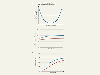
    But BWAS have a perennial, and well-known, problem of low replicability: two studies on the same topic can come to different conclusions. Much of the problem is that some BWAS studies need huge sample numbers to reflect effects accurately. Small sample sizes can exaggerate the relationship of a certain brain feature to a behaviour or trait. In the similar field of genome-wide association studies — which seek to relate differences in DNA with traits in health or disease — the problem of unreliability is being overcome by gathering data sets with tens of thousands of samples from participants. However, in the case of the brain, this is much more difficult, especially for researchers outside Europe and the United States. One hour of scanning in a molecular resonance imaging (MRI) machine costs about US$1,000. The US National Institutes of Health distributes around $2 billion for neuroimaging research each year, but few other countries have this level of resource.

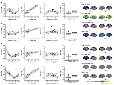
    Vandekar and his colleagues suggest that concentrating on quality, rather than quantity, could be one answer. They analysed more than 100,000 MRI scans from healthy adults and healthy children, as well as scans from children with mental-health conditions.

    Their aim was to explore how factors such as age, sex, cognitive function and mental health are associated with brain structure and function across diverse study designs. For example, one study explored how brain volume changes with age. Vandekar and his co-authors found that, compared with one-off scans of multiple people — cross-sectional studies — repeated MRI scans of the same people over time yielded more-robust results (see R. J. Chauvin and N. U. F. Dosenbach Nature https://doi.org/10.1038/d41586-024-03650-5; 2024).

    Such longitudinal studies have long proved their worth in areas of science such as identifying biomarkers for chronic or degenerative diseases (Y. Guo et al. Nature Aging 4, 247–260; 2024). Although they don’t work for some types of question for which cross-sectional studies are needed, longitudinal studies are good at ruling out irrelevant factors that looked as if they might be implicated during small cross-sectional studies.

    There are caveats, however: researchers conducting longitudinal studies must, for instance, take care to leave long enough gaps between measurements in any one individual if they are to capture meaningful and statistically significant differences over time. Vandekar and his colleagues also emphasize that researchers must account both for changes that happen in individuals over time and for differences between individuals.

    All research needs to be planned. For BWAS, selecting participants in such a way as to achieve robust results and using the right statistical models can improve the trustworthiness of findings without an automatic need for massive sample sizes. The benefits of statistical rigour, in turn, highlight the need for more collaboration between statisticians and neuroscientists, as they use more-sophisticated data-handling methods in their research. These findings will be valuable to the neuroscience community, and deserve wider attention.

    Many fields of science are plunging into data-driven discovery, increasingly assisted by the pattern-seeking abilities of artificial-intelligence algorithms. As they do so, questions of correlation and causation, and of ensuring that findings are statistically significant and reproducible, are becoming ever more pertinent. That means researchers must not skate over experimental design, whether they are reading a paper or writing one.

    Greater attention to research methods, and to how a study obtains the signal of its effect, is the way to make sure that results stand the test of time.

    [ad_2]

    Source link

  • Brainwave experiment shows minke whales have ultrasonic hearing

    Brainwave experiment shows minke whales have ultrasonic hearing

    [ad_1]

    New Scientist. Science news and long reads from expert journalists, covering developments in science, technology, health and the environment on the website and the magazine.

    The minke whale is a smaller species of baleen whale

    Kerstin Meyer/Getty Images

    Brainwave testing of two young baleen whales has revealed they can hear higher frequency sounds than previously thought, forcing researchers to rethink how the ocean’s largest animals respond to predators and human noises.

    “This is truly groundbreaking work,” says Susan Parks at Syracuse University in New York, who wasn’t involved in the new study. “Directly measuring the hearing of a wild baleen whale is something the researchers in the field have been working towards for decades… This is, to my knowledge, the first successful test of this method with a baleen whale.”

    But baleen whales include the largest animals on Earth, and the study method of temporarily restraining them for a hearing test isn’t easy. “The body size of most baleen whales is too large for the approach to be effective,” says Dorian Houser at the National Marine Mammal Foundation, a nonprofit organisation based in California. So Houser and his colleagues turned to a relatively small baleen species called the minke whale.

    The researchers examined the migratory route of minke whales along the coast of Norway and found a natural channel between two islands, where they used net barriers and boats to guide two whales – each about 3 to 5 metres in length – into a fish farm enclosure with a drop-down net door. Researchers then used a roller system to pull up a net and hold the teenage animals partially submerged at the water’s surface.

    The hearing test involved placing two gold-plated electrodes with silicone suction cups on each whale’s skin near its blowhole and dorsal fin, which enabled the researchers to record brainwave signals. They measured how the whales’ brains responded to sounds played from an underwater speaker for about 30 minutes for one whale and 90 minutes for the other.


    Such experiments revealed that the whales’ auditory brainstems respond to ultrasonic sounds, which are beyond those the human ear can detect, at frequencies as high as 45 to 90 kilohertz – a much broader hearing range than previously believed possible based on ear anatomy and vocalisations.

    The corralling and restraining of wild marine mammals is “quite controversial” because of the potential for “significant stress” in the animals, says Oliver Boisseau at Marine Conservation Research, a nonprofit organisation based in the UK. But he described the findings as “very important” in helping understand how baleen whales may evade predators such as killer whales, which hunt using high-frequency echolocation clicks.

    Researchers should also rethink how baleen whales are affected by military sonar and commercially available echo sounders used for mapping the seafloor, says Boisseau. “It seems the more we study the hearing of marine mammals, the more we confound our initial assumptions,” he says.

    Topics:

    [ad_2]

    Source link

  • our cells learned to handle the stress that comes with size

    our cells learned to handle the stress that comes with size

    [ad_1]

    Sagittal slice through a human brain.

    Large brains place extra demands on nerve cells.Credit: Science Pictures Ltd/SPL

    Humans have evolved disproportionately large brains compared with our primate relatives — but this neurological upgrade came at a cost. Scientists exploring the trade-off have discovered unique genetic features that show how human brain cells handle the stress of keeping a big brain working. The work could inspire new lines of research to understand conditions such as Parkinson’s disease and schizophrenia.

    The study, which was posted to the bioRxiv preprint server on 15 November1, focuses on neurons that produce the neurotransmitter dopamine, which is crucial for movement, learning and emotional processing.

    By comparing thousands of laboratory-grown dopamine neurons from humans, chimpanzees, macaques and orangutans, researchers found that human dopamine neurons express more genes that boost the activity of damage-reducing antioxidants than do those of the other primates.

    The findings, which are yet to be peer-reviewed, are a step towards “understanding human brain evolution and all the potentially negative and positive things that come with it”, says Andre Sousa, a neuroscientist at the University of Wisconsin–Madison. “It’s interesting and important to really try to understand what’s specific about the human brain, with the potential of developing new therapies or even avoiding disease altogether in the future.”

    Stressed-out neurons

    Just as walking upright has led to knee and back problems, and changes in jaw structure and diet resulted in dental issues, the rapid expansion of the human brain over evolutionary time has created challenges for its cells, says study co-author Alex Pollen, a neuroscientist at the University of California, San Francisco. “We hypothesized that the same process may be occurring, and these dopamine neurons may represent vulnerable joints.”

    Using an imaging tool, Pollen and his colleagues showed that two dopamine-demanding regions of the brain are considerably bigger in humans than in macaques. The prefrontal cortex is 18 times larger, and the striatum nearly seven times bigger.

    Yet humans have only around twice as many dopamine neurons as their primate relatives, says Pollen. These neurons therefore have to stretch further and work harder — each forming more than two million synapses — in the larger, more complex human brain.

    “The dopamine neurons are real athletes,” says Nenad Sestan, a developmental neuroscientist at Yale University in New Haven, Connecticut. “They are constantly activated.”

    To understand how human dopamine neurons might have adapted to cope with the demands of a large brain, Pollen and his colleagues grew versions of these cells in the lab.

    They combined stem cells — which can develop into many cell types — from eight humans, seven chimpanzees, three macaques and one orangutan and grew them into miniature, brain-like structures called organoids. After 30 days, these structures started producing dopamine, mimicking a developing brain.

    The team then genetically sequenced the dopamine neurons to measure which genes were switched on and how they were controlled.

    In an analysis of human and chimpanzee neurons, the researchers found that the human neurons expressed higher levels of genes that manage oxidative stress — a type of cell damage that can be caused by the energy-intensive process of producing dopamine. These genes encode enzymes that break down and neutralize toxic molecules, called reactive oxygen species, that can harm cells.

    To investigate whether human dopamine neurons might have have evolved unique stress responses, the authors applied a pesticide that causes oxidative stress to the organoids. They found that neurons that had developed from human cells increased their production of a molecule known as BDNF, which is reduced in people with neurodegenerative disorders such as Parkinson’s disease. They did not see the same response in chimpanzee neurons.

    Boosting resilience

    Understanding these protective mechanisms could aid the development of therapies that boost cellular defences in people at risk of Parkinson’s disease. “Some of these protections might not be present in everyone due to mutations,” says Sousa. “That creates an extra vulnerability in those individuals.”

    “There are some candidate targets that might be very interesting to perturb and then transplant in [animal] models of Parkinson’s disease to see whether these endow the neurons with more resilience,” says Pollen.

    The organoids in the study represent developing neurons, equivalent to those that are present in an embryo, and do not fully capture the complexity of adult neurons. Future research will need to explore how such protective mechanisms hold up in mature and ageing neurons, says Sousa, because “degenerative diseases that affect these cells are usually at a late age”.

    [ad_2]

    Source link

  • How stress can disrupt memory and lead to anxiety

    How stress can disrupt memory and lead to anxiety

    [ad_1]

    A little brown mouse runs on a white wheel within its enclosure.

    Neurotransmitters in the amygdala might be why we have anxious responses in harmless situations.Credit: Owen Franken/Corbis via Getty

    Stress makes mice form big bundles of neurons in the brain that disrupt memory formation, making them fearful of harmless situations1 — which might help to explain why stressed people often feel threatened in safe environments.

    Researchers have long known that stress or trauma can lead people to fear harmless situations. For instance, after burning a finger on a hot pan, a stressed individual might subsequently avoid not only hot pans but the kitchen or cooking entirely. This kind of generalized fear is common in people with post-traumatic stress disorder (PTSD) and generalized anxiety disorder.

    A study, published in Cell today, describes how stress disrupts memory formation and, in particular, recollections of fearful events. The results could inform the development of therapies for people with PTSD and anxiety.

    “This paper is really a tour-de-force,” says Ryuichi Shigemoto, a neuroscientist at the Institute of Science and Technology Austria, in Klosterneuburg. “They used so many different methods and techniques to prove this long pathway.”

    Memory packages

    Memories are packaged into groups of neurons, called engrams, which are active when a memory is being formed. Sheena Josselyn, a neuroscientist at the Hospital for Sick Children in Toronto, Canada, and her colleagues looked at whether stress disrupts engram formation and focused on a region of the brain called the amygdala, which is involved in stress and emotion response.

    The study involved an elaborate three-step experiment in mice. First, they put some adult mice in a stressed state by injecting them with the stress hormone corticosterone or restraining them in a small tube for 30 minutes, which increased their corticosterone levels.

    They then placed mice — some stressed, and others not — in a chamber and played a medium-pitched sound for 30 seconds, a neutral event. After a break, the mice went back into the chamber and experienced a high-pitched whistling sound for 30 seconds, which ended with a 2-second shock to the foot, to mimic a fearful event.

    To test how the mice had stored the memories of these experiences, the researchers put the mice in a new environment and played the two tones — watching for how they responded.

    The unstressed mice froze mostly when they heard the high-pitched whistling, whereas the stressed mice froze in response to both sounds, suggesting that they couldn’t distinguish between the neutral and fearful events.

    Exclusive club

    The researchers used various techniques to visualize neural activity in the rodents. They found that, during memory formation, the unstressed mice formed small engrams in response to the whistle and foot shock, and these were only reactivated when exposed to the whistle. But the stressed mice formed larger engrams, which were reactivated when exposed to both sounds.

    Further experiments uncovered the chain of events in the brain that created the larger engram in stressed mice. Under normal conditions, specific neurons in the amygdala block neuronal activity through the release of chemical messengers known as gamma-aminobutyric acid (GABA). This ensures that a small engram is created in response to a negative memory. “It’s sort of like the velvet rope at a nightclub: it only lets certain neurons into the nightclub,” says Josselyn. But under stress, excitatory neurons pump the brain with a neurotransmitter known as endocannabinoid, which binds to glucocorticoid receptors on those inhibitory neurons and prevents them from releasing GABA, resulting in larger engrams. In other words, the velvet rope drops, “and many neurons can get into this exclusive club”, says Josselyn.

    The team were able to reverse the effects of stress on memory formation with two drugs, one of which is approved for terminating early pregnancy, mifepristone. The drugs either block the glucocorticoid receptors or the production of endocannabinoids, and the stressed mice recalled memories in the way that unstressed mice did. But researchers caution that the medications have side effects beyond the brain, and work only if they are given at the time the memory is formed, so are unlikely to be useful in people.

    Josselyn and her colleagues are now trying to investigate whether engrams can be altered after a memory has formed, or whether there are other ways of mitigating the effects of stress on memory.

    [ad_2]

    Source link

  • Exquisite bird fossil provides clues to the evolution of avian brains

    Exquisite bird fossil provides clues to the evolution of avian brains

    [ad_1]

    The skeleton of Navaornis hestiae, an 80-million-year-old bird fossil

    S. Abramowicz/Dinosaur Institute/Natural History Museum of Los Angeles County

    An 80-million-year-old fossil bird has been discovered with a skull so exquisitely preserved that scientists have been able to study the detailed structure of its brain.

    In both age and evolutionary development, the new species, named Navaornis hestiae, is almost midway between the earliest known bird-like dinosaur, Archaeopteryx, which lived 150 million years ago, and modern birds. It lived in the Cretaceous Period alongside dinosaurs such as Tyrannosaurus and Triceratops.

    The fossil, which bears a superficial resemblance to a pigeon, was found near Presidente Prudente, Brazil, in 2016 and was immediately recognised as significant because of the rarity of a full bird skeleton, particularly one of that age.

    But Daniel Field at the University of Cambridge says it wasn’t until 2022 that he and his colleagues realised the skull was so intact that they could possibly scan it and create a 3D model of its brain.

    High-resolution CT scanning allows palaeontologists to peer inside fossils. “This involves careful ‘digital dissection’: separating out each individual component of the skull and then reassembling them into a complete, undeformed three-dimensional reconstruction,” says Field.

    “The new fossil provides unprecedented insight into the pattern and timing by which the specialised features of the brain of living birds evolved.”

    Based on the team’s reconstruction of the brain, Field says the cognitive abilities and flying capacity of Navaornis were probably inferior to those of most living birds.

    Artist’s impression of Navaornis hestiae

    J. d’Oliveira

    The portions of the brain responsible for complex cognition and spatial orientation aren’t as enlarged as those of modern birds, he says.

    “Although the cerebrum of Navaornis is greatly expanded relative to the condition in a more archaic bird relative like Archaeopteryx, it is not as expanded as what we see in living birds.”

    The enlarged brains of modern birds support a huge range of complex behaviours, says Field, but understanding how their brains evolved has been challenging due to a lack of adequately complete and well-preserved fossil bird skulls from early bird relatives.

    Navaornis fills a roughly 70-million-year-long gap in our understanding of how the distinctive brains of modern birds evolved.”

    Topics:

    [ad_2]

    Source link

  • Should Alzheimer’s be diagnosed without symptoms? Proposal to rely on blood tests roils scientists

    Should Alzheimer’s be diagnosed without symptoms? Proposal to rely on blood tests roils scientists

    [ad_1]

    A woman holds a piece of paper with a blue cube-shaped drawing on it next to an elderly woman who is writing on a sheet of paper

    Cognitive tests would be used less often to diagnose Alzheimer’s disease under a proposal by some scientists.Credit: Burger/Phanie/Science Photo Library

    Controversy has erupted among researchers over an effort to adopt blood tests and brain scans for diagnosing Alzheimer’s disease, rather than the cognitive screening that has been used for decades.

    Proponents of the change say that new biomarker tests can detect Alzheimer’s at a very early stage — the best time to apply any treatments that are developed to reverse the disease. But critics say that, although the effort is well-intentioned, it means that people can be diagnosed with a single test, even if they have no symptoms of cognitive decline — and might never develop them.

    “There’s a risk of misunderstanding and distress that individuals who are asymptomatic will have if we tell them they have Alzheimer’s, whereas nothing will happen in their lifetime in a majority of cases,” says Nicolas Villain, a neurologist at Sorbonne University in Paris, who co-wrote a paper1 published on 1 November in JAMA Neurology criticizing the new diagnostic criteria.

    Plaques and tangles

    The brains of people with Alzheimer’s have two key features: plaques of sticky amyloid-β proteins and tangles formed from tau proteins. The neurodegeneration linked with the development of these plaques and tangles is irreversible, so researchers have been searching for treatments to give to healthy people to ward off this damage entirely.

    In the past few years, companies have started marketing drugs that slow Alzheimer’s-related cognitive decline by clearing amyloid from the brain, and scientists have been perfecting highly accurate tests for both amyloid and tau protein.

    “It’s this confluence of the possibility of widespread clinically available, accurate diagnosis with the ability to do something about the disease that prompted us to update the criteria,” says Clifford Jack, a specialist in clinical Alzheimer’s and dementia research at Mayo Clinic in Rochester, Minnesota, who co-led the effort to revise the diagnosis criteria. Jack and his colleagues in a working group for the Alzheimer’s Association, a non-profit research and advocacy organization in Chicago, Illinois, published their guidelines2 in the journal Alzheimer’s & Dementia in June.

    The criteria say that any one abnormal result on a core set of biomarker-based tests is sufficient to diagnose Alzheimer’s. These tests include measurements of amyloid and tau-protein levels in blood or cerebrospinal fluid, and a positron-emission tomography (PET) brain scan, which helps to quantify amyloid plaques.

    Devastating diagnosis

    But Villain and his colleagues point out in their critique that a large swathe of people diagnosed in this way would never develop any cognitive symptoms: a 65-year-old man who is amyloid-biomarker positive has a lifetime risk of developing Alzheimer’s dementia of about 22%, which is only roughly 1.7 times higher than the risk for a similar individual who is amyloid-biomarker negative.

    The critics also argue that people who test positive for a single biomarker and are cognitively unimpaired should be informed that they are at risk of the disease but should not be given an official Alzheimer’s diagnosis. A person without symptoms who either tests positive on multiple biomarker tests or has a gene variant known to significantly increase the risk of developing Alzheimer’s dementia could be given a diagnosis of ‘presymptomatic’ Alzheimer’s, the critics write.

    Jack acknowledges that biomarker testing makes it possible for asymptomatic individuals to be diagnosed with the disease — but the guidelines state that biologically-based diagnoses are intended to “assist rather than supplant” clinical evaluations. And the working group does not recommend Alzheimer’s biomarker tests for healthy people, so a hypothetical positive diagnosis for someone without symptoms should not come to pass, he says.

    However, the new criteria might expand eligibility for clinical trials, which could help to develop treatments for asymptomatic individuals, Jack says. “The reality is that every person who ultimately becomes demented due to Alzheimer’s went through a period of time when they were asymptomatic with the disease,” he says. “Medicine needs to focus its future on how to prevent the onset of symptoms, because by the time someone becomes symptomatic, extensive irreversible damage has already occurred.”

    Nothing in the cupboard

    But medications for biomarker-positive, asymptomatic people are currently non-existent except in clinical trials, says Andrea Bozoki, a cognitive neurologist at the University of North Carolina School of Medicine in Chapel Hill who co-wrote the JAMA Neurology critique. This would leave such people with the mental anguish of having a diagnosis for an incurable disease but lacking treatment options, she says.

    The new drugs that that slow the cognitive decline caused by the disease are approved in the United States only for people who are already experiencing mild cognitive impairment.

    Bozoki worries that the new criteria will spur healthy people who fear that they are at risk for Alzheimer’s, or who have a family history of the disease, to find a doctor who will order a biomarker test for them. And if they’re diagnosed, she says, they might be prescribed the new Alzheimer’s drugs. These haven’t been shown to be effective in asymptomatic populations, cost tens of thousands of US dollars a year and carry a risk of brain bleeding and fatal seizures.

    This will make it even more important for researchers and doctors to ensure that they are properly communicating risk and uncertainty as Alzheimer’s tests and drugs become more available, says Winston Chiong, a neurologist and ethicist at the University of California, San Francisco, who was not involved with either workgroup.

    [ad_2]

    Source link

  • Why do wet dogs shake themselves dry? Neuroscience has an answer

    Why do wet dogs shake themselves dry? Neuroscience has an answer

    [ad_1]

    A wet Golden Retriever dog sprays water as it shakes to dry itself.

    Experiments with mice have revealed the neuroscience of why dogs shake their wet fur.Credit: Nat NT/Getty

    When a dog shakes water off its fur, the action is not just a random flurry of movements — nor a deliberate effort to drench anyone standing nearby.

    This instinctive reflex is shared by many furry mammals including mice, cats, squirrels, lions, tigers and bears. The move helps animals to remove water, insects or other irritants from hard-to-reach places. But underlying the shakes is a complex — and previously mysterious — neurological mechanism.

    Now, researchers have identified the neural circuit that triggers characteristic ‘wet dog’ shaking behaviour in mice — which involves a specific class of touch receptors, and neurons that connect the spinal cord to the brain. Their findings were published in Science on 7 November1.

    “The touch system is so complex and rich that [it] can distinguish a water droplet from a crawling insect from the gentle touch of a loved one,” says Kara Marshall, a neuroscientist at Baylor College of Medicine in Houston, Texas. “It’s really remarkable to be able to link a very specific subset of touch receptors to this familiar and understandable behaviour.”

    Sensitive skin

    The hairy skin of mammals is packed with more than 12 types of sensory neuron, each with a unique function to detect and interpret various sensations. A team led by Dawei Zhang, a neuroscientist then at Harvard University in Cambridge, Massachusetts, focused on a type of ultra-sensitive touch detecting receptors called C-fibre low-threshold mechanoreceptors (C-LTMRs), which wrap around hair follicles.

    In humans, these receptors are associated with pleasant touch sensations, such as a soft hug or a soothing stroke. But in mice and other animals, they serve a protective role: alerting them to the presence of something on their skin, whether it’s water, dirt or a parasite. When these stimuli cause hairs on the skin to bend it activates the C-LTMRs, says Marshall, “extending the sensibility of the skin beyond just the surface”.

    To get laboratory mice to shake their fur like wet dogs, the researchers applied drops of sunflower oil to the backs of the mice’s necks. Nearly all the animals shook off these drops within ten seconds. The team then genetically modified some of the mice to remove most of their C-LTMRs. These animals showed a 50% reduction in shakes when oil droplets landed on their neck, compared with unmodified control mice.

    The researchers also wanted to explore how signals from C-LTMRs travel through the nervous system to orchestrate the wet dog shakes. They traced the pathway to a group of neurons in the spinal cord; this connects to an area in the brain called the parabrachial nucleus, which is involved in processing pain, temperature and touch.

    Using optogenetics, a technique that engineers neurons so that they can be switched on and off in response to light, the researchers blocked the activity of the spinal neurons. These mice showed a 58% reduction in shakes compared with control mice. Blocking activity in the parabrachial nucleus produced similar results. The mice still scratched, groomed and moved normally, suggesting that the neural circuit is specific to wet dog shakes.

    Specialized circuit

    The discovery opens up avenues for future research. “The wet dog shake is a very coordinated motor response,” says Thomas Knöpfel, a neuroscientist at Hong Kong Baptist University in Kowloon Tong, who adds that the study is a good starting point to study how the brain sends commands to control the movement. “Wet dog shake is triggered in many animals by psychedelic drugs,” he says. The response to psychedelics involves serotonin receptors, which also play a part in pleasurable touch. “That gives inspiration for some more work connecting the dots.”

    Zhang says that future research could also investigate whether overactive C-LTMRs contribute to conditions such as twitch-skin syndrome in cats, which involves sudden rippling of the skin and excessive twitching, or to other kinds of skin hypersensitivity in humans.

    [ad_2]

    Source link

  • The brain summons deep sleep for healing from life-threatening injury

    The brain summons deep sleep for healing from life-threatening injury

    [ad_1]

    A woman lies in bed asleep with sunlight shining on her face

    Ample sleep after a heart attack dampens inflammation in the organ, aiding recovery.Credit: Getty

    Immune cells rush to the brain and promote deep sleep after a heart attack, according to a new study1 involving both mice and humans. This heavy slumber helps recovery by easing inflammation in the heart, the study found.

    The findings, published today in Nature, could help to guide care for people after a heart attack, says co-author Cameron McAlpine at the Icahn School of Medicine at Mount Sinai in New York City, who studies immune function in the cardiovascular and nervous systems. “Getting sufficient sleep and rest after a heart attack is important for long-term healing of the heart,” he notes.

    The implications of the study go beyond heart attack, says Rachel Rowe, a specialist in sleep and inflammation at the University of Colorado Boulder. “For any kind of injury, your body’s natural response would be to help you sleep so your body can heal,” she says.

    The heart needs its sleep

    Scientists have long known that sleep and cardiovascular health are linked. People who sleep poorly are at a higher risk of developing high blood pressure, for example, than are sound sleepers. But how cardiovascular disease affects sleep has been less explored.

    To learn more, the authors induced heart attacks in mice and investigated the animals’ brainwaves. The researchers found that these mice spent much more time in slow-wave sleep — a stage of deep sleep that has been associated with healing — than did mice that hadn’t had a heart attack.

    Next, the authors sought to understand what was causing that effect. One obvious place to look was the brain, which controls sleep, notes McAlpine. After a heart attack, immune cells trigger a massive burst of inflammation in the heart, he says, and the researchers wondered whether these immune changes also occurred in the brain.

    The team found that, after a mouse’s heart attack, immune cells called monocytes flooded its brain. These cells produced large amounts of a protein called tumour necrosis factor (TNF), which is an important regulator of inflammation and also promotes sleep.

    To confirm that these cells were linked to the increased sleep, researchers prevented monocytes from accumulating in the rodents’ brains. As a result, “the mice no longer had this increase in slow-wave sleep after their heart attack,” McAlpine says, supporting the theory that the influx of monocytes to the brain contributes to the post-heart-attack sleep boost. Similar experiments confirmed TNF’s role as a messenger to sleep-inducing brain cells.

    Slumbering toward recovery

    To understand the purpose of the extra sleep, the researchers repeatedly interrupted slow-wave sleep in mice that had had a heart attack. The team found that these mice had more inflammation in both the brain and the heart, and had a much worse prognosis than mice that were allowed to sleep undisturbed after a heart attack.

    The authors also studied humans who had experienced acute coronary syndrome, a term for conditions, including heart attack, that are caused by a sudden reduction of blood flow to the heart muscle. Those who reported poor sleep in the weeks following such an episode had a higher risk of developing heart attacks and other serious cardiovascular problems over the next two years than did those who were good sleepers.

    Given the findings, “clinicians need to inform patients of the importance of a good night’s sleep” after a heart attack, says Rowe. This should also be considered at the hospital, where tests and procedures would ideally be conducted during the daytime to minimize sleep interruptions.

    She adds that the findings highlight the bidirectional relationship between sleep and the immune system. “When your grandma says, ‘if you don’t get enough sleep, you’ll get sick’, there’s a lot of truth to that.”

    [ad_2]

    Source link

  • We're starting to understand what being bullied does to the brain

    We're starting to understand what being bullied does to the brain

    [ad_1]

    Being bullied when young seems to alter your brain structure for years to come – with different changes seen in males and females

    [ad_2]

    Source link

  • Separating the “woo” from the work of manifesting in two new books

    Separating the “woo” from the work of manifesting in two new books

    [ad_1]

    New Scientist. Science news and long reads from expert journalists, covering developments in science, technology, health and the environment on the website and the magazine.

    If you manifest it hard enough, might you find yourself here?

    David Hornback/Millennium Images

    Mind Magic
    James Doty (Yellow Kite (UK); Avery (US))

    The Neuroscience of Manifesting
    Sabina Brennan (Orion Spring (ebook and audio))

    Earlier this year, my daughter moved into college for her first year of university. Amid the boxes lining the hallways, I noticed a bulletin board covered in photos of scrub-clad physicians and inspirational quotes. When I stopped to take a closer look, the mother of the student it belonged to came out to say hello.

    “I told my daughter to put her vision board where she can see it every time she…

    [ad_2]

    Source link