Tag: cancer

  • Why an Offline Nuclear Reactor Led to Thousands of Hospital Appointments Being Canceled

    Why an Offline Nuclear Reactor Led to Thousands of Hospital Appointments Being Canceled

    [ad_1]

    “I remember in Geneva, two months ago, we said, ‘Pay attention, because on this specific week there is a risk of shortage if there is any problem with one of the active reactors’—and that’s what happened,” recalls David Crunelle, a spokesman for Nuclear Medicine Europe (NMEU), an industrial association.

    Because of their very nature, it’s impossible to stockpile these radioactive substances—they are fleeting. Technetium-99m works as a radioactive tracer because, as it decays, it flings out gamma rays with a photon energy of 140 KeV. This is “fairly ideal” for detection using a gamma ray camera, says Cathy Cutler, chair of isotope research and production at Brookhaven National Laboratory in the US.

    But technetium-99m has a very short half-life, just six hours or so. Hence why radioisotope-producing facilities send miniature generators containing molybdenum-99 out to hospitals. These generators, sometimes called “moly cows,” produce the desired technetium-99m as the molybdenum-99 decays—a bit like a portable vending machine for technetium-99m, which runs out after about two weeks, once the molybdenum-99 has completely decayed.

    Glenn Flux, head of radioisotope physics at London’s Royal Marsden Hospital and Institute of Cancer Research, says the thing that makes a technetium-99m scan different to, say, a CT or MRI scan, is that it reveals how patients’ organs or a tumor are functioning—for example by revealing blood flow to the area in question.

    “The CT will show you if there’s a tumor, but the technetium or other isotopes will tell you whether it’s active or aggressive,” explains Flux.

    The recent radioisotope shortage caused a few thousand appointment cancellations in the UK alone, estimates Stephen Harden, vice president of clinical radiology at the Royal College of Radiologists (RCR). Health care staff swung into action to distribute the remaining radioisotope supplies around the UK, in order to ensure that the most urgent patients—those with cancer, for instance—were still able to attend their scans. “If there hadn’t been a nationally coordinated policy, there would have been significant regions in the country with no supply at all,” says Harden.

    Crunelle and colleagues at NMEU continually monitor medical radioisotope production at key reactors around the world. They learn about maintenance schedules well in advance, and, as such, NMEU will often advise reactor chiefs to push these dates back slightly—for example, in order to help minimize the risk of multiple shutdowns occurring at the same time. NMEU staff use software, a kind of reactor maintenance calendar, that allows them to forecast production levels. But sometimes unpredictable events occur, such as the problem with the pipe in Petten.

    [ad_2]

    Source link

  • Toxicity and costs of cancer treatment reduced by deferring CDK4/6 inhibitor use

    Toxicity and costs of cancer treatment reduced by deferring CDK4/6 inhibitor use

    [ad_1]

    • CLINICAL BRIEFINGS

    The SONIA study shows that early use of inhibitors that target both CDK4 and CDK6 (hereafter, CDK4/6) in metastatic breast cancer prolongs time on treatment but does not improve patient outcomes. Postponing innovative treatments to a later stage of the disease could thus safely reduce the time on treatment, the number of adverse effects for people with breast cancer and the burden on health-care resources.

    [ad_2]

    Source link

  • Québec’s catalyst for cancer research and innovation

    Québec’s catalyst for cancer research and innovation

    [ad_1]

    Oncopole drives cancer research in Québec, uniting partners to accelerate innovation in treatment, prevention, and patient care.

    Oncopole, pôle cancer du Fonds de recherche du Québec – secteur Santé (FRQ), is a key player in the fight against cancer in Québec. Thanks to the financial contribution of its partners, the Ministère de l’Économie, de l’Innovation et de l’Énergie, its founding partner Merck Canada, and its partners GSK and Pfizer Canada, it acts as a catalyst to harness the full potential of the research and innovation ecosystem.

    A model of collaboration and innovation

    Oncopole places a strong emphasis on collaboration between multiple stakeholders: patients, researchers, clinicians, industry and other strategic public and private partners. These collaborations enable the development and commercialisation of innovative medical technologies and help position the province as a world leader in cancer research.

    Our funding programmes and collaborations enable us to support cutting-edge research in three complementary and interrelated research pillars:

    Basic and translational research

    Basic research advances knowledge, while translational research aims to translate that knowledge into concrete applications. Thanks to Oncopole’s support, Québec researchers can accelerate the discovery of new therapeutic targets and the development of innovative treatments (e.g. EMC2).

    Clinical research

    Through its partnerships, Oncopole ensures that Québec has a state-of-the-art clinical research infrastructure, giving patients access to innovative therapies and allowing researchers and companies to demonstrate the effectiveness of their innovations in a real-world setting (e.g. PROVEM).

    Evaluative research and population health

    This pillar focuses on the impact of treatments and health policies. It includes studies on the relevance of treatments, population health and access to care. Through this approach, Oncopole contributes to prevention and screening policies while seeking to improve equity in access to oncology care (e.g. Priorité patient).

    Strength in research and innovation

    Québec boasts a world-class university network, a universally accessible healthcare system and a community of highly experienced oncology researchers in an environment that fosters innovation. Key players include leading groups and organisations such as the Centre de recherche du Centre hospitalier de l’Université de Montréal (CRCHUM), the Centre de recherche du CHU de Québec (CRCHU) and the Centre de recherche de l’Université Laval (CRC), as well as the Réseau de recherche sur le cancer, which are profiled here.

    cancer research

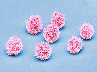
    With one in two Canadians developing cancer in their lifetime, CRCHUM’s Cancer Axis aims to better understand the molecular mechanisms that turn a normal cell into a cancerous one. The aim is to develop cutting-edge tools for personalised medicine and improved patient care. The Axis favours a multidisciplinary approach involving basic researchers and clinicians and is characterised by its expertise in cancers of epithelial origin.

    The Cancer Axis employs 21 core scientists and 107 clinician-scientists. Its development priorities for the coming years are focused on precision medicine, immunotherapy and microbiota. Cancer Axis researchers have acquired multi-omics technologies and stored patient samples in several biobanks, making them ideally positioned for the discovery of new biomarkers and therapeutic targets against cancer.

    Between 2020 and 2024, Axis researchers published more than 1,317 articles, gave more than 265 invited lectures, and filed more than a dozen patents.

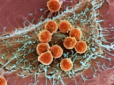
    Highlights of recent years include major publications on cellular senescence by the teams of Drs Rodier and Ferbeyre, on the role of microbiota in cancer by the teams of Drs Routy, Santos and Elkrief, and on precision medicine with the microfluidic culture of tumour explants for personalised screening of anticancer treatments by the teams of Drs Mes-Masson and Gervais. In immunotherapy, important discoveries on new control points of the immune system have been published by Dr Stagg, and anticancer vaccine designs have been designed by Dr Bourgeois-Daigneault. In 2023, Dr S Turcotte treated the first patient in Québec with tumour infiltrating lymphocytes (TIL).

    Finally, Dr Saad published the results of a clinical trial that is changing the treatment of patients with advanced prostate cancer, while Dr Liberman and colleagues established the role of preoperative chemotherapy in the management of gastric cancer.

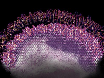
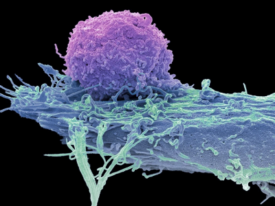
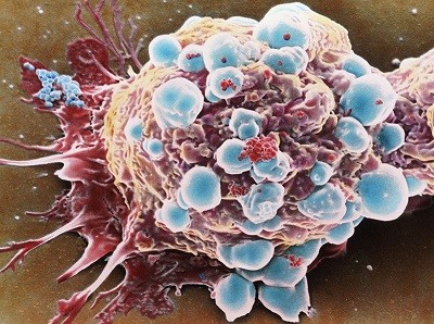
    Revolutionising cancer treatment: The game-changing promise of biomimetic 3D models

    The challenge: Cracking cancer’s complexity

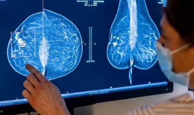
    Precision oncology is transforming cancer care by tailoring treatments to individual patients. However, cancer is not one disease – it’s a complex, dynamic and heterogeneous condition. Tumours vary within patients, shaped by factors such as cellular diversity, the physical environment, immune interactions, and blood supply. To refine personalised treatments, scientists must first understand how these variables drive cancer evolution.

    Outdated models are holding us back

    Current research relies heavily on animal models and organoids, but both have limitations. Animal studies are costly, ethically controversial, and often fail to accurately reflect human biology. Organoids, though useful, lack the complexity of real tumours, making it difficult to replicate the heterogeneity between patients. To unlock the full potential of precision oncology, researchers need models that mimic the intricate realities of cancer.

    A breakthrough in 3D cancer modelling

    Biomimetic 3D cancer models are cutting-edge technology that mimics the spatial organisation, cellular interactions, and mechanical properties of tumours. They replicate essential biological processes like cell migration, proliferation, and differentiation, which are crucial for understanding cancer’s behaviour. Using patient-derived cancer cells, these models can also simulate personalised drug responses, providing a more accurate way to predict treatment outcomes.

    ICE Programme: Québec City’s Beacon of Cancer Research Innovation

    For over 40 years, Québec City has been a leader in cancer research, thanks to trailblazers like Dr Luc Bélanger, who founded the Université Laval Cancer Research Center (CRC) in 1984, establishing a hub for pioneering cancer studies. Around the same time, Dr. François Auger launched the Laboratoire d’organogénèse expérimentale (LOEX), a leader in tissue engineering.

    With the opening of a new Precision Oncology Center (see picture), LOEX and CRC are now side by side within the CHU de Québec-Université Laval research centre, adjacent to one of Canada’s largest cancer treatment facilities, the Centre intégré de cancérologie. This integration, part of a $2bn hospital complex, has sparked unique collaborations across oncology, physics, tissue engineering, and molecular biology.

    The result? The launch of the Innovative Cancer Engineering (ICE) facility, backed by a $6m investment from the Canadian Innovation Funds. ICE features cutting-edge tools like 3D printing, flow cytometry, and spatial transcriptomics to build sophisticated 3D models that replicate tumour complexity – pushing the frontiers of personalised cancer research.

    Collaborative innovation at the ICE facility

    The ICE facility serves as a collaborative platform for researchers, clinicians, and industry experts. By offering unique tools and resources, ICE enhances cancer understanding and supports personalised treatments. We welcome partners to leverage our advanced infrastructure, developing innovative solutions to cancer care challenges. This initiative creates opportunities for researchers and organisations to engage with our state-of-the-art facilities and advance cancer treatment.

    A blueprint for personalised cancer treatment

    The future of cancer treatment lies in precision. By combining tissue engineering with cutting-edge cancer research, the ICE programme offers a powerful new way to understand and combat cancer. With these 3D biomimetic models, we are closer than ever to providing every patient with the best possible treatment – one tailored to their individual needs and biology.

    RRCancer: Uniting Québec’s Oncology Research

    The Québec Cancer Research Network (RRCancer.ca) was formed more than 20 years ago under the leadership of Dr Anne-Marie Mes-Masson (CRCHUM) and is presently co-lead by Drs Jean-Yves Masson (CHU de Québec) and Sonia del Rincon (LDI).

    The network has grown from a few dozen members, mostly fundamental and clinician scientists, with the vision to create an infrastructure of high-quality biobanking to catalyse translational oncology research in the province of Québec. The network has expanded its activities by supporting more than 40 biobanks and now regroups more than 750 members of multidisciplinary expertise. Researchers, clinicians, professionals, students/fellows, patients and strategic partners are working toward the same goal: improving the diagnosis, prognosis, as well as care and services for cancer patients across the province.

    In essence, the mission of RRCancer echoes the ‘Conquering Cancer: Mission Possible’ statement of the Horizon Europe (HE) Programme to deepen cancer understanding, prevent and optimise diagnosis and treatment, support quality of life, and ensure equitable access across Europe.

    Strategic Axes: Catalysts of Innovation with potential impacts on Excellent Science (HE-Pillar I) and Global Challenges (HE-Pillar II)

    RRCancer recognises the importance of aligning its research efforts with those of Horizon Europe, especially since Canada’s announcement in Summer 2024 as a formal decision-maker within the programme. Cancer research within the network is articulated across four main axes focusing on precision health.

    The Translational Research and Bank for Tissue and Data in Solid Cancers (BTD) axis, co-led by Drs Anne-Marie Mes-Masson (CRCHUM) and Morag Park (GCI) focus on biobanking biological material and comprehensive data collection for solid cancers, including hereditary breast and ovarian cancer. Aligned with HE-Pillar I, the BTD develops and optimises live biobanking strategies, uses innovative research infrastructure for biomarker discovery, and consolidates immuno-oncology efforts. The Québec Leukemic Cell Bank and Hematological Cancer Research (BCLQ) axis, led by Dr Josée Hébert (CR-HMR), has a primary objective of understanding the mechanisms involved in haematological malignancies, bringing expertise in haematology, oncology, and immunology.

    The BCLQ develops state-of-the-art tools to facilitate blood cancer diagnosis, prognosis and treatments, as well as develop new therapies for these cancers. The Experimental therapy axis (TE), co-led by Drs Gerald Batist (LDI) and Shirin A Enger (LDI), uses AI- and molecular-based studies to advance personalised medicine. An integral part of this axis is to form a bridge with the bio-pharmaceutical industry for the purpose of rapidly developing new cancer treatments.

    Finally, the newly established Learning and Equitable Health System (SSAE) axis, co-led by Drs Lise Gauvin (CRCHUM) and Sophie Marcoux (CHU de Québec), is well aligned with HE-Pillar II, aiming to foster collaboration amongst researchers in implementation, clinical sciences, with the population and government.

    Additionally, it focuses on accelerating the adoption of innovative therapies and promoting evidence-based cancer practices to enhance the system’s effectiveness and sustainability. For this, they are working toward understanding Québec’s perception of cancer, based on the Baromètre Cancer programme developed in France.

    High-quality biobanking: A research infrastructure supporting Excellent Science (HE-Pillar I)

    The RRCancer is a founding member of the Canadian Tissue Repository Network (CTRNet), which has been providing the research community with educational material, certification processes, policies, and standard operation procedures to support high-quality biobanking aligned with international best practices.

    Furthermore, along with its longstanding partners, the RRCancer supports the development of the ATiM data management system which provides a comprehensive solution to track all biobanking activities including consent, clinical data, inventory management and administrative tasks.

    The strength of this infrastructure is that it favours cohesive interoperability across institutions, which is important as the biobanks and researchers within RRCancer are participating in several multi-institutional initiatives. As an example, researchers of all axes participate and occupy leadership positions within the Marathon of Hope Cancer Centres Network (MOHCCN), an initiative of the Terry Fox Research Institute. This collaborative initiative aims to close the gap between research in the lab and patient care in the clinic.

    To date, more than RRCancer 4,500 biobanked specimens from 1500 participants have had their whole genomes and transcriptomes sequenced, and numbers are growing. These multi-omics data and their associated clinical data will be available to the research community..

    Another RRCancer initiative, Leucegene arising from our BCLQ axis, aims to improve the genomic classification of acute myeloid leukaemia (AML) and develop new, effective precision therapies for AML. Whole genome, exome and/or transcriptome were generated from 452 participants, creating a richly annotated dataset.

    These are only two of the numerous examples of the impact of having this powerful research infrastructure, which has the potential to help overcome the Global Challenge of Cancer (HE-Pillar II).

    Supporting the next generation of scientists: Ensuring Open Science practices

    At RRCancer, we emphasise training the next generation of scientists and integrating them into the research ecosystem. Through our focused initiatives, we support graduate students in disseminating their research locally, nationally and internationally.

    Aligned with the Open Science philosophy, such international interactions are crucial for our trainees to learn the importance of transparent collaboration and will shape well-rounded researchers who are knowledgeable in global challenges in the health research field.

    Furthermore, the RRCancer facilitates the integration of newly recruited early career scientists within the multicentre structuring research projects, favoring interaction, and accelerating collaboration and establishment of their research programme.

    Creating opportunities for increased research impact and visibility through citizen involvement

    In September 2024, we held our biennial two-day Symposium, celebrating the network’s 20th anniversary, attended by over 420 participants. The event featured Dr Aaron Newman from Stanford, who presented computational methods for understanding cancer complexities. Scientific sessions included ten rising star early-career investigators and 20 trainees from across Québec.

    Notably, members of Québec’s RRCancer patient partner group co-chaired the sessions, offering impactful personal testimonies, which made the event especially memorable. The Symposium concluded with two special sessions: one on the ‘Learning and Equitable Health System’ axis, followed by Dr Carol Jabet presenting FRQ-Oncopole’s strategic vision in Québec oncology.

    Please note, this article will also appear in the 20th edition of our quarterly publication.

    [ad_2]

    Source link

  • Why tumour geography matters — and how to map it

    Why tumour geography matters — and how to map it

    [ad_1]

    To people with cancer, tumours can seem like amorphous clumps of defective cells, relentlessly focused on unconstrained growth and invasion. But this does not mean that they’re homogeneous. Cancerous cells have a broad spectrum of mutations, and growths contain healthy host cells, blood vessels and microscale fronts at which immune cells wage war with malignant tissue.

    Until around a decade ago, researchers were ill-equipped to explore this tumour microenvironment. But the emergence of tools that can spatially map large numbers of biomolecules, such as RNA and protein, has caused something of a revolution. Indeed, researchers are increasingly weaving these layers of information together to create rich ‘multiomic’ spatial maps that can classify diverse cell types and probe their activities throughout a tumour.

    “We’re not just talking about tumour heterogeneity any more — we can see it,” says Arutha Kulasinghe, a cancer biologist at the University of Queensland in Brisbane, Australia. “We can see pockets of drug resistance, sensitivity and different biology directly on the tissue.” The spatial factors that contribute to carcinogenesis and disease progression are also increasingly visible, revealing potential vulnerabilities in the process. Such capabilities could transform cancer research and pathology, making it possible to model, interpret and perhaps predict tumour biology with unprecedented sophistication.

    But the barriers to entry are high. There are many technology platforms for spatial omics analyses, and the experiments can be costly and complicated. Even with data in hand, cancer researchers can face a computational odyssey before they can make sense of their results. “Everybody wants what I like to call the ‘blender theory’ of multiomics, which is where you throw all the data sets together and it will tell you the answer as to what’s in them” says Elana Fertig, a bioinformatician at Johns Hopkins Medicine in Baltimore, Maryland. “I’ve become less and less convinced that’s possible, because everybody has a different question that they want to ask.”

    Welcome to the neighbourhood

    For more than a decade, biologists have been studying tumour microenvironments by breaking tissue samples into individual cells and characterizing their molecular contents. These single-cell omics technologies are fairly simple to use, at least for RNA analysis. Instruments such as the Chromium from 10x Genomics in Pleasanton, California, can survey gene expression across millions of individual cells.

    Some researchers, such as cancer genomicist Dan Landau at the New York Genome Center in New York City, have even extended these tools to perform multiomic experiments — coupling transcription to other biological features, such as genomic mutations or epigenetic signals that directly govern gene expression at the single-cell level. “The vision is to try to start understanding how those layers are talking to one another,” says Landau.

    Such experiments can categorize cell types and reveal which biological processes those cells are engaged in — but they lack essential context. “It was pretty clear early on that we miss a lot of information by dissociating a tumour into single cells,” says Bernd Bodenmiller, a systems biologist at the University of Zurich in Switzerland and the Swiss Federal Institute of Technology. For example, the efficacy of immunotherapy against a given tumour depends not only on which immune cells are present, but also where they are in the tumour.

    In 2014, Bodenmiller helped to pioneer the spatial omics era when he and his colleagues combined a laser ablation technique with mass spectrometry to detect and localize proteins labelled with various metal-tagged antibodies (see Nature 567, 555–557; 2019). They called the approach imaging mass cytometry (IMC), and used it to quantify 32 proteins at subcellular resolution in a breast-tumour specimen. Bodenmiller says that these early experiments demonstrated the importance of spatially localized communities of inter-communicating cells, now known as ‘cellular neighbourhoods’, which would have been invisible using dissociated single cells. “These were the first striking examples for me of how the spatial arrangement of tumour cells — and how they form communities with other cells — is really strongly prognostic for patient outcome,” he says.

    Most spatial experiments today focus on the transcriptome, and there are numerous commercial platforms available. Some are sequencing-based, such as the Visium platform from 10x Genomics, which builds on a method developed in 2016 (see Nature 606, 1036–1038; 2022). Tissue slices are prepared on a slide coated with an array of location-barcoded DNA strands. The RNA is then released from the tissue, captured by these strands and converted to DNA for sequencing; the barcode associated with each sequence reveals where it was on the slide.

    Other methods are imaging-based. For example, the MERSCOPE platform from Vizgen in Cambridge, Massachusetts, is based on the technique MERFISH. First reported1 in 2015, the technique involves the serial labelling of tissue samples with fluorescently tagged probes that enable direct visualization, identification and quantification of transcripts in a specimen.

    The choice of platform involves trade-offs. “Generally, the imaging-based technology can capture a larger piece of tissue area, whereas with the sequencing-based [methods] you capture a lot less,” says Kai Tan, a research oncologist at the Children’s Hospital of Philadelphia in Pennsylvania. Imaging-based methods also tend to offer superior spatial resolution — down to the cellular or even sub-cellular scale — but are more labour-intensive and constrained, typically requiring users to select which genes to probe rather than broadly interrogating the tissue RNA, and profiling a smaller fraction of the transcriptome than dissociated, single-cell methods. Sequencing methods can detect even unexpected transcripts, albeit often at lower spatial resolution. But “those two worlds are converging”, Landau notes.

    For instance, the Slide-tags method offers an inventive alternative, in which the address-defining barcodes for spatial transcriptomics are delivered directly into the confines of the cell nucleus, providing subcellular resolution2. These nuclei can then be isolated and analysed more extensively with a range of single-cell methods.

    Regardless of the platform, spatial transcriptomics is unlocking exciting opportunities for cancer researchers. For example, neurosurgeon Dieter Henrik Heiland at the University of Freiburg in Germany has used these techniques to tease apart the conditions that foster the growth and invasive behaviour of brain tumours, such as glioblastoma — specifically, the impact of certain myeloid bone marrow cells on the activity of immune system T cells. “We could identify defined patterns, defined architectures that we could not do before with any other technologies,” he says.

    A multiplicity of maps

    Increasingly, however, transcriptomics represent not the entirety of the spatial analysis but one component thereof — a ‘baseline layer’, as Heiland puts it. “Then, we think what we can do on top.”

    Often, that’s spatial proteomics. Although all proteins are translated from messenger RNAs, not all mRNAs give rise to proteins. Kulasinghe says that in his experience, spatial patterns of RNA and protein can differ by up to 50% in a given sample, such that transcription levels might not reliably predict protein output. Proteins can also form complexes and undergo chemical modifications that would be impossible to determine from transcriptomic data alone. Proteomic analysis is therefore a crucial component in understanding tumour spatial biology. “People are stuck with proteins forever,” says Garry Nolan, an immunologist at Stanford University in California.

    MERSCOPE data image of human ovarian cancer tissue displaying genes represented by different colours

    Data image created using the MERSCOPE platform of the genes in ovarian cancer tissue.Credit: Vizgen

    Today’s spatial proteomics toolbox includes methods that can profile dozens or even hundreds of proteins at a time. For example, Nolan’s group developed3 the widely used CODEX method (now commercialized by Akoya Biosciences of Marlborough, Massachusetts, as the PhenoCycler system) in 2018. This approach uses DNA-tagged antibodies for up to 100 protein targets, which are sequentially detected with an enzymatic process that specifically adds dye-labelled nucleotides to a subset of those DNA tags; these dyes are then cleaved off before the next imaging round. Similarly, the GeoMx platform from NanoString in Seattle, Washington, allows researchers to image RNA at the same time as several hundred proteins in the same sample.

    Fertig and her team reported the combined power of spatial proteomics and transcriptomics in a study that explored the involvement of cells known as fibroblasts in the progression of premalignant pancreatic growths to cancer4. “With the transcriptomics data, we were able to find the fibroblasts and determine their impact on epithelial cells,” Fertig says. The method lacked the spatial resolution to discriminate between cell types fully, but layering on IMC data revealed how some cancer-associated fibroblasts help to establish a microenvironment that promotes malignant growth.

    Perhaps the most direct readout of what a cell is doing at any given moment is the metabolome — the sugars, lipids, peptides and other biomolecules that act as inputs and outputs of biological processes. Several groups are mapping the metabolome using imaging mass spectrometry, in which a laser is scanned over a specially prepared sample to generate spatially localized chemical signatures. “The beauty of the technology is you basically get a completely different picture of your tissue than what you have in your transcriptomic data,” says Heiland. In one 2022 study, Heiland and his colleagues combined this approach with spatial transcriptomics and imaging mass cytometry to map out patterns of oxygen deprivation in glioblastoma. They found that hypoxic conditions lead to more-severe genomic disruption and abnormal gene expression5.

    Room for error

    Still, spatial omics can be intimidating for newcomers. “Everybody wants to adopt spatial, but it’s overwhelming,” says Jasmine Plummer, a geneticist at St Jude Children’s Research Hospital in Memphis, Tennessee. As head of the hospital’s Center for Spatial Omics core facility, she advises users to “think about a specific question you want to answer, not just a fishing expedition”, and then select the method that provides the necessary resolution, multiplexing or other capabilities.

    Some platforms allow users to directly survey multiple molecular categories at once. For example, the NanoString GeoMx and CosMx instruments can perform both protein and gene-expression analysis, and the Landau group collaborated with 10x Genomics to achieve similar analyses using Visium6. But Bodenmiller cautions that in some cases, “you don’t get the optimum of each method” with simultaneous analyses. For example, the enzymatic digestion steps required to liberate RNA from tissue can damage proteins. But optimized workflows are emerging to serially analyse the same specimen using multiple platforms, with the sample-preparation process modified to minimize loss between steps. “I think researchers have realized that you need optimized technology stacks,” says Kulasinghe.

    Other groups perform parallel analyses on consecutive thin sections derived from the same tumour sample, then align and merge the resulting data sets. This is harder than it sounds, however. “If you go from one tissue section to the next, you only find about 50–60% of the cells in both sections,” says Bodenmiller. Furthermore, different experimental formats can produce radically different data types, confounding integration. For example, an IMC experiment yields an array of pixels denoting different proteins at subcellular resolution, whereas sequencing-based transcriptomic experiments map ‘spots’ that often encompass multiple cells. “Every data set has to be treated on its own, and you have to figure out how we can now integrate those data by some kind of similarity measurement,” says Heiland.

    There is also the fundamental challenge of segmentation: accurately defining and classifying individual cells in the spatial data. “If you cannot accurately segment the boundary of the cell, then everything downstream will be off,” says Tan. Different spatial platforms bring different challenges, and there are no universal solutions — Kulasinghe’s team has tested multiple algorithms for this purpose and observed inconsistent performance. As a solution, his team draws boundaries based on a ‘majority vote’ derived from multiple algorithms. Kulasinghe also emphasizes the importance of using conventional histology stains to fact check algorithmic analyses and establish ‘ground truth’ for a spatial study.

    Above all, careful planning is essential. Spatial omics experiments are expensive — Kulasinghe says that a single imaging-based transcriptomics assay can cost nearly US$10,000 — and can generate terabytes of data. “Getting pilot data in this realm is important,” says Plummer. “I don’t think you want to take a whole deep dive in until you’ve understood your data a little bit first.”

    The final frontier?

    Fortunately, the number of core facilities is growing, giving researchers access to expert guidance as well as the technological capabilities needed to perform spatial analyses.

    In parallel, international and cross-institutional research efforts are leveraging single-cell — and, increasingly, spatial — multiomic analysis at unprecedented scale, including the US National Institutes of Health-backed Human Tumour Atlas Network and global consortium the Human Cell Atlas. These efforts are developing and optimizing analytical pipelines and tools, and, more importantly, generating vast collections of reference data for healthy and diseased tissues that the scientific community can use to interpret future experiments. “Without those large initiatives, we would really not be at the state of technology and possibility where we are now,” says Bodenmiller.

    Meanwhile, some spatial-omics pioneers are looking to new horizons. Bodenmiller’s group reported an alternative to IMC in which antibodies are labelled with isotopic tags that can be detected and distinguished by X-ray imaging rather than mass spectrometry, allowing rapid mapping of many proteins at once throughout the specimen7. He says that the method could be an excellent fit for 3D imaging, and is fast because it avoids the slow scanning process that is typical of IMC. First, however, the team must work out logistical challenges, such as how to efficiently deliver antibodies into the interior of intact tissues.

    The imminent deluge of spatial data will also provide a treasure trove for researchers looking to apply deep-learning methods to cancer. This includes ‘digital pathology’ strategies, in which artificial-intelligence algorithms are trained to correlate features on conventional pathology slides with molecular indicators that are associated with tumour identity, prognosis and susceptibility to treatment. Companies are already entering this space with assays that guide drug selection based on spatial data, and Kulasinghe sees opportunities to assess immune activity in a tumour or predict the likelihood of metastasis without the need for spatial assays in the clinic. “This can give us deeper insights into the tumour microenvironments that ultimately associate with clinical endpoints,” he says.

    For his part, Nolan predicts a post-data world, in which the research priority shifts from generating molecular maps to using them to train AI models that reveal hidden vulnerabilities. “We’re going to be able to create a virtual tissue that looks just like colon cancer,” he says. “Then you can start to change the parameters, and say: ‘OK, how do I stop the following structure from forming?’”

    [ad_2]

    Source link

  • The cells that help the immune system fight lung cancer

    The cells that help the immune system fight lung cancer

    [ad_1]

    Access options

    Rent or buy this article

    Prices vary by article type

    from$1.95

    to$39.95

    Prices may be subject to local taxes which are calculated during checkout

    doi: https://doi.org/10.1038/d41586-024-03857-6

    Subjects

    Latest on:


    [ad_2]
    Source link

  • fresh tools for activating Notch

    fresh tools for activating Notch

    [ad_1]

    From roundworms to humans, signalling through the Notch protein-receptor family drives embryonic development, cellular differentiation and tissue homeostasis. It’s also absolutely essential for the transformation of immature human immune cells into T cells — cellular warriors that target viruses and tumours.

    Yet the activation of Notch signalling has been hard to mimic in the laboratory, making it close to impossible to exploit in the clinic and hampering efforts to generate T-cell therapies. But that could be changing.

    In July, attendees at a conference in Lewiston, Maine, dedicated to Notch signalling were treated to not one, but two new tools to activate the pathway. Unbeknown to each other, two research groups had been working in parallel to develop what they unveiled at the conference.

    “It was a turning point,” says Juan Carlos Zúñiga-Pflücker, a developmental immunologist at the University of Toronto in Canada, whose lab has pioneered many Notch activation techniques. For him, the announcements represented a “sort of a re-emergence” of this work in the Notch field.

    Trouble with 2D

    Most receptors undergo some kind of conformational change after binding to their ligands, leading to a chain of events that alters the cell’s behaviour. But ligand binding alone isn’t enough to activate Notch.

    Once the receptor binds to its ligand (which is tethered to another cell), the ligand-expressing cell reels in the ligand-receptor complex. Like tugging at a loose thread in a sweater, the resulting pulling force unravels part of the Notch receptor, exposing it to enzymes that release the receptor’s intracellular domain and allow it to travel to the nucleus to influence gene expression.

    “The real technological challenge is coming up with a drug that can mimic that pulling effect,” says Vincent Luca, a protein engineer at the Moffitt Cancer Center in Tampa, Florida.

    Current strategies require the co-culture of cells expressing the Notch receptor with cells that express the ligand, or fixing the ligand onto plates or beads. But these ‘2D’ methods can’t be used in vivo, says Luca.

    They’re also a bottleneck for large-scale T-cell production, says George Daley, a haematologist and stem-cell biologist at Harvard Medical School in Boston, Massachusetts, because only the cells at that ligand-presenting interface can be activated. For years, Daley’s lab has struggled with the limitations of 2D methods while trying to coax pluripotent stem cells into T cells.

    In 2021, Daley was approached by Rubul Mout, then a postdoctoral researcher at the University of Washington in Seattle in the lab of David Baker, who won a share of the 2024 Nobel Prize in Chemistry for his work in computer-aided protein design. Mout wanted to apply those design techniques to Daley’s stem-cell-based systems.

    Daley was “tremendously excited” at the idea, and before Mout even officially joined his lab, they discussed engineering a soluble Notch agonist — one that could present a Notch ligand to cells in suspension rather than on a flat surface.

    Using the Rosetta protein-design tool developed in Baker’s lab, Daley, Mout and their colleagues designed potential agonists by attaching multiple copies of the ligand to a protein scaffold. They experimented with designs with as few as two and as many as 120 copies of the ligand, as well as different geometries.

    One arrangement worked particularly well: three copies of the ligand radiating out from the scaffold, like spokes. When added to Notch-expressing cells in suspension, this multivalent ligand linked the cells together in a microscopic pas de deux, with each developing T cell gently pulling on (and activating) the other. Compared with 2D activation, Notch activation using the soluble agonist in a bioreactor created five times more T cells per microgram of agonist1, suggesting that this approach might be useful for large-scale T-cell manufacturing.

    Boosting Notch

    Luca says that his group, by contrast, was searching for a “targeted, focused way of activating Notch”, with an eye to therapeutics. For example, boosting Notch could rejuvenate exhausted T cells trying to destroy a tumour — but this must be done without activating the tumour cells, which also express Notch.

    Luca’s lab generated SNAGs, synthetic Notch agonists (see ‘Mimicking notch’), by fusing a gene for a Notch agonist to one that encodes an antibody fragment targeting cancer cells. The result encodes a bispecific protein that forms what Luca calls a “little molecular bridge” between a Notch-expressing cell and the cancer-antigen-expressing cell. That binding event mechanically activates the receptor in a manner akin to natural Notch signalling2.

    Mimicking Notch: A graphic showing the structure of synthetic notch agonists which, upon binding Notch, mimics the tugging force that induces notch signalling in the cell.

    Source: Adapted from ref. 2

    A very different soluble Notch agonist was unveiled in January. Biophysicist Björn Högberg and his team at the Karolinska Institute in Stockholm study the minuscule, tactile interactions between proteins during cell signalling, which Högberg likens to “a Braille system” that the cells use to talk to one another.

    Working with Luca, the team created a hybrid DNA-protein agonist by attaching multiple copies of the Notch ligand along a piece of DNA that had been folded, origami-style, into a rod. To their surprise, the agonist activated Notch without generating any measurable pulling force3.

    The researchers suggest that their origami agonist might be triggering an alternative mechanism of activation, in which long-term ligand binding is sufficient to cause receptor unravelling, albeit at a much slower pace. But they’re still trying to understand how this might work.

    Reactions from the community about the possibility of a pulling-independent Notch activation mechanism have been mixed, Högberg says: “I think some of them were really intrigued and really, really wanted to know more, and some of them were like, ‘I don’t think this is anything.’”

    For his part, Zúñiga-Pflücker is sceptical that the origami ligand works without any sort of pulling force. But “the work is really nicely done”, he says, adding that “all three papers are quite exciting in different ways”.

    Even just a few years ago, says Mout, de novo protein design wasn’t advanced enough to address meaningful biology. But the field is rapidly evolving, he says, and real problems are being solved. “Everything is coming together now,” he says.

    [ad_2]

    Source link

  • Thandapani, P. et al. Valine tRNA levels and availability regulate complex I assembly in leukaemia. Nature 601, 428–433 (2022).

    Article 
    ADS 
    CAS 
    PubMed 

    Google Scholar
     

  • Taya, Y. et al. Depleting dietary valine permits nonmyeloablative mouse hematopoietic stem cell transplantation. Science 354, 1152–1155 (2016).

    Article 
    ADS 
    CAS 
    PubMed 

    Google Scholar
     

  • Tarlungeanu, D. C. et al. Impaired amino acid transport at the blood brain barrier is a cause of autism spectrum disorder. Cell 167, 1481–1494.e18 (2016).

    Article 
    CAS 
    PubMed 
    PubMed Central 

    Google Scholar
     

  • Chantranupong, L., Wolfson, R. L. & Sabatini, D. M. Nutrient-sensing mechanisms across evolution. Cell 161, 67–83 (2015).

    Article 
    CAS 
    PubMed 
    PubMed Central 

    Google Scholar
     

  • Hu, X. & Guo, F. Amino acid sensing in metabolic homeostasis and health. Endocr. Rev. 42, 56–76 (2021).

    Article 
    PubMed 

    Google Scholar
     

  • He, X. D. et al. Sensing and transmitting intracellular amino acid signals through reversible lysine aminoacylations. Cell Metab. 27, 151–166.e6 (2018).

    Article 
    CAS 
    PubMed 

    Google Scholar
     

  • Wang, S. et al. Lysosomal amino acid transporter SLC38A9 signals arginine sufficiency to mTORC1. Science 347, 188–194 (2015).

    Article 
    ADS 
    CAS 
    PubMed 
    PubMed Central 

    Google Scholar
     

  • Wolfson, R. L. et al. Sestrin2 is a leucine sensor for the mTORC1 pathway. Science 351, 43–48 (2016).

    Article 
    ADS 
    CAS 
    PubMed 

    Google Scholar
     

  • Wolfson, R. L. & Sabatini, D. M. The dawn of the age of amino acid sensors for the mTORC1 pathway. Cell Metab. 26, 301–309 (2017).

    Article 
    CAS 
    PubMed 
    PubMed Central 

    Google Scholar
     

  • Chen, J. et al. SAR1B senses leucine levels to regulate mTORC1 signalling. Nature 596, 281–284 (2021).

    Article 
    ADS 
    CAS 
    PubMed 

    Google Scholar
     

  • Liu, G. Y., Jouandin, P., Bahng, R. E., Perrimon, N. & Sabatini, D. M. An evolutionary mechanism to assimilate new nutrient sensors into the mTORC1 pathway. Nat Commun. 15, 2517 (2024).

  • Gu, X. et al. SAMTOR is an S-adenosylmethionine sensor for the mTORC1 pathway. Science 358, 813–818 (2017).

    Article 
    ADS 
    CAS 
    PubMed 
    PubMed Central 

    Google Scholar
     

  • Jiang, C. et al. PRMT1 orchestrates with SAMTOR to govern mTORC1 methionine sensing via Arg-methylation of NPRL2. Cell Metab. 35, 2183–2199.e7 (2023).

    Article 
    CAS 
    PubMed 

    Google Scholar
     

  • Sivanand, S. & Vander Heiden, M. G. Emerging roles for branched-chain amino acid metabolism in cancer. Cancer Cell 37, 147–156 (2020).

    Article 
    CAS 
    PubMed 
    PubMed Central 

    Google Scholar
     

  • Wang, Z. et al. Methionine is a metabolic dependency of tumor-initiating. cells. Nat. Med. 25, 825–837 (2019).

    Article 
    CAS 
    PubMed 

    Google Scholar
     

  • Pandey, U. B. et al. HDAC6 rescues neurodegeneration and provides an essential link between autophagy and the UPS. Nature 447, 859–863 (2007).

    Article 
    ADS 
    CAS 
    PubMed 

    Google Scholar
     

  • Kawaguchi, Y. et al. The deacetylase HDAC6 regulates aggresome formation and cell viability in response to misfolded protein stress. Cell 115, 727–738 (2003).

    Article 
    CAS 
    PubMed 

    Google Scholar
     

  • Hubbert, C. et al. HDAC6 is a microtubule-associated deacetylase. Nature 417, 455–458 (2002).

    Article 
    ADS 
    CAS 
    PubMed 

    Google Scholar
     

  • Zhang, T. et al. HDAC6 regulates primordial follicle activation through mTOR signaling pathway. Cell Death Dis. 12, 559 (2021).

    Article 
    CAS 
    PubMed 
    PubMed Central 

    Google Scholar
     

  • Liu, Y., Peng, L., Seto, E., Huang, S. & Qiu, Y. Modulation of histone deacetylase 6 (HDAC6) nuclear import and tubulin deacetylase activity through acetylation. J. Biol. Chem. 287, 29168–29174 (2012).

    Article 
    CAS 
    PubMed 
    PubMed Central 

    Google Scholar
     

  • Wang, Z. et al. Genome-wide mapping of HATs and HDACs reveals distinct functions in active and inactive genes. Cell 138, 1019–1031 (2009).

    Article 
    CAS 
    PubMed 
    PubMed Central 

    Google Scholar
     

  • Bertos, N. R. et al. Role of the tetradecapeptide repeat domain of human histone deacetylase 6 in cytoplasmic retention. J. Biol. Chem. 279, 48246–48254 (2004).

    Article 
    CAS 
    PubMed 

    Google Scholar
     

  • Jones, P. A. Functions of DNA methylation: islands, start sites, gene bodies and beyond. Nat. Rev. Genet. 13, 484–492 (2012).

    Article 
    CAS 
    PubMed 

    Google Scholar
     

  • Tahiliani, M. et al. Conversion of 5-methylcytosine to 5-hydroxymethylcytosine in mammalian DNA by MLL partner TET1. Science 324, 930–935 (2009).

    Article 
    ADS 
    CAS 
    PubMed 
    PubMed Central 

    Google Scholar
     

  • Zhang, H. et al. TET1 is a DNA-binding protein that modulates DNA methylation and gene transcription via hydroxylation of 5-methylcytosine. Cell Res. 20, 1390–1393 (2010).

    Article 
    ADS 
    PubMed 

    Google Scholar
     

  • He, Y. F. et al. Tet-mediated formation of 5-carboxylcytosine and its excision by TDG in mammalian DNA. Science 333, 1303–1307 (2011).

    Article 
    ADS 
    CAS 
    PubMed 
    PubMed Central 

    Google Scholar
     

  • Schutsky, E. K. et al. Nondestructive, base-resolution sequencing of 5-hydroxymethylcytosine using a DNA deaminase. Nat. Biotechnol. https://doi.org/10.1038/nbt.4204 (2018).

  • Sun, J. et al. SIRT1 activation disrupts maintenance of myelodysplastic syndrome stem and progenitor cells by restoring TET2 function. Cell Stem Cell 23, 355–369.e9 (2018).

    Article 
    CAS 
    PubMed 
    PubMed Central 

    Google Scholar
     

  • Wang, D. et al. Active DNA demethylation promotes cell fate specification and the DNA damage response. Science 378, 983–989 (2022).

    Article 
    ADS 
    CAS 
    PubMed 
    PubMed Central 

    Google Scholar
     

  • Shukla, V. et al. TET deficiency perturbs mature B cell homeostasis and promotes oncogenesis associated with accumulation of G-quadruplex and R-loop structures. Nat. Immunol. 23, 99–108 (2022).

    Article 
    CAS 
    PubMed 

    Google Scholar
     

  • An, J. et al. Acute loss of TET function results in aggressive myeloid cancer in mice. Nat. Commun. 6, 10071 (2015).

    Article 
    ADS 
    CAS 
    PubMed 

    Google Scholar
     

  • Kafer, G. R. et al. 5-Hydroxymethylcytosine marks sites of DNA damage and promotes genome stability. Cell Rep. 14, 1283–1292 (2016).

    Article 
    CAS 
    PubMed 

    Google Scholar
     

  • Oswald, J. et al. Active demethylation of the paternal genome in the mouse zygote. Curr. Biol. 10, 475–478 (2000).

    Article 
    CAS 
    PubMed 

    Google Scholar
     

  • Fry, D. W. et al. Specific inhibition of cyclin-dependent kinase 4/6 by PD 0332991 and associated antitumor activity in human tumor xenografts. Mol. Cancer Ther. 3, 1427–1438 (2004).

    Article 
    CAS 
    PubMed 

    Google Scholar
     

  • Groelly, F. J., Fawkes, M., Dagg, R. A., Blackford, A. N. & Tarsounas, M. Targeting DNA damage response pathways in cancer. Nat. Rev. Cancer 23, 78–94 (2023).

    Article 
    CAS 
    PubMed 

    Google Scholar
     

  • Thomas, A. et al. Therapeutic targeting of ATR yields durable regressions in small cell lung cancers with high replication stress. Cancer Cell 39, 566–579.e7 (2021).

    Article 
    CAS 
    PubMed 
    PubMed Central 

    Google Scholar
     

  • Harding, S. M. et al. Mitotic progression following DNA damage enables pattern recognition within micronuclei. Nature 548, 466–470 (2017).

    Article 
    ADS 
    CAS 
    PubMed 
    PubMed Central 

    Google Scholar
     

  • Nishihira, T. et al. Anti-cancer therapy with valine-depleted amino acid imbalance solution. Tohoku J. Exp. Med. 156, 259–270 (1988).

    Article 
    CAS 
    PubMed 

    Google Scholar
     

  • Farmer, H. et al. Targeting the DNA repair defect in BRCA mutant cells as a therapeutic strategy. Nature 434, 917–921 (2005).

    Article 
    ADS 
    CAS 
    PubMed 

    Google Scholar
     

  • She, P. et al. Disruption of BCATm in mice leads to increased energy expenditure associated with the activation of a futile protein turnover cycle. Cell Metab. 6, 181–194 (2007).

    Article 
    CAS 
    PubMed 
    PubMed Central 

    Google Scholar
     

  • Ananieva, E. A. & Wilkinson, A. C. Branched-chain amino acid metabolism in cancer. Curr. Opin. Clin. Nutr. Metab. Care 21, 64–70 (2018).

    Article 
    CAS 
    PubMed 

    Google Scholar
     

  • Jegga, A. G., Inga, A., Menendez, D., Aronow, B. J. & Resnick, M. A. Functional evolution of the p53 regulatory network through its target response elements. Proc. Natl Acad. Sci. USA 105, 944–949 (2008).

    Article 
    ADS 
    CAS 
    PubMed 
    PubMed Central 

    Google Scholar
     

  • Knaus, L. S. et al. Large neutral amino acid levels tune perinatal neuronal excitability and survival. Cell 186, 1950–1967.e25 (2023).

    Article 
    CAS 
    PubMed 

    Google Scholar
     

  • Kanarek, N., Petrova, B. & Sabatini, D. M. Dietary modifications for enhanced cancer therapy. Nature 579, 507–517 (2020).

    Article 
    ADS 
    CAS 
    PubMed 

    Google Scholar
     

  • Gao, X. et al. Dietary methionine influences therapy in mouse cancer models and alters human metabolism. Nature 572, 397–401 (2019).

    Article 
    ADS 
    CAS 
    PubMed 
    PubMed Central 

    Google Scholar
     

  • Li, T. et al. Histone deacetylase 6 in cancer. J. Hematol. Oncol. 11, 111 (2018).

    Article 
    PubMed 
    PubMed Central 

    Google Scholar
     

  • Wang, X. et al. AMPK promotes SPOP-mediated NANOG degradation to regulate prostate cancer cell stemness. Dev. Cell 48, 345–360.e7 (2019).

    Article 
    CAS 
    PubMed 

    Google Scholar
     

  • Kulak, N. A., Pichler, G., Paron, I., Nagaraj, N. & Mann, M. Minimal, encapsulated proteomic-sample processing applied to copy-number estimation in eukaryotic cells. Nat. Methods 11, 319–324 (2014).

    Article 
    CAS 
    PubMed 

    Google Scholar
     

  • Wisniewski, J. R., Zougman, A., Nagaraj, N. & Mann, M. Universal sample preparation method for proteome analysis. Nat. Methods 6, 359–362 (2009).

    Article 
    CAS 
    PubMed 

    Google Scholar
     

  • Demichev, V., Messner, C. B., Vernardis, S. I., Lilley, K. S. & Ralser, M. DIA-NN: neural networks and interference correction enable deep proteome coverage in high throughput. Nat. Methods 17, 41–44 (2020).

    Article 
    CAS 
    PubMed 

    Google Scholar
     

  • Nakagawa, T. et al. CRL4VprBP E3 ligase promotes monoubiquitylation and chromatin binding of TET dioxygenases. Mol. Cell 57, 247–260 (2015).

    Article 
    CAS 
    PubMed 

    Google Scholar
     

  • Yang, J. et al. Highly sensitive and selective determination of bisphenol-A using peptide-modified gold electrode. Biosens. Bioelectron. 61, 38–44 (2014).

    Article 
    CAS 
    PubMed 

    Google Scholar
     

  • Su, W. Q., Cho, M., Nam, J. D., Choe, W. S. & Lee, Y. Highly sensitive electrochemical lead ion sensor harnessing peptide probe molecules on porous gold electrodes. Biosens. Bioelectron. 48, 263–269 (2013).

    Article 
    CAS 
    PubMed 

    Google Scholar
     

  • Qin, J., Kim, S., Cho, M. & Lee, Y. Hierarchical and ultra-sensitive amyloid beta oligomer sensor for practical applications. Chem. Eng. J. 401, 126055 (2020).

    Article 
    CAS 

    Google Scholar
     

  • Qin, J., Jo, D. G., Cho, M. & Lee, Y. Monitoring of early diagnosis of Alzheimer’s disease using the cellular prion protein and poly(pyrrole-2-carboxylic acid) modified electrode. Biosens. Bioelectron. 113, 82–87 (2018).

    Article 
    CAS 
    PubMed 

    Google Scholar
     

  • Fang, S. et al. Tet inactivation disrupts YY1 binding and long-range chromatin interactions during embryonic heart development. Nat. Commun. 10, 4297 (2019).

    Article 
    ADS 
    PubMed 
    PubMed Central 

    Google Scholar
     

  • Wu, H., Wu, X., Shen, L. & Zhang, Y. Single-base resolution analysis of active DNA demethylation using methylase-assisted bisulfite sequencing. Nat. Biotechnol. 32, 1231–1240 (2014).

    Article 
    CAS 
    PubMed 
    PubMed Central 

    Google Scholar
     

  • Wu, H., Wu, X. & Zhang, Y. Base-resolution profiling of active DNA demethylation using MAB-seq and caMAB-seq. Nat. Protoc. 11, 1081–1100 (2016).

    Article 
    CAS 
    PubMed 
    PubMed Central 

    Google Scholar
     

  • Qu, J., Zhou, M., Song, Q., Hong, E. E. & Smith, A. D. MLML: consistent simultaneous estimates of DNA methylation and hydroxymethylation. Bioinformatics 29, 2645–2646 (2013).

    Article 
    CAS 
    PubMed 
    PubMed Central 

    Google Scholar
     

  • Canela, A. et al. DNA breaks and end resection measured genome-wide by end sequencing. Mol. Cell 63, 898–911 (2016).

    Article 
    CAS 
    PubMed 
    PubMed Central 

    Google Scholar
     

[ad_2]

Source link

  • Evidence is growing that microbes in your mouth contribute to cancer

    Evidence is growing that microbes in your mouth contribute to cancer

    [ad_1]

    Scientists may have found another reason to prioritise dental hygiene

    skynesher/Getty Images

    Mounting evidence suggests that the microbes in our mouths could be putting us at risk of certain cancers, as well as affecting our prognosis if we do develop them, but the relationship isn’t straightforward.

    Second only to the gut, the mouth is home to a diverse microbial community, with more than 700 species of bacteria alone colonising our teeth, tongues and soft tissues.

    Over the past decade, research has increasingly linked gum disease…

    [ad_2]

    Source link

  • Bone marrow in the skull plays a surprisingly important role in ageing

    Bone marrow in the skull plays a surprisingly important role in ageing

    [ad_1]

    Download the Nature Podcast 13 November 2024

    In this episode:

    00:46 The role of skull bone marrow in ageing

    During ageing, bone marrow in the skull becomes an increasingly important site of blood-cell production. This is in stark contrast to most bones where the ability of marrow to make blood and immune cells declines. Studies in mice and humans showed that ageing results in skull bone-marrow expanding, and in mice this marrow was more resistant to inflammation and other hallmarks of ageing. The team behind the work hope by understanding this process better it may be possible to help organs become more resistant to ageing.

    Research Article: Koh et al.

    08:56 Research Highlights

    Elderly big brown bats show remarkable resistance to age-related hearing loss, and why search-engine algorithms may not be the main driver steering people towards misinformation.

    Research Highlight: No hearing aids needed: bats’ ears stay keen well into old age

    Research Highlight: Don’t blame search engines for sending users to unreliable sites

    11:38 How to make lead a useful material to date the Solar System

    Researchers have overcome a major hurdle preventing the radioactive isotope lead-205 from being used as a ‘clock’ to date the age of the Solar System. Lead-205 is made in some stars and thanks to its half life of around 17 million years has been proposed as a potential way to date ancient astronomical processes. However, exactly how much 205Pb can escape a star were unclear, limiting its dating potential. Now, researchers have mimicked the conditions seen in stars to pin down how much 205Pb can escape into space, paving the way for its use as a clock.

    Research Article: Leckenby et al.

    19:51 Briefing Chat

    How millions of Android smartphones were used to map Earth’s ionosphere, and the ethical implications of a virologist who treated her own cancer.

    Nature: Google uses millions of smartphones to map the ionosphere

    Nature: This scientist treated her own cancer with viruses she grew in the lab

    Subscribe to Nature Briefing, an unmissable daily round-up of science news, opinion and analysis free in your inbox every weekday.

    Never miss an episode. Subscribe to the Nature Podcast on Apple Podcasts, Spotify, YouTube Music or your favourite podcast app. An RSS feed for the Nature Podcast is available too.

    [ad_2]

    Source link

  • This scientist treated her own cancer with viruses she grew in the lab

    This scientist treated her own cancer with viruses she grew in the lab

    [ad_1]

    Coloured transmission electron micrograph of cultured measles virus particles.

    Viruses such as measles (pictured here) can be used to attack cancerous cells. Credit: Eye Of Science/Science Photo Library

    A scientist who successfully treated her own breast cancer by injecting the tumour with lab-grown viruses has sparked discussion about the ethics of self-experimentation.

    Beata Halassy discovered in 2020, aged 49, that she had breast cancer at the site of a previous mastectomy. It was the second recurrence there since her left breast had been removed, and she couldn’t face another bout of chemotherapy.

    Halassy, a virologist at the University of Zagreb, studied the literature and decided to take matters into her own hands with an unproven treatment.

    A case report published in Vaccines in August1 outlines how Halassy self-administered a treatment called oncolytic virotherapy (OVT) to help treat her own stage 3 cancer. She has now been cancer-free for four years.

    In choosing to self-experiment, Halassy joins a long line of scientists who have participated in this under-the-radar, stigmatized and ethically fraught practice. “It took a brave editor to publish the report,” says Halassy.

    Up-and-coming therapy

    OVT is an emerging field of cancer treatment that uses viruses to both attack cancerous cells and provoke the immune system into fighting them. Most OVT clinical trials so far have been in late-stage, metastatic cancer, but in the past few years they have been directed towards earlier-stage disease. One OVT, called T-VEC, has been in approved in the United States to treat metastatic melanoma, but there are as yet no OVT agents approved to treat breast cancer of any stage, anywhere in the world.

    Halassy stresses that she isn’t a specialist in OVT, but her expertise in cultivating and purifying viruses in the laboratory gave her the confidence to try the treatment. She chose to target her tumour with two different viruses consecutively — a measles virus followed by a vesicular stomatitis virus (VSV). Both pathogens are known to infect the type of cell from which her tumour originated, and have already been used in OVT clinical trials. A measles virus has been trialled against metastatic breast cancer.

    Halassy had previous experience working with both viruses, and both have a good safety record. The strain of measles she chose is used extensively in childhood vaccines, and the strain of VSV induces, at worst, mild influenza-like symptoms.

    Portrait of Beata Halassy.

    Halassy’s experience with self-treatment has changed the focus of her research. Credit: Ivanka Popić

    Over a two-month period, a colleague administered a regime of treatments with research-grade material freshly prepared by Halassy, injected directly into her tumour. Her oncologists agreed to monitor her during the self-treatment, so that she would be able to switch to conventional chemotherapy if things went wrong.

    The approach seemed to be effective: over the course of the treatment, and with no serious side effects, the tumour shrank substantially and became softer. It also detached from the pectoral muscle and skin that it had been invading, making it easy to remove surgically.

    Analysis of the tumour after removal showed that it was thoroughly infiltrated with immune cells called lymphocytes, suggesting that the OVT had worked as expected and provoked Halassy’s immune system to attack both the viruses and the tumour cells. “An immune response was, for sure, elicited,” says Halassy. After the surgery, she received a year’s treatment with the anticancer drug trastuzumab.

    Stephen Russell, an OVT specialist who runs virotherapy biotech company Vyriad in Rochester, Minnesota, agrees that Halassy’s case suggests the viral injections worked to shrink her tumour and cause its invasive edges to recede.

    But he doesn’t think her experience really breaks any new ground, because researchers are already trying to use OVT to help treat earlier-stage cancer. He isn’t aware of anyone trying two viruses sequentially, but says it isn’t possible to deduce whether this mattered in an ‘n of 1’ study. “Really, the novelty here is, she did it to herself with a virus that she grew in her own lab,” he says.

    Ethical dilemma

    Halassy felt a responsibility to publish her findings. But she received more than a dozen rejections from journals — mainly, she says, because the paper, co-authored with colleagues, involved self-experimentation. “The major concern was always ethical issues,” says Halassy. She was particularly determined to persevere after she came across a review highlighting the value of self-experimentation2.

    That journals had concerns doesn’t surprise Jacob Sherkow, a law and medicine researcher at the University of Illinois Urbana-Champaign who has examined the ethics of researcher self-experimentation in relation to COVID-19 vaccines.

    The problem is not that Halassy used self-experimentation as such, but that publishing her results could encourage others to reject conventional treatment and try something similar, says Sherkow. People with cancer can be particularly susceptible to trying unproven treatments. Yet, he notes, it’s also important to ensure that the knowledge that comes from self-experimentation isn’t lost. The paper emphasizes that self-medicating with cancer-fighting viruses “should not be the first approach” in the case of a cancer diagnosis.

    “I think it ultimately does fall within the line of being ethical, but it isn’t a slam-dunk case,” says Sherkow, adding that he would have liked to see a commentary fleshing out the ethics perspective, published alongside the case report.

    Halassy has no regrets about self-treating, or her dogged pursuit of publication. She thinks it is unlikely that someone would try to copy her, because the treatment requires so much scientific knowledge and skill. And the experience has given her own research a new direction: in September she got funding to investigate OVT to treat cancer in domestic animals. “The focus of my laboratory has completely turned because of the positive experience with my self-treatment,” she says.

    [ad_2]

    Source link