[ad_1]
In a recent study published in the journal Circulation, researchers investigate the inflammatory response to acute respiratory distress syndrome (ARDS) within the heart.
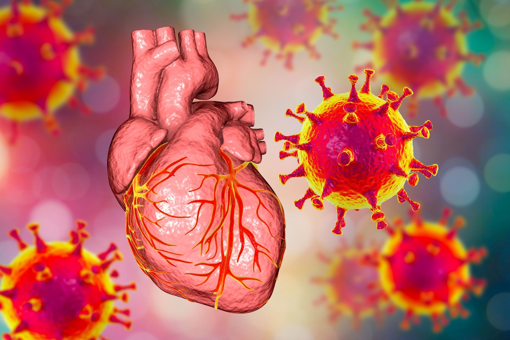 Study: Virus-Induced Acute Respiratory Distress Syndrome Causes Cardiomyopathy Through Eliciting Inflammatory Responses in the Heart. Image Credit: Kateryna Kon / Shutterstock
The link between respiratory viral infections and CVD
Seasonal viral infections can range in severity from mild flu-like symptoms to potentially lethal ARDS. For example, despite being primarily a respiratory tract infection, coronavirus disease of 2019 (COVID-19) can lead to ARDS and other severe cardiovascular disease outcomes with high mortality rates.
Circulating immune cells may respond to COVID-19 by upregulating cytokine release, which can lead to myocardial injury. Cardiac macrophages, immune cells responsible for the myocardial inflammatory response, are increasingly being investigated for their role in ARDS. Recent evidence indicates that macrophage expansion, which can be accompanied by changes in the population size and relative abundances of various cardiac macrophages, is a characteristic feature of ARDS.
The main two types of cardiac macrophages include C-C chemokine receptor type 2 negative (CCR2–) and CCR2+ macrophages. Further research is needed to determine the viral-induced contributions of these macrophages to adverse cardiac outcomes.
These data would allow clinicians to make informed intervention decisions and elucidate whether these outcomes are COVID-19-induced or if observed inflammation is a systemic immune response to viral infection. Furthermore, this information could support the development of future therapies to prevent cardiovascular disease (CVD) following recovery from COVID-19.
About the study
In the present study, researchers investigate the role of viral- and non-viral-induced ARDS-associated immune signals in altering cardiac macrophage populations, thereby impacting CVD parameters, including systemic inflammation.
This study was conducted at Massachusetts General Hospital and involved 33 control samples obtained from patients who died between September and December 2019, prior to the onset of COVID-19, as well as 21 samples obtained between May and July 2020 from patients who died from COVID-19-associated complications. Samples consisted of autopsy tissue excised from the left ventricular or septal region.
Simultaneously, in vivo studies involved a daily intratracheal administration of an ARDS cocktail of immunostimulatory agents to mice, which included resiquimod, imiquimod, lipopolysaccharide (LPS), and angiotensin-converting enzyme 2 (ACE2) inhibitor MLN-4760. This model allowed the researchers to reproduce clinical ARDS features in mice without the severe acute respiratory syndrome coronavirus 2 (SARS-CoV-2).
Patient data included results obtained from electrocardiogram (ECG), echocardiography, lung computed tomography (CT) scan, blood gas analyses, body temperature evaluation, bronchoalveolar lavage fluid (BALF) characterization, blood pressure measurements, and flow cytometry. Both human and murine autopsy samples were processed using ribonucleic acid (RNA) isolation, real-time polymerase chain reaction (PCR) assay, and enzyme-linked immunosorbent assays (ELISAs) for protein and gene expression determinations.
Similar immune responses in non-viral- and SARS-CoV-2-associated ARDS
In the absence of viral infection, mice treated with the ARDS cocktail exhibited significant weight loss over the five-day cocktail treatment period. This was accompanied by hypothermia, a common feature of both ARDS and septic shock, as well as a mortality rate of over 40% by day five.
Mice with ARDS exhibited bilateral opacities and immune cell infiltrations within their lungs, as well as reduced blood oxygenation. Furthermore, increased D-dimer, neutrophil, and monocyte levels were observed, as well as reduced blood pressure and lower heart rates in ARDS mice. Other inflammatory pathways that were activated in ARDS mice included increased levels of interleukin 6 (IL-6), IL-1ß, tumor-necrosis factor α (TNF-α), and interferon y (IFN-y), all of which are also associated with SARS-CoV-2 infection.
In both non-infected ARDS and SARS-CoV-2-infected mice, an increased infiltration of interstitial macrophages and reduced levels of alveolar macrophages were observed. Although both mouse models exhibited increased levels of cardiac macrophages, this immune response was more pronounced in infected mice. Nevertheless, both models’ subsets of cardiac macrophages were altered to similar levels.
Upon comparison of control and COVID-19 patient myocardium samples, SARS-CoV-2 infection recruited a more significant number of CCR2+ CD68+ macrophages, thus indicating that a robust immune response is elicited after severe infection compared to other life-threatening diseases.
“Our findings indicate that systemic and myocardial inflammatory signals elicited by virally induced ARDS may contribute to the cardiovascular complications and high mortality rates of this condition. In addition, our study confirms previous reports that SARS-CoV-2 infection increases overall macrophage numbers in hearts.”
The cardiac benefits of TNF-α immune therapy
TNF-α neutralizing antibodies were also administered to mice to evaluate their effects on immune activation during ARDS. To this end, TNF-α immune therapy reduced weight loss, improved body temperature, increased blood oxygenation, and led to better survival rates. Histological analysis indicated that ARDS mice receiving anti-TNF-α therapy exhibited reduced macrophages, Cxcl2, IL-1ß, and IL-6 expression within the lungs.
TNF-α therapy also improved systolic dysfunction, cardiomyocyte apoptosis, and monocyte infiltration in ARDS mice. Total cardiac macrophage counts and reduced expression of IL-1ß, IL-6, and TNF-α within the myocardium were also observed, thus demonstrating the anti-inflammatory benefits associated with TNF-α immune therapy in the lungs and hearts of mice with ARDS.
Conclusions
The study findings demonstrate that SARS-CoV-2 infection leads to significant alterations in cardiac macrophage subset levels, particularly increased levels of CCR2+ macrophages, in both mice and humans. Even in the absence of SARS-CoV-2 or another virus, the immune response to ARDS-like injury is capable of inducing significant alterations in heart macrophage levels, which may increase the risk of cardiovascular complications and mortality associated with ARDS.
[ad_2]
Source link