[ad_1]
In a recent study published in Nature, researchers developed a treatment to restore the immunological system to a younger condition, with fewer myeloid-biased output-hematopoietic stem cells (my-HSCs), more HSCs, and a balanced generation of myeloid and lymphoid lineage cells (bal-HSCs).
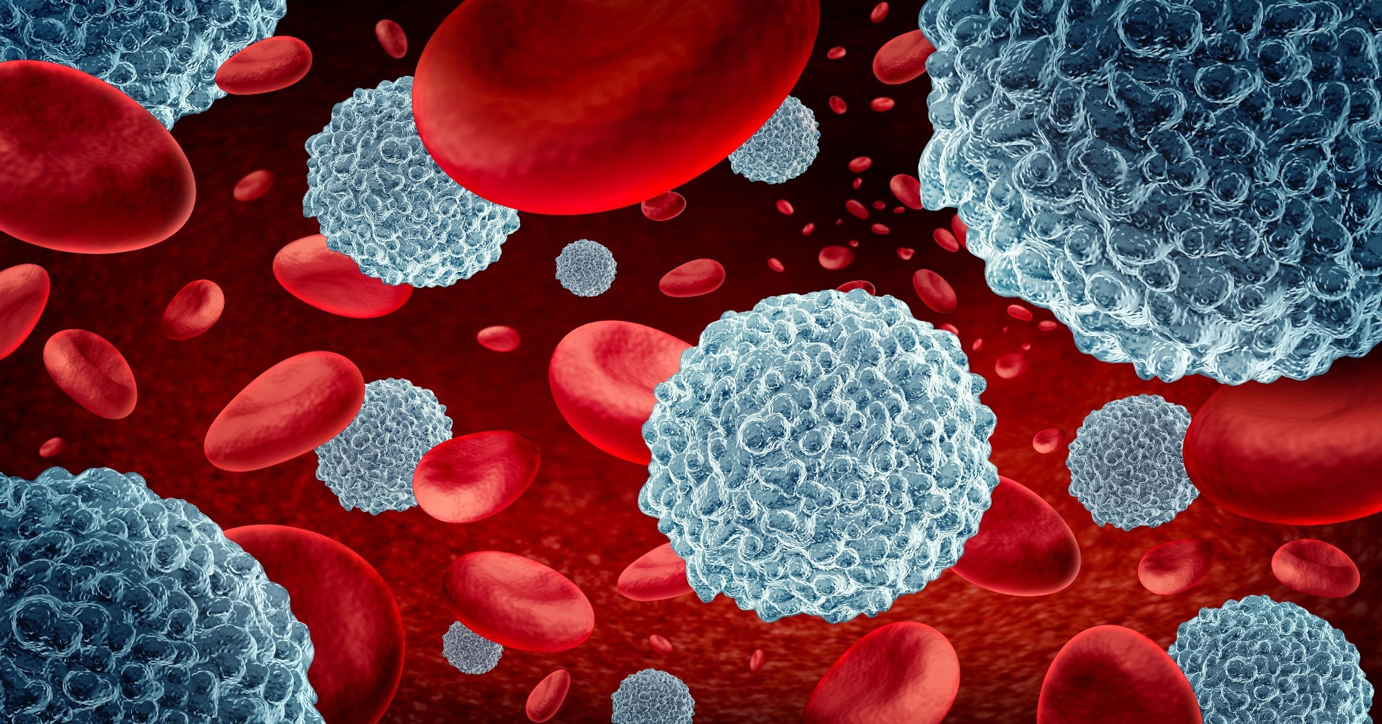 Study: Depleting myeloid-biased haematopoietic stem cells rejuvenates aged immunity. Image Credit: Lightspring / Shutterstock.com
The effects of aging on the immune system
The aging immune system is associated with reduced lymphopoiesis, increased inflammation, and myeloid diseases due to alterations in self-renewing HSCs. During childhood, bal-HSCs predominate, thereby facilitating lymphopoiesis and adaptive immune responses.
Age increases my-HSCs, which reduces lymphopoiesis and enhances myelopoiesis. Myeloid-HSC origin and possible interconversions are unclear; however, removing my-HSCs in aged mice may reverse the aging phenotype.
About the study
The researchers investigated whether antibody-regulated reduction of my-HSCs may cure age-related immunological reductions by restricting myeloid cell-induced inflammation and restoring lymphopoiesis. To this end, the impact of reduced my-HSCs on the hematopoietic system, immunological phenotypes, and functional responses to incident infections was assessed.
Several cell-surface antigen molecules were developed and validated to identify potential targets for therapeutic my-HSC reduction. The levels of my-HSCs and balanced-HSCs were determined using antibodies and flow cytometry.
Several my-HSC antigens, including neogenin 1 (NEO1), cluster of differentiation 62p (CD62p), and CD150, were subsequently targeted to determine their role in reducing my-HSC levels. Separate antibody-conditioning treatments were then developed for my-HSC depletion for each target, with a focus on cell clearance regulators such as anti-phagocytic signals, isotype, and antibody density.
To establish the role of CD150 targeting, the ability of CD150-targeted antibodies to reduce my-HSCs in vivo was assessed. To target CD62p or NEO1, goat anti-mouse NEO1 antisera was mixed with anti-CD47 and anti-KIT antibodies.
Gene expression analysis of pure total HSCs extracted from 11-month-old mice was performed to confirm alterations in HSC composition following my-HSC elimination. Transplant tests using pure HSCs were also performed to compare the myeloid and lymphoid lineage potential in recipient mice.
After antibody conditioning, myeloid and common lymphocyte progenitors (CLPs) were measured in murine bone marrow. These analyses were performed after one week to assess acute effects, as well as after eight and sixteen weeks to determine long-term effects. The impact of this treatment on non-self-renewing progenitors was also evaluated after eight weeks.
T-cell subsets were analyzed using canonical markers or cluster-based analysis. The effects of my-HSC depletion in aged animals on pro-inflammatory mediators and functional immunity to infection were also examined by analyzing mouse immune responses to a live-attenuated virus and subsequent challenge with a pathogenic viral infection using the mouse Friend retrovirus (FV) model.
Study findings
Antibody-mediated reduction of my-HSCs in elderly mice restored young immune system characteristics, such as increased CLPs, naïve T-cells, and B-cells, while lowering immunological decline indicators associated with aging. Depletion of my-HSCs in old mice increased primary and secondary adaptive immune responses to viral infection.
Twelve potential genes that encode cell-surface proteins significantly expressed in aged HSCs and my-HSCs were identified. Moreover, CD150, CD4, CD6, CD62p20, and NEO1 were identified as markers for my-HSCs.
Antibodies to CD41 and NEO1 enhanced the frequency of my-HSC staining, thus indicating myeloid bias. CD62p targeting resulted in the highest my-HSC enrichment.
The most abundant protein molecules on my-HSCs were NEO1, CD41, and CD62p. Flow cytometry analysis did not identify any surface protein strongly expressed by the subgroups, except CD41, which was highly expressed by megakaryocyte progenitor cells.
Anti-CD150 antibodies significantly reduced my-HSCs in mice, thereby increasing naïve T-cell and mature B-cell levels. In aged mice, CD4+ T lymphocytes with an exhausted phenotype (PD1+ CD62L-) grew more than those with a non-exhausted phenotype (PD1- CD62L+).
Antibody training reduced CD4+ PD1+ CD62L- cells as compared to CD4+ PD1- CD62L+. Aged mice also acquired age-associated B-cells associated with impaired humoral immunity.
Antibody training reduced the levels of pro-inflammatory proteins including interleukin-1 alpha (IL-1α), and C‐X‐C motif chemokine ligand 5 (CXCL5), which were higher in elderly animals. Aged animals with my-HSC depletion exhibited higher virus-specific CD8+ T-cell responses in the spleen following vaccination, thus indicating a better initial response to live-attenuated viral infection.
Conclusions
Rising my-HSC levels during aging may result in inadequate adaptive immunological and inflammatory responses. Thus, depleting my-HSCs may improve immune responses by enhancing the synthesis of new T- and B-cells while decreasing the production of inflammatory myeloid cells. In the current study, my-HSC depletion in older animals allowed bal-HSCs to recover youthful immunological characteristics such as enhanced lymphocyte progenitors and naïve cells and decreased lymphocyte dysfunction or exhaustion indicators and inflammatory mediators.
Further research could refine conditioning techniques and examine the impact on differentiated cells, such as regulatory T-cells.
Journal reference:
- Ross, J. B., Myers, L. M., Noh, J. J., et al. (2024). Depleting myeloid-biased haematopoietic stem cells rejuvenates aged immunity. Nature. doi:10.1038/s41586-024-07238-x
[ad_2]
Source link