Tag: Cell

  • Focusing on oncogenic driver mutations and resistance mechanisms in the treatment of NSCLC

    Focusing on oncogenic driver mutations and resistance mechanisms in the treatment of NSCLC

    [ad_1]

    Lung cancer is the leading cause of cancer-related death worldwide. Improved understanding of driver mutations of non-small cell lung cancer (NSCLC) has led to more biomarker-directed treatment for patients with advanced stages. The expanding number of drugs targeting these driver mutations offers more opportunity to improve patient’s survival benefit.

    To date, NSCLCs, especially those with non-squamous histology, are recommended for testing epidermal growth factor receptor (EGFR) mutations, anaplastic lymphoma kinase (ALK) gene rearrangements, ROS proto-oncogene receptor tyrosine kinase 1 (ROS-1) rearrangements, B-raf proto-oncogene (BRAF) mutations, rearranged during transfection (RET) fusions, Met proto-oncogene (MET) amplification and exon 14 skipping alterations, neurotrophic receptor tyrosine kinase (NTRK) gene fusions, and immunohistochemistry (IHC) testing for the programmed death receptor-ligand 1 (PD-L1) expression.

    EGFR-activating mutations are the most common driver mutations in NSCLC. Targeted therapies included first-generation epidermal growth factor inhibitor tyrosine kinase inhibitors (EGFR-TKIs), erlotinib, gefitinib, and icotinib; second-generation pan-human epidermal growth receptor (HER) family inhibitor, afatinib and dacotinib; and third-generation EGFR-TKI, osimertinib, that inhibits both EGFR-sensitive mutations and resistant mutation EGFR T790M. ALK inhibitors included first-generation, crizotinib; second generation, ceritinib, alectinib, and brigatinib; and third-generation, lorlatinib, with increasing capacity for ALK inhibition generation-by-generation. The resistance caused by secondary ALK mutations can be overcome by next-generation ALK-TKIs. Moreover, alectinib, brigatinib, and lorlatinib are all recommended as first-line treatment choice for ALK-rearranged NSCLC for their superior survival compared with crizontinb. Crizotinib, ceritinib, brigatinib, and lorlatinib can also inhibit ROS-1. BRAF inhibitor dabrafenib combined with MEK inhibitor trametinib is now the recommended treatment for BRAF V600E-mutated NSCLC attributing to improved outcomes compared with vemurafenib or dabrafenib monotherapy. Drugs targeting MET aberrations with different binding modes included type Ia, crizotinib; type Ib, tepotinib, capmatinib, and savolitinib; and type II, cabozantinib. Entrectinib and larotrectinib are the current standard treatment for NTRK fusion-positive NSCLC. Kirsten ratsarcoma viral oncogene homolog (KRAS) mutations occur in ~20%–30% of patients with NSCLC. Recently, several small molecules including sotorasib (AMG510), adagrasib (MRTX-849) have been developed to specifically target KRAS G12C.

    Although drugs targeting oncogenic driver mutations have significantly improved survival but their long-term responses are still uncommon for most patients, the emergence of acquired drug resistance is inevitable. Therefore, exploration and understanding of the resistance mechanism of target therapy are important to enhance the clinical outcomes and ultimately increase the treatment rate of NSCLC. Meanwhile, improved understanding of biological characteristics in various molecular subtypes of each driver mutation helps explore new classified treatment strategies. Until recently, multiomics analysis has ushered in a new era of precision targeted therapy in lung cancer and led to a deeper understanding of the underlying resistant mechanism. All these scientific and clinical progresses ultimately lead to improved survival in NSCLC and achieve more refined individualized treatment.

    The discovery of oncogenic driver alterations and target therapy have brought significant clinical benefits and established an individualized treatment approach. The management of advanced NSCLC has shifted from a histology based on a biomarker-driven process.

    The treatment landscape of oncogenic-addicted NSCLC has become complex. On the one hand, more individualized treatment based on fine stratification has become the focus of research nowadays. The choice of optimal treatment strategy should consider gene subtype, concomitant mutation, dynamic gene alternation, and metastasis site. On the other hand, understanding primary and acquired resistance to targeted therapy provides an insight into the molecular evolution of tumor development. The recognition of resistant mechanisms is the basis to design new drugs or combinatorial therapeutic strategies.

    Combination strategies require integration with immunotherapy, and the immunosuppressive microenvironment should be reversed to improve the sensitivity of ICIs by the drug combination. We should also be aware of the possible risk of combined toxicity and thereby explore the optimal timing and combined regimen of immunotherapy. Meanwhile, the interaction between driver mutations and immune-microenvironment is essential to uncover after drug resistance and to establish robust predictive biomarker for the NSCLC with driver mutations, in order to identify specific oncogenic driving patients with NSCLC who can benefit from immunotherapy.

    Source:

    Journal reference:

    Zhong, J., et al. (2023). Treatment of advanced non-small cell lung cancer with driver mutations: current applications and future directions. Frontiers of Medicine. doi.org/10.1007/s11684-022-0976-4.

    [ad_2]

    Source link

  • Household chemicals endanger brain’s myelin-forming cells

    Household chemicals endanger brain’s myelin-forming cells

    [ad_1]

    In a recent study published in the journal Nature Neuroscience, researchers in the United States pinpointed and evaluated environmental chemicals that hinder oligodendrocyte development through varied mechanisms, assessing their neurodevelopmental impacts.

    Study: Pervasive environmental chemicals impair oligodendrocyte development. Image Credit: Lightspring / ShutterstockStudy: Pervasive environmental chemicals impair oligodendrocyte development. Image Credit: Lightspring / Shutterstock

    Background 

    Human exposure to environmental chemicals, especially during the critical developmental stages of children’s central nervous systems, raises significant health concerns. Substances like methylmercury, lead, and polychlorinated biphenyls are linked to disrupting brain development, potentially contributing to the increasing prevalence of neurodevelopmental disorders such as autism and Attention-Deficit/Hyperactivity Disorder (ADHD). These trends suggest that environmental factors play a critical role beyond genetics. Oligodendrocytes, vital for brain functionality through myelination and neuronal support, are particularly susceptible to these chemicals from fetal development into adolescence. Despite their significance, limited research has focused on the impact of environmental toxins on oligodendrocytes. This gap highlights the need for further investigation into how these chemicals affect oligodendrocyte development and identifying ways to counteract their detrimental effects on neurodevelopment.

    About the study 

    The present study adhered to ethical standards set by the International Society for Stem Cell Research and the National Institutes of Health, receiving approval from the Case Western Reserve University Institutional Animal Care and Use Committee. Mouse oligodendrocyte precursor cells (OPCs) were cultured from induced pluripotent stem cells (iPSCs), following established protocols that involved removing iPSCs from a feeder layer, dissociating them, and then cultivating them in a medium conducive to OPC expansion and maturation. The culture medium was switched on the tenth day to promote OPC development, utilizing a specific combination of supplements to enrich OPC populations. Additionally, primary mouse OPCs and astrocytes were derived from dissected mouse brain tissue, with the cells undergoing culture in specially prepared media to encourage the growth of OPCs and astrocytes, respectively.

    Human cortical organoids were generated from embryonic stem cells and iPSCs, following rigorous stem cell research guidelines. These organoids were cultured in a medium optimized for OPC expansion and differentiation, incorporating various growth factors and supplements. Chemical screening on OPCs utilized the United States Environmental Protection Agency (US EPA) Toxicity Forecaster chemical library to identify compounds that disrupt OPC development. 

    Various methods, including immunocytochemistry, high-content imaging, and cell viability assays, were employed to assess the impact of chemicals on OPCs. Additionally, the study explored the effects of specific quaternary compounds on cell viability, employing a range of experimental setups across different cell types to understand the compounds’ toxicity profiles. 

    Study results 

    The present study developed a high-throughput screening method to assess the impact of environmental chemicals on the development of mouse pluripotent stem cells (mPSCs)- derived OPCs into oligodendrocytes. Among the 1,823 chemicals screened, a selection was found to either be cytotoxic to developing oligodendrocytes or impede their generation without inducing cytotoxicity. The screening revealed that a majority of the chemicals had no significant effect on oligodendrocyte development or viability, yet 292 were identified as cytotoxic and 47 as inhibitors of oligodendrocyte generation.

    Further investigation using the MTS assay, which measures metabolic activity as an indicator of cell viability, validated the cytotoxic effects of certain chemicals. Comparison of cytotoxicity profiles across different cell types, including mouse astrocytes and data from the US EPA, identified quaternary compounds as selectively cytotoxic to oligodendrocytes. These compounds, characterized by a central nitrogen with four alkyl groups, demonstrated a specific toxicological sensitivity in developing oligodendrocytes. The study also explored the activation of the integrated stress response (ISR) as a potential mechanism for the cytotoxicity induced by quaternary compounds.

    Quaternary compounds were also tested for their ability to cross the blood-brain barrier and were found to be present in brain tissue at nanomolar concentrations following administration to mice. Furthermore, the study extended to human pluripotent stem cell-derived regionalized neural organoid models, confirming that quaternary compounds could disrupt human oligodendrocyte development, reducing the density of SOX10+ OPCs and oligodendrocytes.

    Additionally, the screening identified organophosphate flame retardants as inhibitors of oligodendrocyte development. These compounds were shown to arrest the progression of early to intermediate and mature oligodendrocytes. The study’s findings were extended to in vivo and in vitro models of human brain development, demonstrating that exposure to organophosphate flame retardants, particularly Tris(1,3-dichloro-2-propyl) phosphate (TDCIPP), significantly reduced the number of SOX10+CC1+ oligodendrocytes in both mouse and human models.

    Lastly, the study utilized data from the National Health and Nutrition Examination Survey (NHANES) to investigate associations between exposure to organophosphate flame retardants and neurodevelopmental outcomes in children. High levels of urinary Bis(1,3-dichloro-2-propyl) phosphate (BDCIPP), a metabolite indicative of TDCIPP exposure, were associated with an increased likelihood of special education needs and gross motor dysfunction, suggesting a strong link between organophosphate flame retardant exposure and adverse neurodevelopmental outcomes. 

    [ad_2]

    Source link

  • Antioxidant-rich diets linked to lower type 2 diabetes risk, supplements less effective

    Antioxidant-rich diets linked to lower type 2 diabetes risk, supplements less effective

    [ad_1]

    Scientists at the Karolinska Institute in Sweden have conducted a systematic review and meta-analysis to determine the relationship between dietary antioxidant intake and type 2 diabetes risk.

    The study is published in the journal Advances in Nutrition

    Review: Vitamins C, E, and beta-carotene and risk of type 2 diabetes: a systematic review and meta-analysis. Image Credit: alicja neumiler / ShutterstockReview: Vitamins C, E, and beta-carotene and risk of type 2 diabetes: a systematic review and meta-analysis. Image Credit: alicja neumiler / Shutterstock

    Background

    Type 2 diabetes is a serious metabolic disease characterized by reduced secretion or impaired functioning of insulin and subsequent increase in blood glucose level (hyperglycemia). Insulin resistance and pancreatic beta-cell dysfunction are two major hallmarks of the disease.

    More than 10% of the global population is currently affected by type 2 diabetes. Its prevalence is sharply increasing worldwide mainly because of an increasing inclination towards unhealthy food habits and sedentary lifestyles.

    Diet plays an important role in regulating the body’s metabolism, among various lifestyle factors. Evidence indicates that an increased adherence to healthy diets, such as the Mediterranean or DASH (Dietary Approach to Stop Hypertension), can significantly reduce the risk of type 2 diabetes. The fundamental characteristic of these diets is a higher intake of plant-based foods rich in antioxidants, including vitamins C, E, and beta-carotene.    

    In this systematic review and meta-analysis, scientists have assessed whether consumption of dietary vitamins C, E and beta-carotene can reduce the risk of type 2 diabetes.

    Vitamins C, E, and beta-carotene and risk of type 2 diabetes: a systematic review and meta-analysis

    Study design

    The scientists searched various electronic databases to identify studies investigating the association between dietary intakes, circulating levels, or supplementation of vitamin C, E, and beta-carotene and type 2 diabetes incidence or insulin resistance/sensitivity and beta cell function in non-diabetic individuals.

    The final screening led to the identification of 25 prospective observational studies and 15 randomized controlled trials. Moderate and serious risks of bias were observed in 21 and 4 observational studies, respectively. Among randomized controlled trials, 13 had a low risk of bias, and 2 had some concerns.

    Important observations

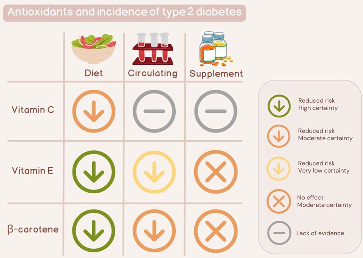
    The study found that moderate intakes of vitamins C, E, and beta-carotene can reduce the risk of type 2 diabetes.

    Vitamin C

    The meta-analysis of observational studies revealed that a vitamin C intake of up to 70 mg per day can reduce type 2 diabetes risk by 24%. However, no further risk reduction was observed for an intake higher than this level.

    An inverse association was observed between dietary intake of vitamin C and insulin resistance. Vitamin C intake also showed a positive impact on beta cell function.

    Vitamin E

    The meta-analysis of observational studies revealed that the vitamin E intake of up to 12 mg per day can reduce the risk of type 2 diabetes by 28%. Similar to vitamin C, no further risk reduction was observed for an intake higher than this level.

    The meta-analysis of randomized clinical trials revealed that vitamin E supplementation does not have any protective effect against type 2 diabetes development. However, vitamin E supplementation showed a positive impact on insulin sensitivity.

    Beta-carotene

    The meta-analysis of observational studies revealed that the beta-carotene intake of up to 4 mg per day can reduce the risk of type 2 diabetes by 22%. No further risk reduction was observed above this level.

    The meta-analysis of randomized controlled trials revealed that beta-carotene supplementation cannot reduce the risk of type 2 diabetes. It was also observed that circulating beta-carotene can reduce insulin resistance and increase insulin sensitivity.

    Study significance

    This systematic review and meta-analysis found an inverse association between dietary and circulating vitamins C, E, and beta-carotene and the risk of type 2 diabetes. However, no protective efficacy of supplementation with these antioxidants has been observed against type 2 diabetes.

    The robust antioxidant properties of these vitamins are mainly responsible for their anti-diabetic effects. Vitamin C is a water-soluble vitamin commonly found in fruits and vegetables. It can remove free radicals in the body’s hydrophilic compartments and regenerate vitamin E from its oxidized form.

    Vitamin E is a fat-soluble vitamin commonly found in nuts, seeds, and vegetable oils. It can prevent lipid peroxidation and protect lipid parts of the body, such as cell membranes. Beta-carotene is a fat-soluble provitamin A carotenoid commonly found in fruits and vegetables. Similar to vitamin E, beta-carotene can protect lipid parts of the body from free radical-mediated damage.

    Article Revisions

    • Mar 27 2024 – Addition of Graphical abstract illustration.

    [ad_2]

    Source link

  • Study reveals genetic variant linked to increased risk of leukemia in Hispanic/Latino children

    Study reveals genetic variant linked to increased risk of leukemia in Hispanic/Latino children

    [ad_1]

    Acute lymphoblastic leukemia (ALL), the most common childhood cancer, disproportionately affects children of Hispanic/Latino origin in the United States. They are 30-40% more likely to get ALL than non-Hispanic white children, but the exact genetic basis and cause of that increased risk are unknown.

    Now, a study from the Keck School of Medicine of USC has revealed a key genetic variant contributing towards the increased risk, as well as details about the biological basis of ALL. The team used genetic fine-mapping analysis, a statistical method that allows researchers to disentangle the separate effects of genetic variants in a region of the genome. They identified a variant found at a relatively high frequency in people of Hispanic/Latino origin that increases ALL risk by around 1.4 times. The study, funded in part by the National Institutes of Health, was just published in the journal Cell Genomics.

    “Combined with the fact that around 30% of Hispanic/Latino people in the United States carry this gene variant, but it’s basically absent in people of predominantly European ancestry, we think it’s an important contributor to the increased ALL risk among this group,” said the study’s lead author, Adam de Smith, PhD, an assistant professor of population and public health sciences and a member of the USC Norris Comprehensive Cancer Center at the Keck School of Medicine, as well as a scholar of the Leukemia & Lymphoma Society.

    The researchers also performed tests to better understand how the variant, located on the IKZF1 gene, which underlies B-cell development, relates to ALL through its influence on the development of B-cells, a type of white blood cell known to be disrupted by the disease.

    Together, the analyses in our study provide the statistical, biological and evolutionary insights behind this increased risk, and may ultimately aid scientists working to develop screening tools and therapies for ALL.”


    Charleston Chiang, PhD, associate professor of population and public health sciences and associate director of the Center of Genetic Epidemiology at the Keck School of Medicine and study’s co-senior author

    The genetic basis of leukemia risk

    To pinpoint the genetic basis of the elevated ALL risk Hispanic/Latino children face, the researchers analyzed genetic data from the California Cancer Records Linkage Project. Their dataset included 1,878 Hispanic/Latino children in California with ALL and 8,411 without the condition; 1,162 non-Hispanic white children with ALL and 57,341 without; and 318 East Asian children with ALL and 5,017 without. 

    The research team focused on the IKZF1 gene, known to relate to ALL but never before linked with ethnic risk disparities. Using genetic fine-mapping analysis, they independently analyzed each position along the gene-;known as a single nucleotide polymorphism (SNP)-;to determine whether having a certain variant increased ALL risk.

    They found three independent SNPs linked to higher ALL incidence, one of which was present in about 30% of people of Hispanic/Latino origin in the U.S. and less than 1% of people of primarily European origin. Although overall risk for the disease is low across all racial/ethnic groups, children with that gene variant, located at SNP rs76880433, were 1.44 times as likely to develop ALL as children without the variant. 

    The genetic ancestry of most Hispanics/Latinos can be traced to Europe, Africa, and Indigenous America. Further investigation revealed that the risk variant was specifically linked with Indigenous American ancestry and may have become more common in this group because it conferred a selective advantage at some point in human history.

    Next, the Keck School of Medicine team partnered with co-senior author Vijay Sankaran, MD, PhD, an associate professor of pediatrics at Harvard Medical School and attending physician at the Dana-Farber/Boston Children’s Cancer and Blood Disorders Center, to conduct a series of experiments to better understand how the genetic variant at IKZF1 increases risk for ALL.

    One experiment analyzed chromatin accessibility, a test which indicates how fully a given gene can be expressed. The researchers found that the risk variant reduced chromatin accessibility, preventing IKZF1 proteins from being fully expressed.

    Sankaran and his team also conducted experiments with stem cells, finding that “knocking out” the IKZF1 gene caused B-cell development to stall in its early stages.

    “Looking at all of this together, we think that the risk variant is reducing IKZF1 expression,” de Smith said. “By doing so, it’s keeping B-cells in a more immature state, which would increase ALL risk by giving the cells more chance to develop mutations that could eventually lead to overt leukemia.”

    Leukemia screening and treatment

    The new insights about IKZF1 bring researchers one step closer to developing effective screening tools to predict who may develop ALL, but more research is needed. In addition, the findings provide important clues about potential ways to treat the disease, for instance by progressing B-cell development after it stalls.

    “We also need to understand whether this variant is associated with different patient outcomes, such as the risk of relapse or chances of survival, and why that might be,” de Smith said.

    He and his colleagues also hope to explore whether the newly identified risk variant helps explain the even higher risk of ALL among Hispanic/Latino adolescents and young adults, who are more than twice as likely to get the disease than people who are non-Hispanic white.

    About this research

    In addition to de Smith, Chiang and Sankaran, the study’s other authors are Soyoung Jeon, Jalen Langie, Tsz-Fung Chan, Steven Gazal, Nicholas Mancuso and Joseph Wiemels from the Center for Genetic Epidemiology and the USC Norris Comprehensive Cancer Center, Keck School of Medicine of USC; Lara Wahlster, Susan Black, Liam Cato, Soumyaa Mazumder and Fulong Yu from Boston Children’s Hospital and Department of Pediatric Oncology, Dana-Farber Cancer Institute; Linda Kachuri from the Stanford University School of Medicine; Nathan Nakatsuka from the New York Genome Center; Guangze Xia from the Guangzhou National Laboratory, Guangzhoi, China; Wenjian Yang and Jun Yang from St. Jude Children’s Research Hospital, Memphis; Celeste Eng, Donglei Hu, Esteban Gonzalez Burchard and Elad Ziv from the Helen Diller Family Comprehensive Cancer Center, University of California, San Francisco; Catherine Metayer from the School of Public Health, University of California, Berkeley; and Xiaomei Ma from the Yale School of Public Health.

    This work was supported by the National Institutes of Health [R01CA262263, R01CA155461, R00CA246076, R35GM142783, R01DK103794, R01CA265726]; the New York Stem Cell Foundation; and the Dana-Farber Cancer Institute Presidential Priorities Initiative.

    Source:

    Journal reference:

    de Smith, A. J., et al. (2024) A noncoding regulatory variant in IKZF1 increases acute lymphoblastic leukemia risk in Hispanic/Latino children. Cell Genomics. doi.org/10.1016/j.xgen.2024.100526.

    [ad_2]

    Source link

  • Bio-Rad launches validated antibodies for rare cell and circulating tumor cell enumeration

    Bio-Rad launches validated antibodies for rare cell and circulating tumor cell enumeration

    [ad_1]

    Bio-Rad Laboratories, Inc. (NYSE: BIO and BIO.B), a global leader in life science research and clinical diagnostics products, today announced the launch of validated antibodies for rare cell and circulating tumor cell (CTC) enumeration. Validated for use with Bio-Rad’s Celselect Slides Enumeration Stain Kits, the antibodies are specific to CTC surface markers to enable the sensitive and specific identification of target cell populations, enhancing the study of tumor heterogeneity and disease progression at various stages. 

    Bio-Rad’s Genesis Cell Isolation System is a customizable benchtop solution that uses unbiased size-based cell selection to gently and efficiently capture a wide range of CTCs and other rare cells from liquid biopsy samples. After capture, the enriched cells can be recovered for downstream analysis, or immuno-stained on-slide for immunofluorescence applications such as enumeration and identification of various CTC types. 

    The enumeration of captured CTCs provides valuable insights into the surface markers that indicate cancer type and progression and is critical to understanding the mechanisms of cancer metastasis. For successful enumeration, the antibody reagents require careful selection to ensure not only sensitivity and specificity to the target cell surface marker, but also compatibility with the staining method. Bio-Rad’s new range of validated primary and secondary antibodies enables accurate immunostaining of captured CTCs, supporting cancer researchers working in this field. 

    CTC analysis is a promising tool for the study of tumor heterogeneity and disease progression, offering real-time data and unique insights into cancer metastasis,” said Stephen Kulisch, Vice President of Marketing for Bio-Rad’s Digital Biology Group. “The introduction of validated antibodies for target cell identification reflects Bio-Rad’s growing single-cell oncology product portfolio and is a testament to our commitment to deliver highly efficient rare cell capture, enrichment, enumeration, and recovery for cancer researchers.”  

    To learn more about the new validated antibodies, visit bio-rad-antibodies.com/val-abs

    BIO-RAD and CELSELECT SLIDES are trademarks of Bio-Rad Laboratories, Inc. in certain jurisdictions.  

    [ad_2]

    Source link

  • Vitamin D receptor presence in breast cancer tumors linked to better survival outcomes

    Vitamin D receptor presence in breast cancer tumors linked to better survival outcomes

    [ad_1]

    In a recent study published in the journal Nutrients, researchers explored the role of vitamin D receptors (VDR) as a potential prognostic marker for breast cancer.

    Their results indicate significant associations between VDR, the mode of detection, and the size of invasive tumors, suggesting a promising avenue for further study.

    Study: The Vitamin D Receptor as a Prognostic Marker in Breast Cancer—A Cohort Study. Image Credit: Peddalanka Ramesh Babu / ShutterstockStudy: The Vitamin D Receptor as a Prognostic Marker in Breast Cancer—A Cohort Study. Image Credit: Peddalanka Ramesh Babu / Shutterstock

    Background

    People with breast cancer often show lower serum levels of vitamin D compared to healthy individuals; some studies have also found links between low vitamin D levels and the probability of an adverse prognosis, while others suggest that supplementation could reduce the risk of developing advanced cancer.

    Calcitriol, an active metabolite of vitamin D, binds to the VDR before it translocates into the nucleus, modifying regulatory genes that control cell signaling, apoptosis, and cell growth.

    This could be a mechanism that underlies the association between VDR and favorable breast cancer prognoses, but further research is needed to understand its potential role as a biomarker for the progression of tumors.

    About the study

    In this study, researchers attempted to validate prior work that found positive associations between breast cancer prognoses and nuclear VDR presence. They additionally explored if prognostic information can be refined by VDR subcellular localization.

    The dataset included tumor samples obtained from individuals who received a diagnosis of primary breast cancer between October 2002 and June 2012. The samples were utilized for tissue microarray (TMA) construction after excluding people who had received a prior diagnosis of any cancer in the previous 10 years.

    During preoperative visits, patients were asked to complete questionnaires that included information on reproductive factors, lifestyles, and medication and supplements consumed during the previous week. Vitamin D consumption was calculated from the product information for supplements.

    Nurses measured hip and waist circumference, height, weight, and breast volume. Pathology reports were used to obtain characteristics of tumors, including size, histologic type, grade, involvement of axillary lymph nodes, and status of progesterone receptors (PR) and estrogen receptors (ER).

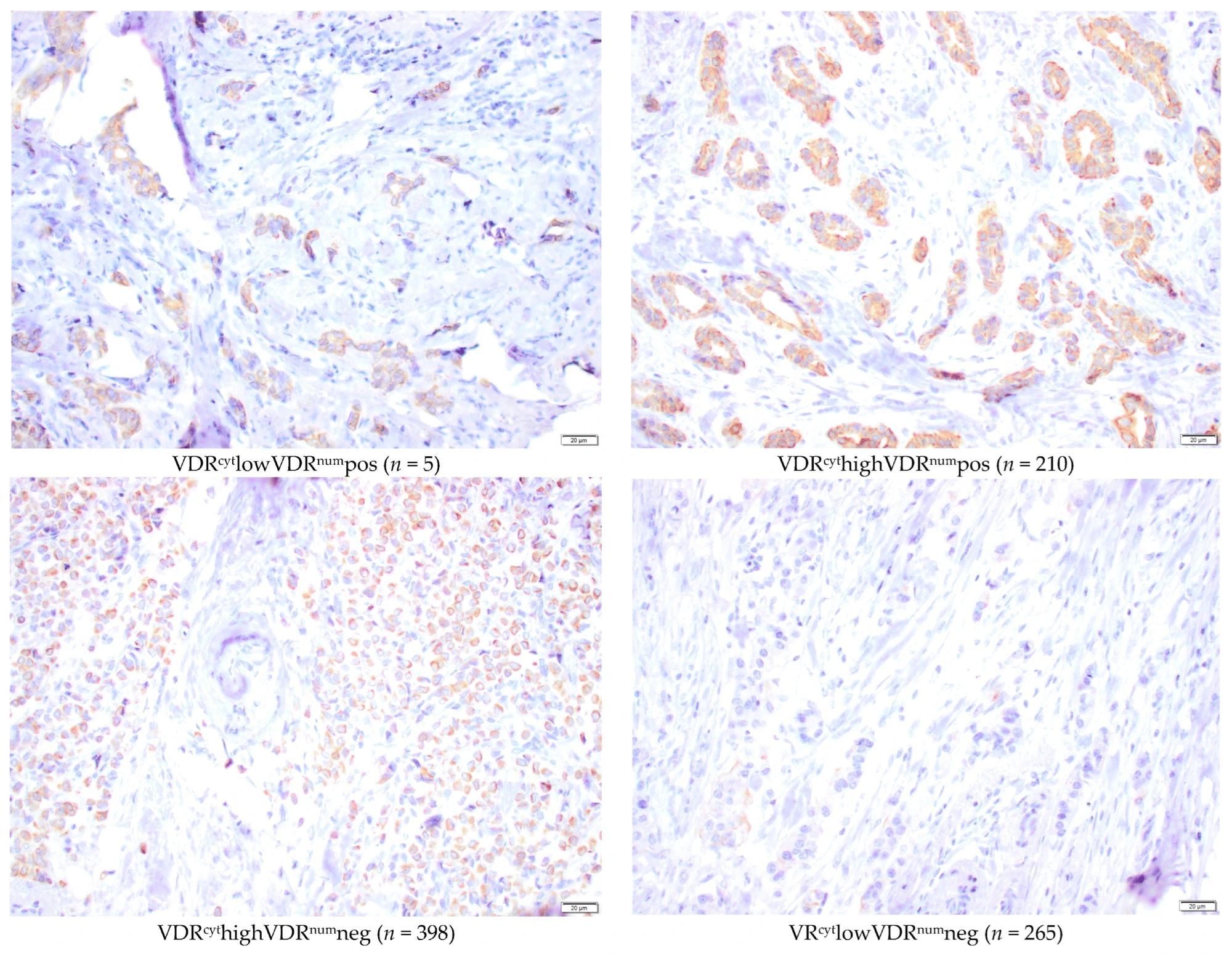
    Pathological assessment of human epidermal growth factor receptor 2 (HER2) status and TMA were assessed jointly using microscopic methods after blinding. In multiple subcellular locations of invasive tumor cells, VDR staining was examined in the nuclear membrane (VDRnum), cytoplasm (VDRcyt), and nucleus (VDRnuc).

    During the final follow-up in 2019, medical records, pathology reports, and national registries for population and tumors were used to calculate the breast cancer-free interval (BCFI) and calculate endpoints (death or last follow-up) for survival analyses.

    Findings

    On average, patients were 61 years old, and VDR was obtained for 878 tumors; cytoplasmic intensity evaluations showed that 7% were VDR-negative, while VDRnum was positive in 25% of patients. Tumors included in the analysis were, on average, large, of a higher grade, and more likely to be lymph node-negative than those that were excluded.

    Microscopic representative images of immunohistochemical staining intensities of nuclear membrane and cytoplasmic VDR (40×) in the TMA. Bar represents 20 µm.Microscopic representative images of immunohistochemical staining intensities of nuclear membrane and cytoplasmic VDR (40×) in the TMA. Bar represents 20 µm.

    Patients with VDRnum-positive tumors had smaller waist circumferences and breast volumes on average and were more likely to have screening-detected tumors. VDRnum-positive tumors were linked with lower tumor grade, positive hormone receptors, and the absence of HER2 amplification.

    After surgery, more VDRnum-negative patients received chemotherapy and trastuzumab, while VDRnum-positive patients received further endocrine therapy. In terms of prognosis, patients with VDRnum-positive tumors had better outcomes in both BCFI and overall survival (OS) in univariable analysis. This effect persisted after adjusting for various factors.

    When VDR localization was considered, VDRnum-positive tumors showed the best prognosis regardless of VDRcyt. Patients with both ER-positive and VDRnum-positive tumors had the best prognosis, while those with ER-negative and VDRnum-negative tumors had the worst. Even among ER-positive tumors, VDRnum-positive tumors were associated with lower tumor grade and longer OS.

    Interaction analyses suggested that VDRnum interacts with the mode of detection and size of tumors in the case of BCFI. Larger VDRnum-positive tumors (>20 mm) were associated with significantly fewer breast cancer events than small ones. Clinically detected VDRnum-positive tumors showed better BCFI than screening-detected ones.

    Conclusions

    This study highlights significant correlations between positive VDR staining in the nuclear membranes of breast cancer cells and favorable characteristics of tumors, OS, and longer BCFI.

    Results indicate that evaluating nuclear membrane VDR levels may be a better prognosis predictor compared to cytoplasmic levels. VDRnum status could refine luminal breast cancer selection with favorable prognoses, especially when considering interactions with tumor size and detection mode.

    VDR expression inversely correlates with aggressiveness, particularly in triple-negative and HER2-amplified cancers, possibly due to mutations in the tumor protein p53. Further research is needed to standardize VDR evaluation methods and explore associations with vitamin D levels.

    The study suggests potential clinical relevance for VDR as a prognostic marker and underscores the need for understanding the interplay between vitamin D, VDR, and breast cancer outcomes. Since most participants in the study were of European descent, heterogeneous populations should be included in future studies to ensure the generalizability of findings.

    Journal reference:

    • The vitamin D receptor as a prognostic marker in breast cancer – a cohort study. Huss, L., Gulz-Haake, I., Nilsson, E., Tryggvadottir, H., Nilsson, L., Nodin, B., Jirström, K., Isaksson, K., Jernström, H. Nutrients (2024). 10.3390/nu16070931, https://www.mdpi.com/2072-6643/16/7/931

    [ad_2]

    Source link

  • Statins show promise in reducing gum disease inflammation

    Statins show promise in reducing gum disease inflammation

    [ad_1]

    Could taking statins benefit your mouth in addition to your arteries? A new study conducted in cell cultures showed that cholesterol-lowering drugs help to dampen the inflammation associated with periodontal disease by altering the behavior of macrophages, a type of immune cell.

    Statins are the most common type of prescription medication in the United States today, taken by over 40 million Americans to lower cholesterol. The study suggests these drugs improve gum health and reduce the risk of heart disease.

    Subramanya Pandruvada, an assistant professor in the College of Dental Medicine at the Medical University of South Carolina, oversaw the work.

    During our study, we replicated specific conditions in periodontal disease and demonstrated that introducing statins to our in vitro model modifies macrophage response. This allowed us to explore how medication like statins can help us treat inflammatory conditions such as periodontal disease.”


    Subramanya Pandruvada, Assistant Professor, College of Dental Medicine, Medical University of South Carolina

    Pandruvada will present the new research at Discover BMB, the annual meeting of the American Society for Biochemistry and Molecular Biology, which is being held March 23–26 in San Antonio. The study’s lead authors are Waleed Alkakhan, a graduate dental resident in periodontology, and Nico Farrar, a dental student at the Medical University of South Carolina.

    Periodontal disease occurs when the growth of bacteria in the gums causes the immune system to mount an inflammatory response, contributing to symptoms such as swelling, bleeding and bone degradation. Untreated, it can lead to tooth loss. Nearly half of adults over age 30 have some form of periodontal disease, according to the U.S. Centers for Disease Control and Prevention.

    Current treatments for advanced periodontal disease include antibiotics, deep cleanings of tooth and root surfaces, and various surgical procedures. Researchers have sought new ways to calm gum disease through less invasive treatment strategies.

    Some previous studies have shown that people taking statins tend to show fewer signs of periodontitis than people who do not take statins. The new study is the first to trace the biochemical pathways through which statins appear to reduce periodontal inflammation.

    “Recent periodontal literature has shown the beneficial effects of statins when used with traditional periodontal therapy,” Pandruvada said. “However, our study highlights a novel approach in which statins affect macrophages specifically, which, through this mechanism, can help treat periodontal disease.”

    Macrophages play an important role in helping the body fight infections; however, they can also worsen inflammation depending on the form they take at different phases of the immune response. The researchers grew macrophages and gum cells together for the study and exposed them to various conditions. They found that exposure to simvastatin, a common statin drug, suppressed the macrophage inflammatory response.

    As a next step, the researchers plan to study the impacts of statins on periodontal disease in animal models, a step toward determining whether this strategy might be a safe and effective approach for future periodontal therapies.

    The new findings build upon the group’s initial results, which were published last year in the journal Cells.

    [ad_2]

    Source link

  • New glycan found to play a key role in nasal colonization of whooping cough bacteria

    New glycan found to play a key role in nasal colonization of whooping cough bacteria

    [ad_1]

    Researchers have identified a new complex-carbohydrate biomolecule, or glycan, that plays a key role in the nasal colonization of the Bordetella bacteria responsible for whooping cough. The discovery could make it possible to create a new drug or vaccine that interferes with the glycan to greatly reduce or even stop ongoing Bordetella transmission.

    Bordetella pertussis is the cause of the respiratory infection pertussis, which is widely known as whooping cough. Today’s pertussis vaccines keep people from getting severely sick, but they don’t eliminate the bacteria because it excels at colonizing, growing and persisting inside the nose. This means that despite more than 99% of people being vaccinated in the U.S., whooping cough continues to spread, leading to infections among vulnerable populations, particularly infants and elderly people.

    Yang Su led the study at the University of Georgia in Athens.

    Our newly discovered glycan is crucial for the bacteria to maintain its ability to efficiently colonize the nose and transmit to a new host. By understanding the biochemical and molecular function of genes and enzymes involved in its formation, we can now intervene in the production of this glycan.”


    Yang Su, doctoral candidate, department of biochemistry and molecular biology, University of Georgia

    Su will present the research at Discover BMB, the annual meeting of the American Society for Biochemistry and Molecular Biology, which will be held March 23–26 in San Antonio. He is co-advised by Maor Bar-Peled and Eric T. Harvill, both from the University of Georgia, and collaborates with Andrew Preston from the University of Bath in the UK and Thomas M. Krunkosky from the University of Georgia.

    “My multidisciplinary approach integrates enzymology, glycan structural analyses, genetics, airway cell models and mouse infection models,” said Su. “To my knowledge, this is the first report of a glycan that is significant for the early colonization in the nose of its host.”

    Glycans are biomolecules made of chains of carbohydrates such as polysaccharides. They are essential in various biological processes, including cell–cell recognition, signaling and immune response modulation.

    In a previous study, the researchers discovered that a glycan known as transmission extracellular polysaccharide (tEPS) was required for Bordetella to spread among hosts. They then discovered that the production of tEPS glycan was related to another group of genes. The investigators suspected that this new group of genes likely produced another glycan, but nothing was known about its function or structure.

    In the new work, the researchers eliminated the genes that expressed this unknown glycan from bacteria to see if they could uncover its function. The resulting Bordetella mutant showed a 70% reduction in its ability to colonize the nose of mice within six hours of inoculation. The mutant also showed a significantly reduced ability to transmit from the original host to a new host.

    The researchers discovered that this new glycan, which they named bordetellea colonization oligosaccharide, or b-Cool, is found in multiple Bordetella species, including those infecting dogs and other animals, as well as in strains of Bordetella pertussis isolated from patients. This suggests that targeting b-Cool could lead to the development of vaccines and medications that would be effective against both animal and human infections.

    The researchers are now working to understand how b-Cool mediates Bordetella colonization in the nose, information that will help develop therapeutics that interfere with colonization. They are also developing a vaccine that targets the b-Cool glycan, which they plan to test in various hosts.

    [ad_2]

    Source link

  • MUTYH gene mutation linked to increased risk of various solid tumors

    MUTYH gene mutation linked to increased risk of various solid tumors

    [ad_1]

    A gene associated with colorectal cancer appears to also play a role in the development of other solid tumors, according to a study of over 350,000 patient biopsy samples conducted by researchers at the Johns Hopkins Kimmel Cancer Center, the Johns Hopkins Bloomberg School of Public Health and Foundation Medicine. 

    Since the early 2000s, scientists have known that inheriting two mutated copies of the gene MUTYH leads to a 93-fold increased risk of colorectal cancer and is a major cause of that cancer in individuals younger than 55. The new study, published online Feb. 23 in JCO Precision Oncology, is the largest analysis to date to investigate whether a single mutated copy of MUTYH also affects one’s risk of developing cancer. 

    We know two missing copies of MUTYH greatly increases the risk of colon cancer, and now it appears that having only one missing copy may lead to a small increased risk of other cancer types.”


    Channing Paller, M.D., study’s lead author, director of prostate cancer clinical research and associate professor of oncology at the Johns Hopkins University School of Medicine

    She co-led the work with Emmanuel Antonarakis, M.D., associate director of translational research at the Masonic Cancer Center and Clark Endowed Professor of Medicine at the University of Minnesota Medical School. He was at Johns Hopkins at the time the research was conducted.

    The gene MUTYH encodes a critical enzyme in the base excision repair (BER) pathway, which fixes DNA damage in human cells. When the BER pathway isn’t working, routine DNA damage is not repaired, leading to additional DNA mutations or cell death. 

    Since 2021, Paller has co-led PROMISE, a genetic registry of patients with inherited mutations in prostate cancer. When one of her patients asked whether his MUTYH mutation, for which he had one defective copy rather than two, affected his aggressive prostate cancer, there was not enough data on MUTYH variants to answer the question, says Paller. Past studies reached conflicting results about whether a single, heterozygous mutation of MUTYH might predispose a person to cancer. 

    In pursuit of an answer, Paller reached out to Foundation Medicine, a Massachusetts-based genomic profiling company that maintains one of the world’s largest cancer genomic databases. With researchers at Foundation Medicine; Alexandra Maertens, Ph.D., of the Center for Alternatives to Animal Testing at the Bloomberg School of Public Health; and others, the team applied an advanced algorithm to analyze the genetic data of 354,366 solid tumor biopsies stored in the Foundation database. 

    Within that population of tumor samples, 5,991 had one working version and one mutated version of MUTYH. Of those, 738 (about 12%) had lost their working copy of the gene, leaving them with just the mutated copy. Those with a single, mutated copy of MUTYH showed a genetic signature, like a fingerprint, of additional genetic mutations and a defective BER pathway. Individuals with that genetic signature had a modest increase in susceptibility to a subset of solid tumors, including adrenal gland cancers and pancreatic islet cell tumors. However, they did not have an increased risk for breast or prostate cancer, resolving the original patient’s question. 

    The results suggests that MUTYH variants might be involved in a broader range of cancers than previously known, Paller says. 

    “The next question is whether this finding has therapeutic implications,” she says. “Can we target the BER pathway for possible drug sensitivities?” If so, doctors might be able to add a new therapeutic approach to their arsenal of tools against solid cancers. 

    Other study co-authors were from Cardiff University School of Medicine in the United Kingdom and the University of Minnesota Masonic Cancer Center in Minneapolis. 

    The research was supported in part by Department of Defense funding from the Congressionally Directed Medical Research Programs (grant W81XWH-22-2-0024), the National Institutes of Health (grant P30CA006973) and Advancing Cancer Treatment.Paller is a consultant or adviser for Dendreon, Omnitura, Exelixis and AstraZeneca; receives research funding from Lilly (Inst); and travel, accommodations and expenses from Bayer. Maertens maintains stock and other ownership interests in Pfizer.

    Source:

    Journal reference:

    Paller, C. J., et al. (2024). Pan-Cancer Interrogation of MUTYH Variants Reveals Biallelic Inactivation and Defective Base Excision Repair Across a Spectrum of Solid Tumors. JCO Precision Oncology. doi.org/10.1200/po.23.00251.

    [ad_2]

    Source link

  • Unraveling epigenetic contributions to autoimmune skin diseases

    Unraveling epigenetic contributions to autoimmune skin diseases

    [ad_1]

    The central dogma proposed that genetic information predominantly transfers from DNA to RNA during gene expression to make a functional product protein. This absolute theory has been debunked because of the influence of the environment on how genes are transcribed. According to the human genome sequencing and analysis, an extremely complex regulatory mechanism is required to regulate gene expression inside the human body, resulting in the activation or inhibition of pathways or molecules that may contribute to health or illness. Epigenetic processes partly account for this regulation. Epigenetics is the structural modification of chromosomal areas to record, signal, or maintain changed activity levels. Since the word “epi” means “above,” epigenetics literally means “above genetics.” Over the past decades, the study of epigenetics has blossomed, exposing an astonishing level of complexity in the way genetic information is stored and retrieved in eukaryotes.

    Autoimmunity is a pathological disease in which immune cells cannot recognize self-antigens; thus, they attack self-tissues and organs, causing inflammation and organ damage, including the skin. As the body’s largest organ, the skin can be affected by overactivated immune cells and autoantibody production. The pathogenetic mechanisms related to autoimmunity in skin diseases remain poorly understood. However, these diseases are widely believed to result from the interaction of genetic susceptibility and environmental stimuli. Although several associated risk loci have been found via genome-wide association analysis, the high prevalence of discordance in the occurrence of autoimmune diseases in homozygous twins cannot fully be explained by genetic factors. A growing body of research has demonstrated that epigenetic modifications have a critical role in the onset and development of autoimmune-related skin diseases. Multiple forms of epigenetic modifications modulate gene expression. The three major features are DNA methylation, histone modifications, and noncoding RNAs (ncRNAs). These epigenetic modifications may substantially affect cell function, defining the phenotype of each cell. Therefore, this review summarizes the most important and current findings of epigenetic contributions to autoimmune diseases in dermatology, including systemic lupus erythematosus (SLE), bullous skin diseases, psoriasis, and systemic sclerosis (SSc).

    Given the development of potent new techniques, studies on epigenetics published over the last few decades have identified multiple epigenetic aberrations in various autoimmune-related skin diseases. These epigenetic alterations have a remarkable impact on the four principal autoimmune-related skin disorders covered in this study, demonstrating a close relationship between epigenetic dysregulation and the pathogenesis of autoimmune-related skin disorders. Specific epigenetic dysregulations in autoimmune diseases may serve as possible disease biomarkers. One of our earlier studies demonstrated that the IFI44L promoter methylation level is a very sensitive and specific diagnostic biomarker for SLE. This research finding was successfully translated into clinical application. Instead of pyrosequencing IFI44L DNA methylation levels, our group has established a high-resolution melting technique that can be quickly accomplished using quantitative polymerase chain reaction. Although various epigenetic regulations present insight into the possible biomarkers for diseases, the goal of using epigenetics to develop a stable diagnostic marker remains alluring. Despite clinical biomarkers, the development of precise epigenetic medicines as treatments for autoimmune skin disorders remains constrained. Striving to transform these epigenetic alterations into therapeutic therapies is worthwhile, notwithstanding the challenges in reaching the goal.

    Source:

    Journal reference:

    Gao, L. & Lu, Q. (2023) The critical importance of epigenetics in autoimmune-related skin diseases. Frontiers of Medicine. doi.org/10.1007/s11684-022-0980-8.

    [ad_2]

    Source link