Tag: Cell

  • Study sheds light on how androgens shape sex differences at the cellular and molecular levels

    Study sheds light on how androgens shape sex differences at the cellular and molecular levels

    [ad_1]

    Sex differences are widespread across human development, physiological processes, and diseases, making it important to characterize the impact of sex differences in these areas. Understanding the regulatory mechanisms associated with these differences, including the role of androgens, is also vital for clinical translation-;especially for diseases more prevalent in one sex.

    To answer these questions, a team led by Prof. GAO Dong and Prof. CHEN Luonan from the Center for Excellence in Molecular Cell Science, Shanghai Institute of Biochemistry and Cell Biology of the Chinese Academy of Sciences, Prof. BAI Fan from Peking University, and Prof. YU Chen from the Shenzhen Bay Laboratory, deeply explored the role of androgens in shaping sex differences at the molecular and cellular levels. Their study was published in Nature. 

    The researchers developed a detailed single-cell transcriptomic map from 17 different tissues of the mouse (Mus musculus). Using this dataset, they analyzed sex differences in depth and investigated how androgens influence these differences through specific molecular pathways and cell types. They also explored the implications of their findings on sex-biased diseases. 

    They then pinpointed the genes (i.e., AASB-DEGs) among these various tissues and cell types whose expression is sex-biased and directly influenced by androgens. These genes, including Egfr, Fos, and Il33, were highlighted as potential targets for precision medicine by modulating the androgen pathway. 

    The researchers also detailed how androgens affect the prevalence of certain cell types across sexes in various tissues, notably within immune cell populations. A key finding was the identification of group 2 innate lymphoid cells (ILC2s), which play a role in inflammation and enhancing PD-1 blockade therapy. Interestingly, ILC2s exhibited the highest androgen receptor (Ar) expression levels among the major immune cell types. The presence of these cells was notably affected by androgen levels, suggesting a mechanism by which androgens influence immune responses and disease susceptibility. 

    By integrating their findings with data from the UK Biobank, the researchers discovered that the most common risk genes for multiple sex-biased diseases were major histocompatibility complex (MHC) genes, some of which showed sex differences or were androgen-responsive. Cross-species analyses based on this atlas also identified associations between cell types and sex-biased diseases. 

    Overall, this study sheds light on the intricate ways in which androgens contribute to sex differences at the cellular and molecular levels and provides the foundation for developing targeted therapies for sex-biased diseases by modulating the androgen pathway. 

    Source:

    Journal reference:

    Li, F., et al. (2024). Sex differences orchestrated by androgens at single-cell resolution. Nature. doi.org/10.1038/s41586-024-07291-6.

    [ad_2]

    Source link

  • How diet and hypertension sway risks for heart disease and cancer

    How diet and hypertension sway risks for heart disease and cancer

    [ad_1]

    In a recent research review published in Nature Reviews Cardiology, researchers reviewed epidemiological studies on shared mechanisms and modifiable risk factors for cardiovascular disease (CVD) and cancer.

    CVD and cancer are leading causes of morbidity and death worldwide, and both illnesses are increasingly understood to be fundamentally linked. Understanding the risk factors and processes that link CVD and cancers allows for the prediction, prevention, and treatment of both, which is critical for advancing the area of cardio-oncology and improving the standard of care.

    In the present review, researchers reviewed existing data on the association between CVD and cancer.

    Cardiovascular disease and cancer: shared risk factors and mechanisms. Image Credit: ESB Professional / ShutterstockCardiovascular disease and cancer: shared risk factors and mechanisms. Image Credit: ESB Professional / Shutterstock

    Shared modifiable factors contributing to cardiovascular disease and cancer risk

    Hypertension contributes to CVD and several cancer types, including colorectal, breast, and renal cell cancers. Cancer patients and survivors have higher hypertension rates than healthy individuals. Hyperlipidemia is also associated with atherosclerotic CVD and low-density cholesterol (LDL)–lowering treatment can decrease CVD-related and any-cause deaths. Studies indicate that hyperlipidemia increases breast and colorectal cancer risk.

    Obesity, an independent CVD risk factor, exacerbates other risk factors such as diabetes, hypertension, and hyperlipidemia. Diabetes, an established contributory factor for cardiovascular disease, increases colorectal, breast, endometrial, and gallbladder cancer risk. Smoking elevates CVD risk and cancer incidence, increasing cardiovascular morbidities and deaths and malignancies in the upper respiratory organs.

    The link between alcohol intake and CVD risk is ambiguous; however, excessive drinking can increase CVD risk. The Mediterranean diet and increased exercise are dose-dependent and significantly related to a lower risk of cardiovascular disease, tumors, and related deaths. Socioeconomic determinants of health (SDOH) measures are strongly related to worsened cardiovascular health and poorer cancer outcomes.

    The dysregulation of systems regulating cellular aging, proliferation, metabolism, and damage connect cardiovascular disease and cancer. Oxidative stress in CVD raises noncommunicable disease risk, whereas clonal hematopoiesis causes chronic inflammation, which leads to atherosclerosis and inflammation. Microbial dysbiosis in cancer is associated with increased cell turnover, genotoxic metabolite production, inadequate immune surveillance, and chronic inflammation. Metabolic instability in cancer cells can result in circulating oncometabolites and cardiovascular remodeling. Environmental factors such as diet and medication use can influence dysbiosis. Circulating soluble chemicals are potential mediators of accelerated tumor growth and increased cancer risk in CVD patients.  

    Epidemiological evidence concerning shared factors increasing CVD and cancer risk

    Each 5.0 mmHg decrease in systolic blood pressure (SBP) lowers major adverse cardiovascular events [MACE, hazard ratio (HR) 0.9 without prior CVD; HR 0.9 with prior CVD] risk. A 10-mm Hg drop in SBP lowers CVD [relative risk (RR) 0.8] and any-cause mortality (RR 0.9) risks. Hypertension raises the chance of developing kidney, colorectal, and breast cancers.

    Elevated serum triglyceride raises colorectal cancer risk (HR 1.2), but increased high-density cholesterol (HDL) lowers colorectal (adjusted HR 0.8) and breast cancer incidences (RR 0.9). A 5.0 kg/m2 rise in body mass index (BMI) increases CVD risk factor risk, including hypertension, heart failure, ischemic stroke, atrial fibrillation, rectal cancer, and biliary tract cancers with RR values of 1.5, 1.4, 1.4, 1.2, 1.1, and 1.6, respectively. Elevated BMI is also associated with coronary artery diseases (HR, 1.2) and CVD-related deaths (HR, 1.5).

    Diabetes mellitus is associated with increased cardiovascular and any-cause deaths (HR 1.2). Smoking raises significant CVD risk (RR 1.6) and related deaths (HR 2.8). Quitting cigarette smoking within five years lowers the incidence of new-onset CVD (HR 0.6). Low-level drinking (1.3–5.0 g of alcohol daily) reduces coronary heart disease-related death risk compared to non-drinkers (RR 0.8); however, drinking >50 g of alcohol daily increases the risk of oropharyngeal, oesophageal, colorectal, laryngeal, and breast cancers.

    The Mediterranean diet, which includes olive oil and mixed nuts, lowers the incidence of CVD (HR 0.7). Mediterranean diets reduce the risk of nonfatal MI (RR 0.5), CVD mortality [odds ratio (OR) 0.6], all-cause mortality (OR 0.7), colorectal and breast cancers, and cancer death (RR 0.9). Low cardiorespiratory fitness raises all-cause mortality and CVD events (HR 1.7). High leisure-time physical activity lowers the incidence of 13 malignancies, with the most robust relationships seen in esophageal, lung, and kidney cancers (HR 0.6). The presence of at least one SDOH increases 90-day mortality after heart failure hospitalization (HR 2.8). Three or more SDOHs raise the likelihood of fatal events (CVD HR 1.5) and cancer-related mortality (HR 1.3 for those over 65 years).

    Based on the findings, CVD and cancer have a bidirectional link, with shared processes and risk factors producing both conditions. CVD raises the risk of certain types of cancer and cancer-related mortality, whereas cancer raises the risk of certain types of CVD and CVD-related death. Common risk factors include hypertension, high cholesterol, diabetes, obesity, smoking, nutrition, physical activity, and SDOH. Addressing shared risk factors for CVD and cancer has far-reaching public health consequences, as technological discoveries have made cancer a chronic illness, and an increasing population of aging adult survivors may acquire comorbid CVD.

    [ad_2]

    Source link

  • Gut bacteria play a pivotal role in obesity’s impact on body fat metabolism

    Gut bacteria play a pivotal role in obesity’s impact on body fat metabolism

    [ad_1]

    In a recent review published in the journal Nutrients, researchers explored how the dysregulation of gut microbiota in obesity impacts adipose tissue (AT) metabolism through direct and indirect effects on the mitochondria within white (WAT) and brown adipose tissue (BAT).

    Study: The Crosstalk between Gut Microbiota and White Adipose Tissue Mitochondria in Obesity. Image Credit: KateStudio / ShutterstockStudy: The Crosstalk between Gut Microbiota and White Adipose Tissue Mitochondria in Obesity. Image Credit: KateStudio / Shutterstock

    Background 

    Obesity, affecting 13% of the global population as of 2016, has reached epidemic levels, challenging both developed and developing nations. By 2039, it is projected that more than 30% of adults in Europe and even more in the United States of America (USA) will be obese. This condition arises from a complex interplay of genetic, lifestyle, and environmental factors, leading to excessive energy storage in AT. This storage exceeds the tissue’s capacity for oxygenation, causing inflammation, insulin resistance, and increased cardiometabolic and cancer risks. Despite extensive study, the role of cellular and mitochondrial metabolism in obesity, especially the influence of gut microbiota on AT, needs clearer understanding. Identifying how gut microbiota affects AT mitochondria could lay the groundwork for novel obesity treatments, highlighting the need for further research.

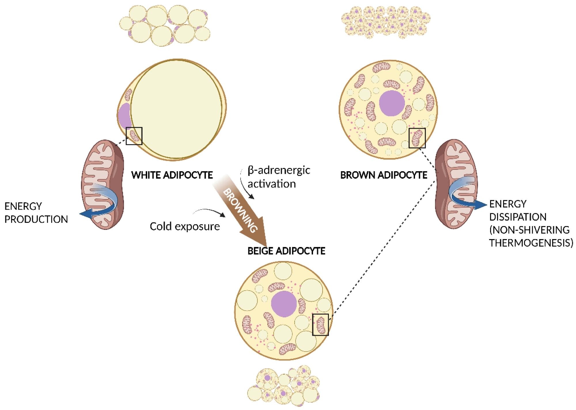
    WAT, BAT, and WAT browning. White adipocyte has one large droplet in the centre of the cell that compresses nucleus and mitochondria at one pole. Brown adipocyte has multiple small lipid droplets and more mitochondria, spread out between the droplets. Beige adipocyte has intermediate characteristics. Cold exposure and β-adrenergic activation determine the browning of WAT. Both brown and beige mitochondria are involved in non-shivering thermogenesis.  Abbreviations: BAT, brown adipose tissue; WAT, white adipose tissue.WAT, BAT, and WAT browning. White adipocyte has one large droplet in the centre of the cell that compresses nucleus and mitochondria at one pole. Brown adipocyte has multiple small lipid droplets and more mitochondria, spread out between the droplets. Beige adipocyte has intermediate characteristics. Cold exposure and β-adrenergic activation determine the browning of WAT. Both brown and beige mitochondria are involved in non-shivering thermogenesis.  Abbreviations: BAT, brown adipose tissue; WAT, white adipose tissue.

    AT: An active endocrine organ

    AT has transcended its traditional view as a mere energy reservoir and insulator and is now recognized as an active endocrine organ instrumental in metabolic regulation. This shift is attributed to its secretion of hormones like leptin and adiponectin and a variety of cytokines termed adipokines, marking its profound impact on metabolism. Within this tissue, adipocytes and other cell types, such as pre-adipocytes and immune cells, form a complex cellular environment underlying its multifaceted functions.

    Diverse functions of WAT and BAT

    AT, classified into WAT for energy storage and BAT for thermogenic energy expenditure, plays vital roles in metabolic health. WAT’s large cells store fat, contributing to mechanical protection and metabolic regulation, while BAT’s smaller, lipid-rich cells generate heat through non-shivering thermogenesis, offering potential in treating metabolic disorders.

    The metabolic and endocrine role of AT

    Ectopic fat deposition and the activation of ATs highlight the complexity of their roles in health and disease. While ectopic fats are associated with metabolic complications, the process of “browning” in WAT, wherein cells adopt BAT-like characteristics, offers therapeutic prospects for metabolic diseases. The endocrine functions of WAT further elucidate its role in energy and metabolic homeostasis, with adipokines like leptin and adiponectin playing critical roles. BAT’s contribution to energy expenditure through non-shivering thermogenesis represents a fundamental aspect of metabolic health, distinguishing the unique contributions of WAT and BAT to the body’s energy balance and metabolic regulation.

    Mitochondrial function in AT

    Mitochondria play a critical role in energy metabolism within both WAT and BAT, driving adenosine triphosphate (ATP) production through nutrient oxidation and regulating lipid metabolism. In WAT, they support lipid synthesis and breakdown, influencing adipocyte differentiation and metabolic health. Dysfunctional mitochondria in WAT are linked to metabolic diseases due to impaired regulation of adipokines and fatty acid oxidation. Conversely, BAT mitochondria facilitate non-shivering thermogenesis via uncoupling protein-1 (UCP-1), showcasing their essential role in energy expenditure. This distinct mitochondrial function in BAT versus WAT underscores their significance in metabolic regulation and the potential for therapeutic targets in obesity and related conditions.

    Mitochondrial dysfunction and obesity: A close connection

    Obesity’s impact on mitochondrial metabolism in AT, particularly in WAT and BAT, underscores a critical aspect of its pathophysiology. Mitochondrial dysfunction, characterized by altered bioenergetics and impaired lipid and glucose metabolism, plays an important role in intensifying obesity-related metabolic complications. Studies have revealed significant mitochondrial alterations in obesity, including diminished expression of mitochondrial proteins, reduced mitochondrial deoxyribonucleic acid (mtDNA) copy numbers, and decreased activity of oxidative phosphorylation complexes. These changes not only contribute to inefficient energy utilization and storage but also foster a shift towards adipocyte hypertrophy, further promoting inflammation and insulin resistance. The compromised mitochondrial function in WAT affects fatty acid oxidation and adipocyte differentiation, while in BAT, it impairs thermogenic efficiency, potentially shifting its metabolism towards a WAT-like phenotype. This connection between mitochondrial dysfunction and obesity highlights the urgent need for therapeutic strategies aimed at restoring mitochondrial health, offering a promising avenue for obesity management and the reduction of its associated metabolic disorders.

    The role of gut microbiota in obesity and AT mitochondrial function

    The interplay between gut microbiota and AT mitochondria significantly influences obesity management and metabolic health. The diverse gut ecosystem plays a fundamental role in metabolic processes, impacting lipid and glucose metabolism through the production of microbial metabolites like short-chain fatty acids (SCFAs). These metabolites affect mitochondrial function in ATs, modifying fatty acid oxidation and adipocyte differentiation, which are critical in obesity progression. Dysbiosis, characterized by an imbalance in gut microbial composition, has been linked to obesity-related metabolic impairments, suggesting that modulating gut microbiota could offer therapeutic avenues for improving mitochondrial function and combating obesity. This highlights the necessity for continued exploration into the gut microbiota- AT mitochondria axis to develop targeted interventions for obesity and its associated metabolic disorders.

    Journal reference:

    • Colangeli L, Escobar Marcillo DI, Simonelli V, Iorio E, Rinaldi T, Sbraccia P, Fortini P, Guglielmi V. The Crosstalk between Gut Microbiota and White Adipose Tissue Mitochondria in Obesity. Nutrients. (2023). DOI – 10.3390/nu15071723, https://www.mdpi.com/2072-6643/15/7/1723 

    [ad_2]

    Source link

  • Tumor microbiomes offer new insights for enhancing cancer therapies

    Tumor microbiomes offer new insights for enhancing cancer therapies

    [ad_1]

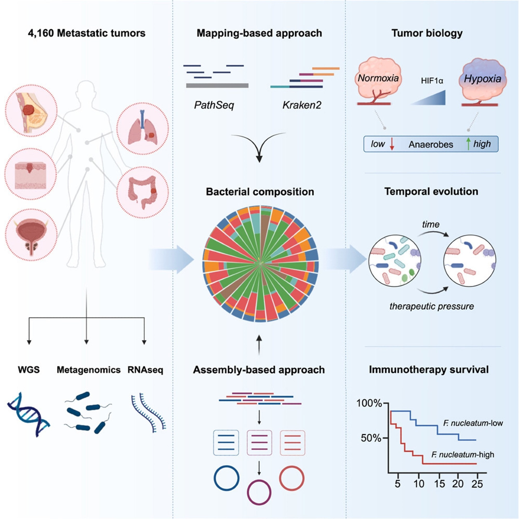
    In a recent study published in the journal Cell, researchers used metagenomics, genomics, and transcriptomics to examine microbiome genomes in over 4,000 metastatic tumor tissues. They analyzed the tumor microbiome and tumor microenvironment (TME), offering biological information and influencing the development of bacteria-focused techniques to supplement and improve cancer treatments.

    Microbial communities play a crucial role in the human body, influencing the immune system and anticancer therapies. They are present in primary tumors and interact with the commensal microbiota. The gut microbiota can modulate immune checkpoint blockers (ICB) and conventional chemotherapies. Fecal microbial transplants may improve clinical responsiveness to ICB agents. Understanding how tumor-resident bacteria shape tumor biology, immune infiltration, and treatment responsiveness is essential for understanding tumor response to ICB.

    Study: A pan-cancer analysis of the microbiome in metastatic cancerStudy: A pan-cancer analysis of the microbiome in metastatic cancer

    About the study

    In the present study, researchers used bioinformatics to investigate the microbiota in metastatic malignancies, evaluating 4,160 specimens from diverse cancer types.

    The researchers used mapping and assembly-based metagenomics, genomes, transcriptomics, and clinical data to develop a pan-cancer repository that might help advance treatment techniques. They used two distinct computational approaches, PathSeq and Kraken2, to define tumor-resident microbiome communities at the genus level and a metagenomic assembly-based approach at the species level. The team then shaped the metastatic tumor microbiome by identifying the elements that influence its makeup and evaluating cancer-type-specific microbial communities. They used the characteristic hypoxia gene profile to assess the degree of hypoxia in metastatic cancers and then performed gene set enrichment analysis (GSEA). They also investigated whether microbial communities may affect host immunity and the TME.

    The researchers investigated the relationship between gram-negative bacteria in metastases and Toll-like receptor (TLR) expression and whether lipopolysaccharide (LPS), obtained from dead or active bacteria, plays a primary role in TLR4 signaling in metastases. They additionally examined the relationship between bacterial makeup and tumor gene expression and the relationships between particular bacteria and immune cells.

    To further understand the impact of metastatic heterogeneity and the durability of tumor-resident microorganisms over time, the team examined 185 pairs of 370 repeated tumor specimens obtained from 173 different individuals. They examined bacterial enrichment changes before and after tumor treatment with immunotherapy, targeted therapy, or hormone therapy. They also investigated bacterial count reductions following immunotherapy in responsive patients and whether these germs were more prevalent in non-responsive individuals before treatment. Lastly, they examined pre-treatment bacterial communities associated with a lack of response to immunosuppressive medication in an ICB-monotherapy cohort of NSCLC patients.

    Results

    The researchers detected tumor-resident bacteria deoxyribonucleic acid (DNA) in a pan-cancer metastasis cohort, and assembling tumor-derived bacterial DNA provided species-level genomic characterization. Bacterial diversity correlated with cellular and molecular tumor immunity characteristics. In an NSCLC cohort, high levels of fusobacterium DNA imply a poor immunotherapy response. Researchers found organ-specific microbe tropisms, anaerobic bacteria enrichments in hypoxic tumors, links between microbial diversity and tumor-infiltrating neutrophils, and Fusobacterium’s relationship with resistance to ICB therapy in lung cancer.

    Using mapping-based techniques and screening genera to eliminate technical contamination and seldom-seen genera, the team cataloged 165 microbial genera from 3,526 specimens, with 68% facultative/anaerobic and 49% gram-negative anaerobes. They built 514 metagenomic-assembled genomes (MAGs) of medium- to almost high-quality using tumor-derived microbial sequences. The most common tumor types were colorectal, breast, prostate, lung, and melanoma, with the lymph node, liver, and lung being the most common metastatic locations for tumor samples.

    The quantity of bacterial-derived reads expressed as a human-mapped genetic read proportion varied by cancer type, with higher fractions in renal and uterine malignancies and lower burdens in tumors originating from the brain and spinal cord. Renal and colorectal metastases were the most diverse, but head and neck metastatic tumors showed more dominant microbial genera.

    Tumor-resident microbial communities were associated with tumor biology, with a strong correlation between LPS load and TLR4 signaling but not gram-positive lipoteichoic acid (LTA) load.  Multivariate Cox proportional-hazards modeling showed lower overall survival (OS) and progression-free survival (PFS) rates significantly correlated with continuous Fusobacterium abundance, considering the genome-wide mutational load. Using the pan-cancer dataset, the researchers classified all tumors as Fuso-high or Fuso-low based on an upper quartile relative abundance cutoff similar to previously established criteria. Fuso-high tumors showed considerably decreased cytotoxic, interferon-gamma (IFN-γ), and major histocompatibility complex (MHC) class II gene expression profiles.

    The study provides the first large-scale pan-cancer map of intratumor microbiomes in metastatic malignancies, examining diversity across anatomical regions, initial tumor type, and treatment responses, including immunotherapy. The study showed that the metastatic microbiome partially comprises anaerobic bacteria that may get altered during treatment. The study also discovered links between intra-tumoral microorganisms and the activation of innate immune sensing pathways, indicating that the tumor microenvironment alters via direct identification of bacterial ligands.

    [ad_2]

    Source link

  • A balanced take on microbes and disease outcomes

    A balanced take on microbes and disease outcomes

    [ad_1]

    In a recent review published in the journal PNAS, researchers examined the germ theory from a non-centric perspective on infection outcome, considering the variables that influence illness severity while drawing on a known understanding of microbial pathogenesis, evolutionary biology, and pathogen-host interactions.

    The germ theory revisited: A noncentric view on infection outcome. Image Credit: ImageFlow / ShutterstockThe germ theory revisited: A noncentric view on infection outcome. Image Credit: ImageFlow / Shutterstock

    Background

    The germ-disease theory, created in the nineteenth century, is a critical medical paradigm claiming that pathogenic microbes cause infectious diseases. However, this explanation is microorganism-centric, failing to explain varying disease severity and symptomatic profiles among individuals. While the microorganism is necessary to produce illness, it cannot solely determine the infection outcome. As a result, the theory must consider the host’s status fluctuations, which considerably impact infection outcomes.

    Recent research questions the theory, giving rise to the full-blown host theory, which holds that infectious illnesses are caused by inherited or acquired immunodeficiencies in the host. According to this hypothesis, viruses are passive environmental triggers, and disease development results from pre-existing host immunodeficiencies. These inadequacies can be covert or overt, depending on diagnostic procedures used to identify critical diseases.

    About the review

    In the present review, researchers investigated the relative relevance of pathogenic microbes and hosts in determining infection outcomes, revealing a non-centric perspective that acknowledges essential roles for both the pathogenic microorganism and the host in defining infection outcomes.

    Pathogens and virulence

    Pathogens are germs that can cause illness among immunocompetent and healthy hosts; nevertheless, asymptomatic carriership occurs when pathogens infect or colonize hosts without causing symptoms. Examples include Helicobacter pylori and severe acute respiratory syndrome coronavirus 2 (SARS-CoV-2), which can cause asymptomatic infections in 40% to 80% of cases. Mycobacterium tuberculosis is the most prominent example of pathogens producing asymptomatic carriership, with one-fourth of the worldwide human population latently infected but exhibiting no symptoms of illness.

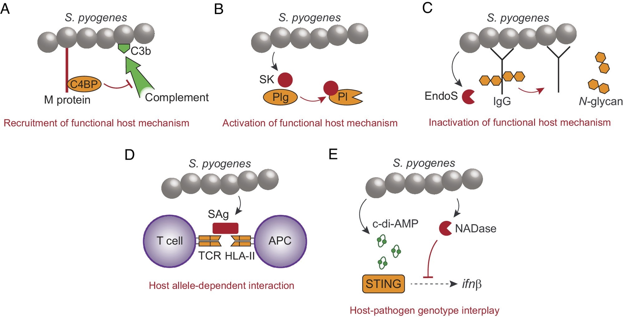
    Examples demonstrating the evolved ability of S. pyogenes to promote disease in immunocompetent individuals. (A) The surface M protein recruits human C4BP to inhibit complement opsonization (C3b) of the bacterial surface. (B) Secreted streptokinase (SK) binds to human Plg, which causes a conformational change of Plg into a plasmin (Pl) active state. (C) The secreted endoglycosidase EndoS inactivates effector functions of IgG by cleaving off N-glycans from the Fc-region. (D) Secreted superantigen (SAg) causes antigen-independent T cell activation by cross-linking the TCR with HLA-II on antigen presenting cells (APC). SAgs have different affinity for different fully functional HLA-II haplotypes. (E) The STING responds to S. pyogenes-derived c-di-AMP to induce transcription of the interferon β gene, which is inhibited by the enzymatic activity of bacterial NADase. Human STING and S. pyogenes NADase exhibit polymorphisms affecting their relative ability to respond to c-di-AMP and to suppress interferon transcription, respectively.Examples demonstrating the evolved ability of S. pyogenes to promote disease in immunocompetent individuals. (A) The surface M protein recruits human C4BP to inhibit complement opsonization (C3b) of the bacterial surface. (B) Secreted streptokinase (SK) binds to human Plg, which causes a conformational change of Plg into a plasmin (Pl) active state. (C) The secreted endoglycosidase EndoS inactivates effector functions of IgG by cleaving off N-glycans from the Fc-region. (D) Secreted superantigen (SAg) causes antigen-independent T cell activation by cross-linking the TCR with HLA-II on antigen presenting cells (APC). SAgs have different affinity for different fully functional HLA-II haplotypes. (E) The STING responds to S. pyogenes-derived c-di-AMP to induce transcription of the interferon β gene, which is inhibited by the enzymatic activity of bacterial NADase. Human STING and S. pyogenes NADase exhibit polymorphisms affecting their relative ability to respond to c-di-AMP and to suppress interferon transcription, respectively.

    In contrast, opportunistic microbes cannot cause illness in usual settings but can be pathogenic in the case of compromised host steady-state conditions. For example, Clostridium difficile, a spore-forming bacteria, can cause symptomatic illness when treated with antibiotic medications, which reduces the microbiota and allows Clostridium difficile germination to develop a footing. Commensal Candidal fungi can also cause illness in regular settings, but only in the case of immunosuppression, as observed in individuals with HIV-associated immunodeficiency syndrome (AIDS).

    Virulence, a pathogen’s capacity to generate illness symptoms or pathology, is vital to infection outcomes. According to evolutionary theory, a pathogen’s Darwinian fitness is often maximized at an intermediate degree of virulence, resulting in fitness costs and benefits. This idea gains support by controlled laboratory tests and epidemiological research on human illnesses such as AIDS.

    Host-pathogenic microbe interactions and genetic variability

    Streptococcus pyogenes, a bacterium often present in humans, can cause asymptomatic carriership and symptomatic illnesses such as septic shock and necrotizing infections in soft tissues. Streptococcus pyogenes’ surface M proteins are polymorphic virulence factors that impart phagocytic resistance and prevent complement opsonization, recruiting the serum C4b-binding protein (C4BP) and enabling, allowing rapid bacterial proliferation in blood.

    A secreted Streptococcus pyogenes protein, streptokinase, preferentially binds with and stimulates human plasminogen (Plg), promoting proteolytic actions and bacterial spread. Transgenic mice infection revealed the importance of this relationship in virulence, revealing that Streptococcus pyogenes activates fibrinolytic pathways to cause illness.

    The association of bacterial virulence characteristics with the genetic diversity of the host is critical. Superantigens (SAgs) of streptococcal species form cross-links with T-cell receptors (TCRs) to human major histocompatibility complex (MHC) class II molecules, causing antigen-independent T-lymphocyte activation. Fully functional human leukocyte antigen II (HLA-II) variations considerably affect infection outcomes. Type I interferon (IFN) signaling protects Streptococcus pyogenes-infected mice against host-damaging inflammation.

    Uncommon mutations that profoundly impact infection outcomes cause Casanova’s inborn immunity defects. More frequent alleles with lesser effects play a primary role in the phenotypic variance in infection outcomes, maintained by balanced selection. In addition to host genetic diversity, pathogens exhibit enormous genetic variation, and the combination of host and pathogen genotypes considerably impacts disease severity.

    Based on the review findings, microbes have evolved to flourish in extreme conditions such as polar ice, scorching vents, sulfur-acidic lakes, and immunocompetent hosts. Pathogens have developed methods to avoid host defenses, allowing them to infect and produce clinical illness in immunocompetent humans. The review stresses that the host and the pathogen’s genetic diversity can impact infection outcomes, implying that both species play active roles. The researchers’ proposed full-fledged host theory is disproven in favor of a non-centric approach, recognizing crucial roles for the causal bacterium and the host in determining infection outcome.

    [ad_2]

    Source link

  • Study shows GHRH antagonist’s role in AML drug resistance

    Study shows GHRH antagonist’s role in AML drug resistance

    [ad_1]

    A new research paper was published in Oncotarget’s Volume 15 on April 8, 2024, entitled, “Exploring the role of GHRH antagonist MIA-602 in overcoming Doxorubicin-resistance in acute myeloid leukemia.”

    Acute myeloid leukemia (AML) is characterized by the rapid proliferation of mutagenic hematopoietic progenitors in the bone marrow. Conventional therapies include chemotherapy and bone marrow stem cell transplantation; however, they are often associated with poor prognosis. Notably, growth hormone-releasing hormone (GHRH) receptor antagonist MIA-602 has been shown to impede the growth of various human cancer cell lines, including AML. In this new study, researchers Simonetta I. Gaumond, Rama Abdin, Joel Costoya, Andrew V. Schally, and Joaquin J. Jimenez from the University of Miami, Florida Atlantic University and Veterans Affairs Medical Center, Miami examined the impact of MIA-602 as monotherapy and in combination with Doxorubicin on three Doxorubicin-resistant AML cell lines, KG-1A, U-937, and K-562. 

    “Given the role of GHRH in multiple cancer types, it is possible that GHRH antagonists may offer an alternative treatment approach for AML as well as drug-resistant AML, which may circumvent the side effects associated with standard chemotherapy.”

    The in vitro results revealed a significant reduction in cell viability for all treated wild-type cells. Doxorubicin-resistant clones were similarly susceptible to MIA-602 as the wild-type counterpart. Their in vivo experiment of xenografted nude mice with Doxorubicin-resistant K-562 revealed a reduction in tumor volume with MIA-602 treatment compared to control. 

    “Our study demonstrates that these three AML cell lines, and their Doxorubicin-resistant clones, are susceptible to GHRH antagonist MIA-602.”

    Source:

    Journal reference:

    Gaumond, S. I., et al. (2024). Exploring the role of GHRH antagonist MIA-602 in overcoming Doxorubicin-resistance in acute myeloid leukemia. Oncotarget. doi.org/10.18632/oncotarget.28579.

    [ad_2]

    Source link

  • Three early-phase clinical studies show promising initial data for patients with lymphoma, gastric cancers

    Three early-phase clinical studies show promising initial data for patients with lymphoma, gastric cancers

    [ad_1]

    Three early-phase clinical studies presented by researchers from The University of Texas MD Anderson Cancer Center at the American Association for Cancer Research (AACR) Annual Meeting 2024 show promising initial data for patients with lymphoma, gastric or gastroesophageal junction cancers, and specific molecularly selected tumors. The studies were featured in a clinical trials minisymposium highlighting novel agents and emerging therapeutic strategies. Information on all MD Anderson AACR Annual Meeting content can be found at MDAnderson.org/AACR.

    Novel combination with evorpacept demonstrates promising results for patients with B-cell non-Hodgkin lymphoma (Abstract CT037)

    Data from an investigator-initiated Phase I trial of the novel CD47 blocker evorpacept (ALX148) in combination with lenalidomide and rituximab (R2), presented by Paolo Strati, M.D., assistant professor of Lymphoma and Myeloma, showed promising early results for patients with relapsed or refractory B-cell non-Hodgkin lymphoma.

    While historical complete response rates (CRRs) with R2 monotherapy are 30%, the new combination achieved an 80% CRR with an overall response rate of 90%. After a median follow-up of 16 months, the one-year progression-free survival rate was 70% and one-year overall survival rate was 90%.

    Previous work from Strati demonstrated that specific white blood cells known as SIRPα+ macrophages may mediate resistance to the R2 combination, leading to the hypothesis that adding evorpacept to the R2 combination could have synergistic results. Blocking CD47 prevents the ‘don’t eat me signal’ that results from the interaction of SIRPα and CD47.

    The addition of evorpacept to R2 seems to significantly increase the efficacy of these therapies, with no synergistic toxicity, in patients with indolent B-cell lymphomas. We look forward to continuing studies with this novel combination in order to bring effective options to our patients in need.”


    Paolo Strati, M.D., assistant professor of Lymphoma and Myeloma

    The trial included 20 patients with four different types of B-cell non-Hodgkin lymphoma, 18 of whom had indolent, or slow-growing, disease. All patients previously received an anti-CD20 monoclonal antibody, and 73% received prior chemotherapy.

    The most common grade 3 or higher adverse events were consistent with those typically seen with R2 therapy, such as neutropenia, infections, elevated liver enzymes, skin rash and anemia, and were not increased by the addition of evorpacept. No dose-limiting toxicities were observed, and the recommended Phase II dose was identified.

    A Phase II study investigating the efficacy of this combination in previously untreated patients is now enrolling.

    This study was funded by ALX Oncology, the Leukemia & Lymphoma Society Scholar in Clinical Research Award, the Gilead and Kite Scholar Award and the Andrew Sabin Family Fellowship Program. 

    Antibody-drug conjugate SKB264 shows potential for durable responses in heavily pretreated patients with advanced gastric or gastroesophageal junction cancers (Abstract CT038)

    Preliminary data from a Phase I/II study evaluating the antibody-drug conjugate (ADC) SKB264, presented by Jordi Rodon, M.D., Ph.D., associate professor of investigational cancer therapeutics showed that pretreated patients with gastric or gastroesophageal junction cancers could have durable responses and potentially longer overall survival with SKB264 monotherapy.

    Of the 41 patients able to be evaluated for response, SKB264 achieved an objective response rate of 22%, a disease control rate of 80.5%, and a median duration of response of 7.5 months.

    “It is interesting to note the change in antitumor activity and safety profile that results from changing payloads and linkers, even among ADCs aiming at the same target,” Rodon said. “One of the big results of this trial is that, by using a different linker-payload combination, we did not see the interstitial lung diseases associated with other ADCs.”

    SKB264 is an ADC that targets TROP2, a factor associated with poor prognosis in advanced gastric cancers. It uses a novel linker to connect the antibody with the payload, a belotecan-derivate topoisomerase I inhibitor.

    Forty-eight patients were evaluable for safety, with a follow-up of at least nine weeks at the data cutoff. All received previous therapy, with 50% having received multiple prior lines of therapy. Treatment-related adverse events higher than grade 3 were seen in 52.1% of patients, with the most common being anemia, decreased white blood cell or neutrophil counts, and neutropenia. Only 18.8% of patients had to decrease dosage, and 33.3% had to delay dosing due to adverse events. No adverse events led to discontinuation of the trial or death.

    Based on these results, a Phase III global study currently is being planned to evaluate SKB264 in comparison to the current standard of care in patients with at least three prior lines of therapy in gastric or gastroesophageal junction adenocarcinomas.

    This study was funded by Klus Pharma and Merck & Co. (MSD). 

    Combining PARP and PI3K inhibitors shows encouraging results in Phase Ib trial for molecularly selected patients (Abstract CT042) 

    Results from a biomarker-driven, tumor-agnostic Phase Ib trial of copanlisib plus olaparib showed the combination was safe and well tolerated, with a clinical benefit in 36% of molecularly selected patients who had advanced cancers harboring PIK3CA hotspot, PTEN, or DNA damage response (DDR) mutations. 

    Trial results were presented by Timothy Yap, M.B.B.S., Ph.D., professor of Investigational Cancer Therapeutics, and vice president and head of clinical development in the Therapeutics Discovery division. 

    Of 22 patients able to be evaluated for efficacy, six achieved partial responses and two achieved stable disease for at least six months. Notably, a patient with advanced triple-negative breast cancer with PIK3CA H1047R, PTEN and ARID1A mutations achieved a confirmed radiological partial response and was on trial for 42 months with minimal side effects. 

    Preclinical studies had shown potential for the combination of PI3K and PARP inhibition in cancers with DDR and PI3K pathway alterations, prompting this investigator-initiated trial conducted through National Cancer Institute Cancer Therapy Evaluation Program (NCI CTEP), which partnered the pan-PI3K inhibitor copanlisib with the PARP inhibitor olaparib. 

    The trial enrolled 28 patients, representing 12 cancer types and 11 mutation types. Ten patients had more than one qualifying mutation. This was a heavily pretreated patient population, with 61% having received three or more prior lines of therapy. The combination was safe and generally well tolerated, with the most common adverse events of any grade being mucositis, nausea, diarrhea and vomiting. 

    “We observed promising antitumor activity in patients with a range of cancers with different DDR and/or PI3K pathway alterations, which may serve as novel predictive biomarkers of response,” Yap said. “Comprehensive translational analyses of longitudinally collected patient samples from the clinical trial are currently ongoing.” 

    This study was funded and sponsored by the National Cancer Institute, part of National Institutes of Health. MD Anderson led the study with participation from the NCI Experimental Therapeutics Clinical Trials Network (ETCTN) (4UM1CA186688-07). AstraZeneca Pharmaceuticals and Bristol-Myers Squibb Company provided support to this study through Cooperative Research and Development Agreement between NCI and each company.

    [ad_2]

    Source link

  • Deciphering cancer plasticity:Insights from MSK research

    Deciphering cancer plasticity:Insights from MSK research

    [ad_1]

    About 90% of deaths from cancer are a result of metastasis -; that is, from cancer’s ability to spread from an initial primary tumor to seed new tumors throughout the body, often in the lungs, liver, and brain.

    And metastasis relies on cancer cells’ ability to adapt to different tissue environments throughout the body by gaining improper access to a variety of playbooks stored in our genetic code -; including gene programs that are generally available only during early stages of human development.

    Today, researchers at Memorial Sloan Kettering Cancer Center (MSK) are using innovative approaches like single-cell sequencing technology and sophisticated computational tools to illuminate cancer cells’ ability to take on new traits. And they’re applying those findings toward treating or preventing metastasis.

    In a plenary presentation at the 2024 American Association for Cancer Research (AACR) Annual Meeting, Dana Pe’er, PhD, Chair of the Computational and Systems Biology Program at MSK’s Sloan Kettering Institute, highlighted three recent research collaborations between her lab and other labs at MSK that have shed new light on the ability of cancer cells to adapt and spread.

    “It’s not genetic mutations that are critical here, but the ability to access gene programs that normally are associated with other cell types -; including early developmental and embryonic programs that should not be accessed by adult cells,” Dr. Pe’er said in a recent interview. “We call this ability for cells to run new programs ‘plasticity.’ So cancer doesn’t reinvent the wheel; it exploits gene programs that exist for other biological purposes.”

    In general, she notes, plasticity isn’t a bad thing. It’s important for early development and for regeneration after injury. And the body also has built-in barriers to limit the scope of plasticity so that cells can’t just run amok.

    But in cancer, these cells can wreak havoc because they’ve lost these natural barriers.”


    Dr. Dana Pe’er, Howard Hughes Medical Institute Investigator

    Plasticity in colorectal cancer

    In the first study Dr. Pe’er presented at AACR, she teamed up with MSK physician-scientist Karuna Ganesh, MD, PhD, to look at the differences in gene programs active in the primary tumors and metastatic tumors of patients with advanced colorectal cancer. Samples of both types of tumors were collected at the same time from 31 patients -; the largest cohort of its kind -; some of whom had undergone chemotherapy and some of whom had not.

    They also created organoids from the patients’ cells -; three-dimensional clusters of cells that act more like human tissue than traditional laboratory cell cultures.

    The study found that primary tumor cells largely run programs still associated with intestinal cells, while metastases often shed their heritage as intestinal cells and take on the characteristics of squamous cells or of neuroendocrine cells, which helps them invade and survive in new tissue contexts and makes them more resistant to treatment. The team also found that chemotherapy exacerbates these transformations. The findings were published as a preprint on bioRxiv in August 2023.

    “The metastatic organoids were very different from the primary tumor organoids,” Dr. Pe’er says. “And it matters what environment they’re in, too. If you put the metastatic organoids into the liver of a mouse, they will adapt their identity in ways that primary tumor organoids are not able to -; so they’re a lot more plastic.”

    The team additionally found that a gene known as PROX1 restricted the ability of cells from the primary tumor to stray too far from their lineage as intestinal cells. But when this factor is removed, the cells gain access to many more types of cell lineages -; which scientists call noncanonical. (For reasons that aren’t fully understood, metastatic cells that lose the restrictions imposed by Prox1 are already primed to go in these noncanonical directions.)

    “I call it a mix-and-match buffet,” Dr. Peer says. “Metastatic cells have this awesome power to combine gene programs across many different types of cells, endowing them with new abilities that allow them to adapt themselves to take advantage of different conditions and environments throughout the body.”

    Plasticity in pancreatic cancer

    The second study Dr. Pe’er presented looked at about a dozen metastases collected from a single patient with pancreatic cancer, who donated their body for research under MSK’s Last Wish Program. A collaboration with physician-scientist Christine Iacobuzio-Donahue, MD, PhD, Director of the David M. Rubenstein Center for Pancreatic Cancer Research at MSK, the research used advanced single-cell and computational approaches to look at the differences in active gene programs in genetically identical cancer cells -; called clones -; that had spread to different locations in the body. The findings of the study have not yet been published.

    “What we see is that these clones are able to adapt to the pressures and metabolic demands of very different environments,” Dr. Pe’er says. “And we see that they’re able to access different gene programs that allow them to thrive in different places, different organs.”

    Moreover, even genetically different cancer cells tend to adapt to specific situations by accessing the same gene programs.

    “The big question of a cancer cell is, ‘Are you plastic or not?’ And once you are, you can acquire all these different traits. The environment is what really determines what traits will be most advantageous,” she says.

    For example, the research showed that cells that metastasize to the peritoneum -; the tissue that lines the abdominal cavity -; are able to adapt their metabolism to take advantage of the lipid-rich environment and exploit it as an energy source, she notes.

    Moving plasticity research from the lab to the clinic

    Lastly, Dr. Pe’er highlighted a third collaboration -; this time with neuro-oncologist Adrienne Boire, MD, PhD, a member of MSK’s Human Oncology and Pathogenesis Program -; which showed how plasticity can be turned against cancer cells.

    The research led to a clinical trial for patients with leptomeningeal metastasis, which is when cancer has spread to the fluid and tissues of the spinal cord and brain. The team showed that cancer cells were able to survive in this challenging environment by reprogramming themselves to outcompete other cells for iron; this fuels their growth while also preventing immune defenders in the area from getting enough iron.

    “It’s an elegant solution on the part of the cancer cell,” Dr. Boire says. “It’s really unique biology that allows them to win the competition.”

    Based on that discovery, doctors at MSK are now determining whether a drug called deferoxamine could be an effective treatment for leptomeningeal metastases by helping to remove iron from the cerebrospinal fluid.

    “The plasticity of these cells allowed several patients with several different cancers to overexpress the same two genes that are typically only expressed in myeloid cells,” Dr. Pe’er says. “And the cells not only got aberrant access to these gene programs, but they also expressed the genes at 100 times the levels seen in their normal counterparts.”

    By injecting the study drug into the spinal fluid, the idea is to prevent the cancer cells from getting the iron they need to thrive. And so far, Dr. Pe’er told the audience, initial results from the trial have been extremely promising.

    Only at a place like MSK

    “The ultimate goal, though we’re not there yet, would be to be able to target plasticity directly -; to restore some of the biological barriers or inhibit plasticity with drugs,” Dr. Pe’er says.

    And MSK is uniquely poised to pursue that aim, with strong collaborations between laboratory and clinical research; a high volume of patients that provides a wide variety of clinical samples from a wide variety of cancers, including rare ones; access to state-of-the art sequencing tools combined with some of the world’s top computational expertise; and a significant number of physician-scientists who focus both on caring for patients and finding new ways to treat their disease.

    “Not every cancer center would see enough patients with leptomeningeal metastasis to set up a clinical trial like this,” Dr. Pe’er notes. “Or have someone like Dr. Boire, who not only cares for patients with metastasis to the central nervous system but who also runs a lab dedicated to studying the underlying molecular mechanisms.”

    [ad_2]

    Source link

  • Enhancer-gene mapping with SCENT method offers insights into disease mechanisms

    Enhancer-gene mapping with SCENT method offers insights into disease mechanisms

    [ad_1]

    Genetic studies of diseases map segments of the genome driving disease. But to understand how those changes contribute to disease progression, it is important to understand how they may alter gene regulation of disease genes in cell populations assumed to be driving disease. “Enhancer-gene maps” link genomic regulatory regions to genes and are essential for understanding disease. But constructing them poses challenges due to limitations in current experimental methods, that make it difficult to apply the technique to rare cell populations and genes that only regulate specific cell types.

    Researchers from Brigham and Women’s Hospital, a founding member of the Mass General Brigham healthcare system, have developed a statistical method called SCENT (single-cell enhancer target gene mapping). This method uses multimodal single-cell data to establish links between regulatory elements and genes, allowing them to pinpoint probable causal gene loci for both common and rare diseases. These insights might assist the development of treatments for various conditions.

    The research team applied SCENT to nine multimodal single-cell datasets representing various human tissues, including immune, neuronal, and pituitary cells, aiming to understand the intricacies of DNA regulation in each specific cell type. With these data, they developed 23 distinct gene-enhancer maps, to investigate genetic variants and expression patterns associated with 1,143 diseases and traits. Notably, they discovered that, for immune diseases, crucial insights emerged not only from immune cells but also from cells within the affected tissues themselves.

    For most autoimmune diseases, people assume that we need a general map of immune cells. But we find that the enhancer-gene maps of immune cells are different in affected disease tissues. We demonstrate how such a map can be used to interpret genetic data from rheumatoid arthritis and other autoimmune diseases.”


    Soumya Raychaudhuri, MD, PhD, of the Brigham’s Division of Rheumatology, Immunology and Inflammation

    Source:

    Journal reference:

    Sakaue, S., et al. (2024). Tissue-specific enhancer–gene maps from multimodal single-cell data identify causal disease alleles. Nature Genetics. doi.org/10.1038/s41588-024-01682-1.

    [ad_2]

    Source link

  • Bio-Techne surpasses 10,000 peer-reviewed publications citing RNAscope technology

    Bio-Techne surpasses 10,000 peer-reviewed publications citing RNAscope technology

    [ad_1]

    Bio-Techne Corporation today announced that Advanced Cell Diagnostics (ACD), part of Bio-Techne’s Spatial Biology Division, has surpassed 10,000 peer-reviewed publications referencing the use and application of RNAscope ISH technology. Over 50% of these publications were released in the past 3 years, as increasing global customer awareness and expanded market adoption solidify ACD’s leadership in spatial biology applications.

    As a pioneer in spatial biology, Bio-Techne’s broad portfolio of over 50,000 unique RNAscope ISH probes across over 400 species enables its biopharma and academic customers to reveal ground-breaking biologic discoveries, accelerate the development of next generation therapeutics, and deliver life-changing diagnostics. Representing the most referenced spatial biology technology in the industry for RNA biomarkers, RNAscope offers best-in-class single-molecule sensitivity with unrivaled specificity at subcellular resolution. This landmark portfolio of 10,000 scientific publications encompasses applications in cancer and neuroscience, as well as additional areas of research including immunology, infectious disease, cell and gene therapy, and regenerative medicine.

    “We are thrilled to celebrate the achievement of over 10,000 publications referencing RNAscope ISH technology,” said Matt McManus, President of Bio-Techne’s Diagnostics & Genomics Segment. “We are honored by the trust the scientific community has placed in RNAscope to uncover new spatial insights and enable significant breakthroughs across a wide range of diseases.”

    [ad_2]

    Source link