Tag: DNA

  • Google DeepMind’s Groundbreaking AI for Protein Structure Can Now Model DNA

    Google DeepMind’s Groundbreaking AI for Protein Structure Can Now Model DNA

    [ad_1]

    Google spent much of the past year hustling to build its Gemini chatbot to counter ChatGPT, pitching it as a multifunctional AI assistant that can help with work tasks or the digital chores of personal life. More quietly, the company has been working to enhance a more specialized artificial intelligence tool that is already a must-have for some scientists.

    AlphaFold, software developed by Google’s DeepMind AI unit to predict the 3D structure of proteins, has received a significant upgrade. It can now model other molecules of biological importance, including DNA, and the interactions between antibodies produced by the immune system and the molecules of disease organisms. DeepMind added those new capabilities to AlphaFold 3 in part through borrowing techniques from AI image generators.

    “This is a big advance for us,” Demis Hassabis, CEO of Google DeepMind, told WIRED ahead of Wednesday’s publication of a paper on AlphaFold 3 in the science journal Nature. “This is exactly what you need for drug discovery: You need to see how a small molecule is going to bind to a drug, how strongly, and also what else it might bind to.”

    AlphaFold 3 can model large molecules such as DNA and RNA, which carry genetic code, but also much smaller entities, including metal ions. It can predict with high accuracy how these different molecules will interact with one another, Google’s research paper claims.

    The software was developed by Google DeepMind and Isomorphic labs, a sibling company under parent Alphabet working on AI for biotech that is also led by Hassabis. In January, Isomorphic Labs announced that it would work with Eli Lilly and Novartis on drug development.

    AlphaFold 3 will be made available via the cloud for outside researchers to access for free, but DeepMind is not releasing the software as open source the way it did for earlier versions of AlphaFold. John Jumper, who leads the Google DeepMind team working on the software, says it could help provide a deeper understanding of how proteins interact and work with DNA inside the body. “How do proteins respond to DNA damage; how do they find, repair it?” Jumper says. “We can start to answer these questions.”

    Understanding protein structures used to require painstaking work using electron microscopes and a technique called x-ray crystallography. Several years ago, academic research groups began testing whether deep learning, the technique at the heart of many recent AI advances, could predict the shape of proteins simply from their constituent amino acids, by learning from structures that had been experimentally verified.

    In 2018, Google DeepMind revealed it was working on AI software called AlphaFold to accurately predict the shape of proteins. In 2020, AlphaFold 2 produced results accurate enough to set off a storm of excitement in molecular biology. A year later, the company released an open source version of AlphaFold for anyone to use, along with 350,000 predicted protein structures, including for almost every protein known to exist in the human body. In 2022 the company released more than 2 million protein structures.

    [ad_2]

    Source link

  • The US Is Cracking Down on Synthetic DNA

    The US Is Cracking Down on Synthetic DNA

    [ad_1]

    The White House has issued new rules aimed at companies that manufacture synthetic DNA after years of warnings that a pathogen made with mail-order genetic material could accidentally or intentionally spark the next pandemic.

    The rules, released on April 29, are the result of an executive order signed by President Joe Biden last fall to establish new standards for AI safety and security, including AI applied to biotechnology.

    Artificially generated DNA allows researchers to do all sorts of things—develop diagnostic tests, make beneficial enzymes to eat up plastic, or engineer potent antibodies to treat disease—without having to extract natural sequences from organisms. Need to study a rare type of bacteria? Instead of going out into the field to collect a sample, its genetic sequence can simply be ordered from a DNA synthesis company instead.

    Synthesizing DNA has been possible for decades, but it’s become increasingly easier, cheaper, and faster to do so in recent years thanks to new technology that can “print” custom gene sequences. Now, dozens of companies around the world make and ship synthetic nucleic acids en masse. And with AI, it’s becoming possible to create entirely new sequences that don’t exist in nature—including those that could pose a threat to humans or other living things.

    “The concern has been for some time that as gene synthesis has gotten better and cheaper, and as more companies appear and more technologies streamline the synthesis of nucleic acids, that it is possible to de novo create organisms, particularly viruses,” says Tom Inglesby, an epidemiologist and director of the Johns Hopkins Center for Health Security.

    It’s conceivable that a bad actor could make a dangerous virus from scratch by ordering its genetic building blocks and assembling them into a whole pathogen. In 2017, Canadian researchers revealed they had reconstructed the extinct horsepox virus for $100,000 using mail-order DNA, raising the possibility that the same could be done for smallpox, a deadly disease that was eradicated in 1980.

    The new rules aim to prevent a similar scenario. It asks DNA manufacturers to screen purchase orders to flag so-called sequences of concern and assess customer legitimacy. Sequences of concern are those that contribute to an organism’s toxicity or ability to cause disease. For now, the rules only apply to scientists or companies that receive federal funding: They must order synthetic nucleic acids from providers that implement these practices.

    Inglesby says it’s still a “big step forward” since about three-quarters of the US customer base for synthetic DNA are federally funded entities. But it means that scientists or organizations with private sources of funding aren’t beholden to using companies with these screening procedures.

    Many DNA providers already follow screening guidelines issued by the Department of Health and Human Services in 2010. About 80 percent of the industry has joined the International Gene Synthesis Consortium, which pledges to vet orders. But these measures are both voluntary, and not all companies comply.

    [ad_2]

    Source link

  • Chemists Have Created the Functional Synthetic Cells That Act Like Real Ones

    Chemists Have Created the Functional Synthetic Cells That Act Like Real Ones

    [ad_1]

    Synthetic Cells Illustration

    Synthetic cells created with programmable peptide-DNA technology that directs peptides, the building blocks of proteins, and repurposed genetic material to work together to form a cytoskeleton, shown in fuscia. Credit: UNC-Chapel Hill

    Researchers employ cutting-edge methods to create functional cells, closing the divide between synthetic and biological materials.

    In a new study published in Nature Chemistry, UNC-Chapel Hill researcher Ronit Freeman and her colleagues describe the steps they took to manipulate DNA and proteins — essential building blocks of life — to create cells that look and act like cells from the body. This accomplishment, a first in the field, has implications for efforts in regenerative medicine, drug delivery systems, and diagnostic tools.

    “With this discovery, we can think of engineering fabrics or tissues that can be sensitive to changes in their environment and behave in dynamic ways,” says Freeman, whose lab is in the Applied Physical Sciences Department of the UNC College of Arts and Sciences.

    Cells and tissues are made of proteins that come together to perform tasks and make structures. Proteins are essential for forming the framework of a cell, called the cytoskeleton. Without it, cells wouldn’t be able to function. The cytoskeleton allows cells to be flexible, both in shape and in response to their environment.

    Without using natural proteins, the Freeman Lab built cells with functional cytoskeletons that can change shape and react to their surroundings. To do this, they used a new programmable peptide-DNA technology that directs peptides, the building blocks of proteins, and repurposed genetic material to work together to form a cytoskeleton.

    Innovative DNA Programming

    “DNA does not normally appear in a cytoskeleton,” Freeman says. “We reprogrammed sequences of DNA so that it acts as an architectural material, binding the peptides together. Once this programmed material was placed in a droplet of water, the structures took shape.”

    The ability to program DNA in this way means scientists can create cells to serve specific functions and even fine-tune a cell’s response to external stressors. While living cells are more complex than the synthetic ones created by the Freeman Lab, they are also more unpredictable and more susceptible to hostile environments, like severe temperatures.

    “The synthetic cells were stable even at 122 degrees Fahrenheit, opening up the possibility of manufacturing cells with extraordinary capabilities in environments normally unsuitable to human life,” Freeman says.

    Instead of creating materials that are made to last, Freeman says their materials are made to task — perform a specific function and then modify themselves to serve a new function. Their application can be customized by adding different peptide or DNA designs to program cells in materials like fabrics or tissues. These new materials can integrate with other synthetic cell technologies, all with potential applications that could revolutionize fields like biotechnology and medicine.

    “This research helps us understand what makes life,” Freeman says. “This synthetic cell technology will not just enable us to reproduce what nature does, but also make materials that surpass biology.”

    Reference: “Designer peptide–DNA cytoskeletons regulate the function of synthetic cells” by Margaret L. Daly, Kengo Nishi, Stephen J. Klawa, Kameryn Y. Hinton, Yuan Gao and Ronit Freeman, 23 April 2024, Nature Chemistry.
    DOI: 10.1038/s41557-024-01509-w


    [ad_2]

    Source link

  • Mechanism of single-stranded DNA annealing by RAD52–RPA complex

    [ad_1]

    Purification of recombinant human RAD52

    Human RAD52 cDNA was codon optimized for expression in E. coli and cloned into pET100 (GeneArt, Thermo Fisher Scientific). Inverse PCR (primers: RAD52_tag_remove_F and RAD52_tag_remove_R) was performed to remove the 6×His, T7 and Xpress tags. The plasmid was transformed into BL21 Star (DE3) (Thermo Fisher Scientific) cells and a single colony was inoculated into an overnight culture using Luria broth (LB) supplemented with 0.8% glucose and 100 μg ml−1 ampicillin. An aliquot was diluted into 2 l of LB containing glucose and ampicillin, to an optical density at 600 nm (OD600) of 0.1, and incubated in an orbital shaker at 37 °C and 180 rpm. When the culture reached an OD600 of 0.8, IPTG (0.5 mM; Thermo Fisher Scientific) was added to induce RAD52 expression and incubation continued for a further 3 h. The culture was collected by centrifugation at 3,300g for 15 min, and the cell pellet was resuspended in 1 vol of PBS and centrifuged again. The pellet was then resuspended in lysis buffer (25 mM MES pH 6.5, 0.5 M NaCl, 10% glycerol and 1 mM EDTA) supplemented with Halt protease inhibitor (Thermo Fisher Scientific) and 0.25 mM TCEP, and lysed with Emulsiflex C5 (Avestin) at 4 °C. The lysate was clarified by centrifugation at 60,000g and 4 °C for 10 min. The supernatant was collected and diluted dropwise with the same lysis buffer without NaCl to reach 300 mM NaCl. The lysate was then clarified again by centrifugation at 60,000g and 4 °C for 20 min and loaded onto a HiTrap SP column (Cytiva) connected to an ÄKTA pure system at 4 °C. The column was washed with 3 column volumes (CV) of buffer containing 25 mM MES pH 6.5, 0.3 M NaCl, 1 mM EDTA, 10% glycerol and 0.25 mM TCEP, and eluted with 10 CV of a linear gradient of the same buffer containing 0.3–1 M NaCl. Peak fractions were diluted 3× with buffer containing 100 mM HEPES pH 7.0, 0.25 mM TCEP and Halt protease inhibitor and loaded onto a HiTrap Q column (Cytiva), that was eluted with 10 CV of a linear gradient (0.1–1 M NaCl) of HEPES buffer containing 0.5 mM EDTA and 0.25 mM TCEP. The HiTrap Q flow-through fraction was collected as crude purified RAD52.

    To separate the two RAD52 conformations, RAD52 was loaded onto a Resource S column (Cytiva). Chromatography was performed using buffer containing 25 mM HEPES pH 7.0, 0.25 mM TCEP and various concentrations of NaCl. The Resource S column was (1) washed with 3 CV of 150 mM NaCl buffer; (2) eluted with 5 CV of linear gradient of 0.2–0.278 M NaCl buffer (until the conductivity was equivalent to 24.4 mS cm−1); (3) washed with 5 CV of 0.278 M NaCl buffer; and (4) eluted with 10 CV of 0.278–0.6 M NaCl buffer. The peak fractions of the two RAD52 forms were collected separately. RAD52-OR and RAD52-CR were loaded onto a Superose 6 Increase 10/300 GL column (Cytiva) using buffer containing 25 mM HEPES pH 8.0, 200 mM KOAc, 10% glycerol and 0.25 mM TCEP. The peak fractions were collected, aliquoted, snap-frozen in liquid nitrogen and stored at −80 °C. RAD52 concentrations were measured at a wavelength of 280 nm using the Nanodrop (Thermo Fisher Scientific) system and calculated as an 11-subunit ring (RAD52-CR and RAD52 NTD) or 10-subunit ring (RAD52-OR) with the exception that protomer concentration was used for circular dichroism (CD) analyses.

    For the RAD52 NTD, inverse PCR (primers: RAD52_NTD_F and RAD52_NTD_R) was used to remove the C terminus (amino acids 210–418). The RAD52 NTD was purified using the same method as the full-length protein except that a linear gradient of 0.2–0.6 M NaCl was used for the Resource S column.

    For RAD52(∆RID), RAD52(RQK/AAA) and RAD52(∆C), inverse PCR was used to remove the RPA-interacting domain (primers: RAD52_RID_F and RAD52_RID_R), extreme C terminus (primer: RAD52_NTD_F and RAD52_C_18D_R) and introduce the R260A, Q261A and K262A mutations (primer: RAD52_RQKAAA_F and RAD52_RQKAAA_R). All mutants were purified using the same method as for the full-length protein.

    Purification of Flag–RAD52 from Sf9 insect cells

    Human RAD52 cDNA was codon optimized for expression in Sf9 insect cells and cloned into pFastBac1 baculovirus expression vector with an N-terminal Flag tag (GeneArt, Thermo Fisher Scientific). The plasmid was transformed into DH10Bac (Thermo Fisher Scientific), and the bacmids were isolated with PureLink HiPure Plasmid Miniprep kit (Thermo Fisher Scientific). Overall, the generation and handling of the baculovirus was performed according to the Invitrogen Bac-to-Bac Baculovirus Expression System user manual with some modifications. In brief, recombinant bacmids were transfected into Sf9 cells with FuGENE HD, and P1 viruses were collected 66–72 h after transfection. The baculovirus titre was determined by isolating the viral DNA with High Pure Viral Nucleic Acid Kit (Roche), and quantitative PCR using Platinum qPCR supermix UDG (Thermo Fisher Scientific) and BaculoQUANT kit (Oxford Expression Technologies). The P2 baculovirus was amplified by infecting Sf9 cells at a multiplicity of infection (MOI) of 0.01 and 2 million cells per ml, and collected at 66–72 h after infection.

    P2 baculovirus (MOI = 1) was used for recombinant Flag–RAD52 expression. Sf9 cells were grown in Sf-900 III SFM (Gibco, Thermo Fisher Scientific) at 27 °C in an orbital shaker at 140 rpm. The Sf9 cells were infected for 66–72 h. Cells were collected by centrifugation at 500g for 5 min and washed once with PBS. The cell pellet was resuspended in lysis buffer (25 mM MES pH 6.5, 600 mM NaCl, 10% glycerol and 1 mM EDTA) supplemented with Halt protease inhibitor (Thermo Fisher Scientific) and 0.25 mM TCEP, and sonicated in ice/water slurry at 25 amplitude for 150 s (with 1 s intervals to prevent warming) using a qSonica Q700 sonicator. The lysate was clarified by centrifugation at 60,000g for 30 min at 4 °C.

    Pre-equilibrated anti-Flag M2 agarose beads (Merck) were added to the lysate, and the mixture was incubated on a rotator at 4 °C for 1.5 h. The beads were pelleted by centrifugation at 500g for 5 min at 4 °C and transferred to a gravity flow chromatography column. The column was washed extensively with the lysis buffer, and subsequently with buffer containing 25 mM HEPES pH 7.0, 450 mM NaCl, 10% glycerol, 1 mM EDTA, 0.25 mM TCEP and Halt protease inhibitor. The last wash was performed with the same buffer at 300 mM NaCl. Flag–RAD52 was then eluted with the buffer containing 450 mM NaCl and 0.5 mg ml−1 Flag peptide. Elution was performed twice by incubating the beads with an equal volume of elution buffer for 1 h at 4 °C. The eluates were combined, and diluted 4× using the same elution buffer at 100 mM NaCl without Flag peptide to lower the NaCl concentration to 150 mM. Resource S chromatography was performed as described above.

    Purification of recombinant human RPA

    Human RPA1, RPA2 and 10×His-RPA3 were synthesized and cloned into the pFastBac1 baculovirus expression vector (GeneArt, Thermo Fisher Scientific). RPA1 (2 copies), RPA2 and 10×His-RPA3, together with their polyhedrin promoters, were then assembled into pBIG1a (biGBac multigene baculovirus expression vector)49 using Gibson assembly (NEB). Bacmids and baculovirus were generated as described above. P2 baculovirus (MOI = 1) was used for recombinant RPA expression. Sf9 cells were grown in Sf-900 III SFM (Gibco, Thermo Fisher Scientific) at 27 °C in an orbital shaker at 140 rpm, and infected for 66–72 h. Cells were collected by centrifugation at 500g for 5 min and washed once with PBS. The cell pellet was resuspended in buffer containing 25 mM HEPES pH 8.0, 0.5 M NaCl, 10% glycerol, 0.01% Tween-20, 20 mM imidazole, Halt protease inhibitor and 0.25 mM TCEP, and sonicated in ice/water slurry at 25 amplitude for 150 s (with 1 s interval to prevent warming) with a qSonica Q700 sonicator. The lysate was clarified by centrifugation at 60,000g for 30 min at 4 °C.

    Pre-equilibrated Ni-NTA beads (Qiagen) were added to the lysate and the mixture was incubated on a rotator at 4 °C for 1 h. The beads were pelleted by centrifugation at 500g for 5 min at 4 °C and transferred to a chromatography column. The column was washed extensively with lysis buffer excluding Tween-20 while gradually decreasing the NaCl concentration from 0.5 to 0.2 M. Recombinant RPA was eluted with buffer containing 25 mM Tris-HCl pH 8.0, 0.2 M NaCl, 10% glycerol, 250 mM imidazole, Halt protease inhibitor and 0.25 mM TCEP. The RPA eluate was diluted 2× with the same elution buffer, without NaCl and imidazole, to lower the NaCl concentration to 100 mM. The diluted eluate was then loaded onto a Resource Q column (Cytiva) and eluted with linear gradient of buffer containing 0.1–0.4 M NaCl, 25 mM HEPES pH 8.0, 10% glycerol and 0.25 mM TCEP. Peak fractions containing RPA were loaded onto a Superdex 200 Increase 10/300 GL column (Cytiva) using buffer containing 25 mM HEPES pH 8.0, 200 mM KOAc, 0.5 mM EDTA, 10% glycerol and 0.25 mM TCEP. The protein was collected, aliquoted, snap-frozen in liquid nitrogen and stored at −80 °C.

    For RPA1(∆FAB) and RPA2(∆WHD), inverse PCR was used to remove the DBD-F, DBD-A and DBD-B of RPA1 (amino acids 2–440) (primers: DBDC_F and DBDC_R) and the WHD of RPA2 (amino acids 207–270) (primers: RPA2_WHD_F and RPA2_WHD_R). Both deletion mutants were purified using the same method as described for the full-length protein.

    Oligonucleotides

    All DNA oligonucleotides were HPLC purified (Merck and Integrated DNA Technologies). The names and sequences of the oligos were as follows where FAM is 6-carboxyfluoroscein: RAD52_tag_remove_F (5′-AGCGGCACCGAAGAAGCAATTTTAGG-3′), RAD52_tag_remove_R (5′-CATATGTATATCTCCTTCTTAAAGTTAAACAAAATTATTTCTAGAGGGG-3′), RAD52_NTD_F (5′-TAAAAGGGCGAGCTCAACGATCCGGCTG-3′), RAD52_NTD_R (5′-ACGACAGCTATTATAACGTGCTTCTTCAACGCTCGG-3′), RAD52_RID_F (5′-CCTCCGGCACCGCCTGTTAC-3′), RAD52_RID_R (5′-ATCCTGATCTGCCGGAATAACTGCATG-3′), RAD52_RQKAAA_F (5′-CGCACAGCTGCAACAGCAGTTTCGTGAACGTATGG-3′), RAD52_RQKAAA_R (5′-GCAGCCAGTTTACGCTGATGGGTTGCTTCGCTTTCAACTGCG-3′), RAD52_C_18D_R (5′-ATTACCGGTGGTACGCTGATCTGCGCTATAGG-3′), DBDC_F (5′-AACTGGAAAACCTTGTATGAGGTCAAATCCGAGAACCTGGG-3′), DBDC_R (5′-CATGGATCCGCGCCCGATGGTGG-3′), RPA2_WHD_F (5′-GCGGCCGCTTTCGAATCTAGAGCCTG-3′), RPA2_WHD_R (5′-AGTGAGGCCATTTGCTGGCATGAAGCTATTCC-3′), SSA1 (5′-TATCGAATCCGTCTAGTCAACGCTGCCGAATTCTACAGAGTTTGGGCTCCTCAACCTGCAGGTT-3′), SSA2 (5′-AACCTGCAGGTTGAGGAGCCCAAACCTCACTGGTAAATTCGCAGCGTTGACTAGACGGATTCGATA-3′), FAM-SSA4 (40nt) (5′-FAM-TATCGAATCCGTCTAGTCAACGCTGCCGAATTCTACCAGT-3′), SSA5 (5′-ACTGGTAGAATTCGGCAGCGTTGACTAGACGGATTCGATA-3′), SSA6 (5′-TGACCATCTTAAGCCGTCGCAACTGATCTGCCTAAGCTAT-3′), SSA7 (5′-CGGCAGCGTTGACTAGACGGATTCGATA-3′), gap 1-1 (5′-CGTGAAGTCGCCGACTGAATGCCAGCAATCTCTTTTTGAGTCTCATTTTGCATCTCGGCAATCTCTTTCTGATTGTCCAGTTGCATTTTAGTAAGCTCTTTTTGATTCTCAAATCCGGCG-3′), gap 1-2 (5′-CGCCGGATTTGAGAATCAAAAAGAGCTTAC-3′) and gap 1-3 (5′-GATTGCTGGCATTCAGTCGGCGACTTCACG-3′). Cy3- and Cy5-labelled and biotinylated oligonucleotides were purchased (Merck). To generate FAM-SSA1/SSA2 dsDNA, equimolar concentrations of FAM-SSA1 and SSA2 were mixed in 10 mM Tris-HCl pH 7.5, 100 mM NaCl and 1 mM EDTA, heated to 90 °C and gradually cooled to room temperature. Gapped DNA was annealed as described using gap 1-1, gap 1-2 and gap 1-3. Concentrations were measured using a spectrophotometer using absorbance values at 260 nm. All DNAs were stored at −20 °C.

    Fluorescence anisotropy

    DNA-binding reactions (20 μl) were performed at 25 °C in buffer containing 25 mM HEPES pH 8.0, 0.2 M KOAc, 10% glycerol, 0.25 mM TCEP, 1 mM Mg(OAc)2 and 0.01% Brij-35. Proteins were serially diluted and mixed with 10 nM (final concentration) of FAM-labelled DNA in 384-well microplates (Corning). The plates were measured using the CLARIOstar microplate reader (BMG Labtech). Blank-corrected anisotropy measurements were averaged and plotted against protein concentration. RAD52 binding was curve-fitted using the following quadratic equation in GraphPad Prism 9 to determine KD values:

    $$Y={A}_{\min }+\left({A}_{\max }{-A}_{\min }\right)\times \frac{x+L+{K}_{{\rm{D}}}-\sqrt{{\left(x+L+{K}_{{\rm{D}}}\right)}^{2}-4\times x\times L}\,}{2\times L},$$

    where Y is the fluorescence anisotropy, Amin and Amax are the minimum and maximum fluorescence anisotropy values, L is the ligand concentration (equal to 0.01 µM), x is the protein concentration and KD is the dissociation constant. At least three independent triplicates of technical replicates were performed for each binding condition.

    Single-stranded DNA annealing

    Reactions (15 μl) contained 5′-32P-labelled SSA1 (68 nucleotides) with its complementary strand SSA2 (68 nucleotides)30 in 25 mM HEPES pH 8.0, 0.2 M KOAc, 1 mM Mg(OAc)2, 0.01% Brij-35, 0.25 mM TCEP and 5% glycerol. Two separate 7.5 µl reaction mixtures were set up. One contained 5′-32P-labelled SSA1 (0.33 nM) in buffer, and the second contained SSA2 (0.33 nM). RPA (0.33 nM) was added to both, as indicated. RAD52 (0.33 nM, or as indicated in figure legends) was added to SSA2 and incubated for 10 min at 25 °C. The two tubes were then mixed and incubated for 10 min at 25 °C, before being stopped by deproteinization using 3 µl of proteinase K (20 mg ml−1 proteinase K in 10 mM Tris-HCl pH 7.5 and 1 mM CaCl2) and incubated at 30 °C for 30 min. The samples were supplemented with Ficoll loading buffer and analysed by PAGE with TBE as the running buffer. Gels were dried and exposed to phosphorimaging plates and images acquired using the Typhoon FLA 9500 biomolecular imager (GE) and quantified using ImageJ50,51.

    For reactions using 40-nucleotide ssDNA (5′-32P-labelled SSA4 with complimentary SSA5), the reactions were set up as described above except that the concentration of ssDNA was lowered to 0.13 nM to prevent self-annealing of ssDNA, and 0.13 nM of RPA was used. Concentrations of RAD52 are indicated in figure legends.

    To determine whether DNA ends were required for RAD52-OR mediated annealing, interactions between 0.33 nM circular φX174 virion ssDNA and 0.33 nM 32P-labelled gapped duplex DNA (a 60-nucleotide-long ssDNA that had 30-mers annealed to each end) were analysed. For these experiments, RPA (0.33 nM or 19.9 nM) was premixed with the gapped and circular ssDNAs, respectively (to provide similar coverage). RAD52 was then added to the gapped ssDNA and annealing was measured by electrophoresis through a 1% agarose gel using TAE buffer.

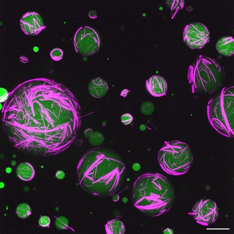
    To analyse ssDNA annealing using size-exclusion chromatography, RAD52-OR (4 µM) was preloaded on SSA2–Cy5 (4 µM, 12.5 µl) before an equal volume of Cy3–SSA1 (4 µM) was added. After 30 min on ice, the reaction was loaded onto the Superdex 200 Increase 3.2/300 column connected to the ÄKTA pure Micro system. Chromatography was performed at 4 °C with buffer containing 25 mM HEPES pH 8.0, 200 mM KOAc, 0.25 mM TCEP and 1 mM Mg(OAc)2.

    Biolayer interferometry analysis

    40-nucleotide (SSA4) ssDNA was biotinylated at either the 5′ or 3′ end (indicated as bio–ssDNA or ssDNA–bio, respectively). 68-nucleotide (SSA1) ssDNA was biotinylated at the 3′ end (indicated as SSA1–bio), and 28 nucleotides of complementary ssDNA was annealed to the 5′ end to protect the 5′ ssDNA end (indicated as ds-ssDNA–bio). The experiments were performed using the Octet R8 system (Sartorius) at 25 °C in buffer containing 25 mM HEPES pH 8.0, 200 mM KOAc, 0.01% Tween-20, 1 mM Mg(OAc)2 and 0.25 mM TCEP. The biotinylated DNA substrates (5 nM) were immobilized onto Octet SA streptavidin biosensors until a 0.05 threshold, and the sensors were then moved to wells containing a range of RAD52 concentrations (20, 10, 5, 2.5, 1.25, 0.625 and 0.312 nM). The association of RAD52 to DNA was recorded for 60 min and the dissociation for 5 min using the Octet BLI Discovery Software. Equilibrium dissociation constants (KD) were obtained by plotting association amplitudes at equilibrium versus protein concentration (Octet Analysis Studio Software; Sartorius) and plotted in GraphPad Prism 9. The following 1:1 binding equation was used to determine KD values: using the following quadratic equation in GraphPad Prism 9 to determine KD values:

    $$Y={B}_{\max }\times X/({K}_{D}+X),$$

    where Y is the association amplitude, Bmax is the maximum amplitude at saturation, X is the protein concentration and KD is the dissociation constant. Three independent triplicates were performed for each binding condition.

    CD analysis

    Far-UV CD measurements were performed on a Jasco J-815 spectropolarimeter fitted with a cell holder temperature-regulated by a CDF-426S Peltier unit. Spectra were recorded at 20 °C at protein concentrations of 3.3 µM (RAD52-OR) and 3.2 µM (RAD52-CR) in 10 mM potassium phosphate buffer pH 8.0, 100 mM NaF and 0.25 mM TCEP. Fused silica cuvettes were used with a 1 mm path length (Hellma). Spectra were recorded at a resolution of 0.2 nm and were baseline corrected by subtraction of the appropriate buffer spectrum. CD intensities are presented as the molar CD extinction coefficient (∆εM) calculated as:

    $${\Delta \varepsilon }_{{\rm{M}}}=\frac{S}{\mathrm{32,980}\times {c}_{{\rm{M}}}\times L}\left({\rm{units:}}{{\rm{M}}}^{-1}{{\rm{cm}}}^{-1}\right),$$

    where S is the signal in millidegrees, cM is the molar concentration and L is the path length (in cm). Secondary structure content was estimated as described52.

    Intact protein MS

    Proteins were diluted to 1 µM with 0.1% (v/v) formic acid and injected onto a C4 BEH 1.7 µm, 1.0 × 100 mm, UPLC column using the Acquity I class LC (Waters) system. Proteins were eluted with a 15 min gradient of acetonitrile (2% (v/v) to 80% (v/v)) in 0.1% (v/v) formic acid using a flow rate of 50 µl min−1. The analytical column outlet was directly interfaced through an electrospray ionization source, with a time-of-flight (TOF) mass spectrometer (BioAccord, Waters). Data were acquired over a m/z range of 300–8,000, in positive-ion mode with a cone voltage of 40 V. Scans were summed together manually and deconvoluted using MaxEnt1 (Masslynx, Waters). The parameters used were as follows; input m/z range (Da): 600–2,000; output mass range (Da): 30000–60000; TOF resolution: 10000.00; and iterate to convergence.

    GuHCl denaturation and renaturation

    RAD52 (purified to the HiTrap Q step) was dialysed into 25 mM HEPES pH 7.0, 6 M GuHCl, 0.5 mM EDTA and 2 mM β-mercaptoethanol overnight at 4 °C. The denatured protein was analysed using a Superose 6 Increase 10/300 GL column, which was run with 6 M GuHCl buffer. Protein was renatured by dialysis in native buffer (25 mM HEPES pH 7.0, 200 mM NaCl, 0.5 mM EDTA and 2 mM mercaptoethanol) for 24 h at 4 °C. The renatured RAD52 was then run on the same column using native buffer. To analyse the percentage of open and closed rings, the renatured RAD52 sample was loaded onto the Resource S column.

    Negative-stain EM sample preparation and data acquisition

    Samples (4 µl, 25 ng µl−1) were applied for 1 min to glow discharged (25 mA, 30 s) 400-mesh carbon-coated copper grids (C400Cu100, EM Resolutions). The grids were sequentially stained in four separate 30 µl droplets of 2% (v/v) uranyl acetate for 10, 15, 20 and 25 s. Excess uranyl acetate was blotted away from the grid using Whatmann paper, allowed to air dry and stored before imaging.

    The grids were imaged on the Tecnai LaB6 G2 Spirit TEM operating at 120 kV equipped with a 2K Gatan Ultrascan 1000 camera. Micrographs were acquired manually using DigitalMicrograph at a nominal magnification of ×30,000 (3.5 Å per pixel) or ×42,000 (2.4 Å per pixel) with defocus values ranging from −0.7 to −1.5 µm.

    Negative-stain EM data analysis

    DM3 files were converted to MRC format using e2proc2d.py (EMAN2)53. Micrographs were imported into Relion 3.1 or 4.154,55, CTF parameters were calculated using CTFFIND456, and particles were picked using crYOLO57 or Topaz58. Particles were extracted and iteratively 2D classified (ignore CTF to first peak = yes, limit resolution E-step = 20 Å, additional arguments = –only-flip-phases).

    Cryo-EM sample preparation

    Recombinant RAD52 and RPA were purified to the Resource S or Resource Q step, and freshly purified on the Superose 6 Increase 10/300 GL or Superdex 200 Increase 10/300 GL column before making the cryo-EM grids. For RAD52-CR, the protein was in a buffer containing 25 mM HEPES pH 7.0, 150 mM NaCl and 0.25 mM TCEP, diluted to 0.3 mg ml−1, and supplemented with 0.00005% Tween-20. A sample (4 μl) was applied to freshly glow-discharged (45 mA, 60 s; Quorum Emitech K100X) Quantifoil R2/1 300 mesh copper grids and vitrified using a Vitrobot Mark IV (Thermo Fisher Scientific) cooled to 4 °C with 95% humidity. Grids were double-side blotted for 0.5 s and plunge frozen in liquid ethane. For RAD52-OR, the grids were prepared as described above except Quantifoil R2/2 200 mesh copper grids were used, and the concentration was 0.25 mg ml−1, the Tween-20 concentration was 0.001%, and blot time was 1.5 s. For RAD52-OR–ssDNA, the protein (0.25 mM) was diluted to 0.5 µM in 25 mM HEPES pH 8.0, 150 mM NaCl, 2 mM Mg(OAc)2 and supplemented with 0.05% octyl-β-glucoside (OG). SSA4 (1 µM) was added and incubated at 25 °C for 10 min. The concentration was determined by Bradford assay (Bio-Rad) and diluted to 0.15 mg ml−1 with the same buffer. Grids were prepared as above except Quantifoil R1.2/1.3 300 mesh copper grids were used and the blot time was 2.5 s. For RPA–ssDNA, the protein (0.25 mM), in 25 mM HEPES pH 8.0, 150 mM NaCl, 2 mM Mg(OAc)2, was diluted to 3 µM, and supplemented with 0.1 mM CHAPSO. SSA7 (6 µM) was added and incubated at 25 °C for 10 min. The concentration was determined using the Bradford assay (Bio-Rad) and diluted to 0.15 mg ml−1 with the same buffer. UltrAuFoil R2/2 200 mesh gold grids (Quantifoil) were prepared as described above and the blot time was 2.5 s. The RAD52-OR–ssDNA–RPA ternary complex was assembled as indicated in the ‘Reconstitution of the RAD52–ssDNA–RPA complex’ section below. The concentration was determined using the Bradford assay (Bio-Rad) and diluted to 0.1 mg ml−1 with buffer supplemented with 0.00075% Tween-20 and 0.075 mM CHAPSO. Quantifoil R2/2 200 mesh copper grids were prepared as described above, except the blot time was 3 s.

    Cryo-EM data collection, image processing and atomic model building

    RAD52-CR and RAD52-OR datasets were collected on a Titan Krios Cryo-TEM equipped with a Falcon III direct electron detector (Thermo Fisher Scientific) at the Francis Crick Institute Structural Biology STP. The RAD52-OR–ssDNA dataset was collected on a Titan Krios G3i (FEI, Thermo Fisher Scientific) equipped with a Gatan K3 direct electron detector at the London consortium for cryo-EM (LonCEM). RPA–ssDNA and RAD52-OR–ssDNA–RPA datasets were collected on a Titan Krios Cryo-TEM (Thermo Fisher Scientific) equipped with a K2 direct electron detector (Gatan) at the Francis Crick Institute Structural Biology STP.

    Single-particle analyses were performed within Relion (v.4.0)54 and CryoSPARC59. The videos were corrected for drift and dose-weighted using RELION’s own implementation of MOTIONCOR260 and subsequent contrast transfer (CTF) parameters were measured using CTFFIND456. Particles were picked automatically using crYOLO57 or Topaz58. Details of image processing are illustrated in Extended Data Figs. 3, 4, 5, 8 and 9. In brief, several rounds of 2D classification were performed to remove particles that cannot be aligned to yield defined 2D averages. Several rounds of 3D classifications were performed to separate different conformations or particles that cannot be aligned to yield high-resolution 3D volumes. 3D auto-refine, Bayesian polishing (minimum two rounds) and CTF refinement (minimum one round) were performed iteratively to achieve high resolution 3D reconstruction in RELION61,62. Polished particles were imported to CryoSPARC59, and refined using non-uniform refinement63. 3D variability64 or 3D classifications were performed to detect heterogeneity within the cryo-EM densities. The cryo-EM maps were sharpened by post-processing in RELION, CryoSPARC or DeepEMhancer65 if there was high variability in local resolution. The overall resolution is reported at FSC = 0.143 (ref. 66).

    All model building was performed using Phenix67,68, COOT69 and ISOLDE70 in ChimeraX71. For RAD52-CR, the crystal structure of the RAD52 NTD (PDB: 1H2I) was placed into a sharpened RAD52-CR cryo-EM map in ChimeraX71 and initially refined using Namdinator72. One RAD52 subunit was removed from RAD52-CR and used for initial refinement in Namdinator for RAD52-OR. ssDNA was built manually in COOT into the RAD52-OR model using RAD52-OR–ssDNA as a starting model. RPA1, RPA2 and RPA3 AlphaFold2 models were used for Dock and rebuild in Phenix73,74 and the ssDNA model was aligned and extracted from the fungal RPA structure (PDB: 4GOP)39. The RAD52-OR–ssDNA model was used as the initial model for RAD52-OR–ssDNA–RPA.

    SEC–MALLS analysis

    SEC–MALLS was used to determine the molar mass composition of RAD52. Purified RAD52-OR (2.0, 1.0 or 0.5 mg ml−1) was loaded onto a Superose 6 Increase 10/300 GL column connected to a Jasco chromatography system. Chromatography was performed at 25 °C with buffer containing 25 mM HEPES pH 7.0, 150 mM NaCl, 0.25 mM TCEP and 3 mM NaN3 at a flow rate of 1.0 ml min−1. RAD52-OR–ssDNA (2 mg ml−1) was analysed in a similar manner using 25 mM Bis-Tris propane pH 8.5, 200 mM NaCl, 5 mM MgCl2, 0.25 mM TCEP and 3 mM NaN3 as the running buffer. The scattered light intensity and protein concentrations of the column eluates were recorded using a DAWN-HELEOS laser photometer and an OPTILAB-rEX differential refractometer (dn/dc = 0.186). The weight-averaged molecular mass of material contained in chromatographic peaks was determined using the combined data from both detectors in the ASTRA software v.7.3.2 (Wyatt Technology).

    Nuclear/chromatin extraction and analysis

    U2OS cells (authenticated and microplasma free, as determined by the Francis Crick Institute) were grown in DMEM (Gibco) supplemented with 10% FBS (Gibco) in humidified incubators at 37 °C and 5% CO2. Cells were collected from four confluent 500 cm2 square dishes and washed once with PBS. The pellet was supplemented with 5× pellet volume of CSK buffer (10 mM PIPES pH 6.8, 100 mM NaCl, 3 mM MgCl2, 300 mM sucrose, 1 mM EGTA, 0.5% Triton X-100 and 0.25 mM TCEP) supplemented with Halt protease and phosphatase inhibitors, incubated on ice for 10 min and centrifuged at 2,000g for 5 min at 4 °C. The supernatant was collected as the first CSK extract. A 3× pellet volume of CSK buffer (containing 0.1% Triton X-100) was added to the pellet, incubated on ice for 10 min and the sample was centrifuged at 3,000g for 5 min at 4 °C. The supernatant was collected as the second CSK extract. An equal volume of benzonase digestion buffer (20 mM HEPES pH 8.0, 2 mM MgCl2, 0.5% Triton X-100, 0.25 mM TCEP and 500 units benzonase/100 µl of buffer) supplemented with Halt protease and phosphatase inhibitors was added to the pellet and incubated on ice for 10 min. A 2× sample volume of high-salt buffer (20 mM HEPES pH 8.0, 600 mM NaCl and 0.25 mM TCEP) supplemented with Halt protease and phosphatase inhibitors was then added, incubated on ice for 10 min, and the sample was centrifuged at 21,000g for 10 min at 4 °C. The supernatant was collected as a nuclear/chromatin extract.

    Glycerol gradients (5 ml, 10–30%) in 25 mM HEPES pH 8.0, 150 mM NaCl, 10–30% glycerol and 0.25 mM TCEP were cast in thin-wall polypropylene tubes (Beckman Coulter) using a Gradient Master (Biocomp) and kept in the cold room overnight to equilibrate to 4 °C. U2OS nuclear/chromatin extracts (200 µl), 200 ng recombinant RAD52-OR or a gel-filtration calibration marker (Cytiva) was loaded gently onto the top of three gradients, which were then centrifuged at 4 °C and 55,000 rpm (368,000g) using SW 55 Ti rotor (Beckman Coulter) for 4 h. The fractions were collected by manual pipetting from the top of the gradients. The U2OS nuclear/chromatin extract (500 µl), 500 ng recombinant RAD52-OR or a gel-filtration calibration marker (Cytiva) were also loaded onto the pre-equilibrated Superose 6 Increase 10/300 GL column (Cytiva). Chromatography was performed with a buffer containing 25 mM HEPES pH 8.0, 150 mM NaCl, 10% glycerol and 0.25 mM TCEP at 4 °C. Fractions were collected and analysed by SDS–PAGE followed by western blotting using antibodies against RAD52 (rabbit monoclonal, 1:500, Abcam, ab124971). Alexa Fluor Plus 800 anti-rabbit secondary antibodies (1:2,000, Invitrogen, A32735) were used and the membranes were imaged using an Odyssey DLx instrument with ImageStudio software (Licor).

    RAD52 Resource S chromatogram peak fitting

    Resource S chromatography was performed as described above except a linear gradient of 0.2–0.6 M NaCl was used. The UV280 absorbance values were imported into GraphPad Prism 9 and curved fitted using a sum of two Gaussians equation to deconvolute open- and closed-ring peaks:

    $$Y={\rm{amplitude}}\times \exp \left(-0.5\times {\left(\frac{X-{\rm{mean}}}{{\rm{s.d.}}}\right)}^{2}\right)+\mathrm{amplitude\; 2}\times \exp \left(-0.5{\left(\frac{X-\mathrm{mean\; 2}}{\mathrm{s.d.\; 2}}\right)}^{2}\right)$$

    RAD52–ssDNA–RPA pull downs

    The RAD52–ssDNA–RPA ternary complex (400 μl) was reconstituted in buffer containing 25 mM HEPES pH 8.0, 200 mM KOAc, 2 mM Mg(OAc)2, 0.01% Tween-20 and 0.25 mM TCEP. Biotin-labelled SSA4 (0.1 μM), with photo-cleavable linker (Integrated DNA Technologies), and recombinant RPA (0.15 μM) were mixed and incubated on ice for 10 min. RAD52-OR (0.15 μM) was then added and incubation continued for a further 10 min. Pre-washed Streptavidin Sepharose Mag beads (10 μl, Cytiva) were then added and incubated for 30 min on a head-to-toe rotator at 4 °C. The beads were washed once with reaction buffer and then with reaction buffer Tween-20. The beads were resuspended in 20 μl reaction buffer, and irradiated with 365 nm UVA on ice/water slurry to cleave the photo-cleavable linker.

    Reconstitution of the RAD52–ssDNA–RPA complex

    RAD52-OR (purified to the Resource S step) and RPA (purified to the Resource Q step) were loaded onto the Superose 6 Increase 10/300 GL (Cytiva) and Superdex 200 Increase 10/300 GL (Cytiva) columns, respectively, and run with buffer containing 25 mM HEPES pH 8.0, 150 mM NaCl, 2 mM Mg(OAc)2 and 0.25 mM TCEP. The reconstitution mixture for cryo-EM was supplemented with 0.00075% Tween-20 and 0.075 mM CHAPSO, whereas the XL-MS sample was supplemented with 0.05% OG. Reconstitution of the RAD52-OR–ssDNA–RPA ternary complex involved two steps: (1) RPA (1 µM final concentration) was added to SSA1 (0.5 µM final concentration) and incubated at 25 °C for 10 min; and (2) RAD52-OR (0.5 µM final concentration) was added and incubated at 25 °C for 30 min. The sample was centrifugated at 21,000g for 1 min at 4 °C before proceeding with cryo-EM grid preparation and XL-MS.

    Protein disorder prediction

    The human RAD52 protein sequence (UniProt: P43351) was uploaded to the ODiNPred75 webserver (https://st-protein.chem.au.dk/odinpred). The predicted disorder probability of each residue was plotted in GraphPad Prism 9.

    Multiple-sequence alignment

    RAD52 protein sequences from different organisms were aligned with Clustal Omega using the default settings76. The alignment was formatted with ESPript3.077.

    XL-MS analysis

    RAD52-OR and RAD52-OR–ssDNA–RPA ternary complexes (0.5 µM, reconstituted as above) were supplemented with a 1:100 molar ratio of disuccinimidyl dibutyric urea (DSBU: 50 µM) for 1 h at room temperature, before the mixture was quenched by the addition of NH4HCO3 to a final concentration of 20 mM (15 min at room temperature). The cross-linked proteins were reduced with 10 mM dithiothreitol and alkylated with 50 mM iodoacetamide. They were then digested with trypsin at an enzyme-to-substrate ratio of 1:100, for 1 h at room temperature and further digested overnight at 37 °C after addition of trypsin at a ratio of 1:20. The peptide digests were then fractionated batch-wise by high pH reverse-phase chromatography on micro spin TARGA C18 columns (Nest Group) into four fractions (10 mM NH4HCO3/10% (v/v) acetonitrile pH 8.0; 10 mM NH4HCO3/20% (v/v) acetonitrile pH 8.0; 10 mM NH4HCO3/40% (v/v) acetonitrile pH 8.0; and 10 mM NH4HCO3/80% (v/v) acetonitrile pH 8.0). The fractions (150 µl) were evaporated to dryness in a CentriVap concentrator (Labconco) before analysis by LC–MS/MS.

    Lyophilized peptides were resuspended in 1% (v/v) formic acid and 2% (v/v) acetonitrile and analysed by nano-scale capillary LC-MS/MS using a Vanquish Neo UPLC (Thermo Fisher Scientific, Dionex) to deliver a flow of approximately 300 nl min−1. A PepMap Neo C18 5 μm, 300 μm × 5 mm nanoViper (Thermo Fisher Scientific, Dionex) trapped the peptides before separation on a 25 cm EASY‐Spray column (25 cm × 75 µm inner diameter, PepMap C18, 2 µm particles, 100 Å pore size, Thermo Fisher Scientific). Peptides were eluted with a gradient of acetonitrile. The analytical column outlet was directly interfaced through a nano-flow electrospray ionization source, with a quadrupole Orbitrap mass spectrometer (Orbitrap Exploris 480, Thermo Fisher Scientific). MS data were acquired in data-dependent mode using a top ten method, where ions with a precursor charge state of 1+ and 2+ were excluded. High-resolution full scans (R = 60,000, m/z 380–1,800) were recorded in the Orbitrap followed by higher-energy collision dissociation (HCD) (stepped collision energy 30 and 32% normalized collision energy) of the ten most intense MS peaks. The fragment ion spectra were acquired at a resolution of 30,000 and a dynamic exclusion window of 20 s was applied.

    For data analysis, Xcalibur raw files were converted into the MGF format using Proteome Discoverer v.2.3 (Thermo Fisher Scientific) and used directly as input files for MeroX78. Searches were performed against an ad hoc protein database containing the sequences of the proteins in the complex and a set of randomized decoy sequences generated by the software. The following parameters were set for the searches: maximum number of missed cleavages: 3; targeted residues K, S, Y and T; minimum peptide length 5 amino acids; variable modifications: carbamidomethylation of cysteine (mass shift 57.02146 Da), methionine oxidation (mass shift 15.99491 Da); DSBU modified fragments: 85.05276 Da and 111.03203 Da (precision: 5 ppm MS and 10 ppm MS/MS); false-discovery-rate cut-off: 5%. Finally, each fragmentation spectrum was manually inspected and validated.

    To compare with the peptide array experiments, the number of cross-links detected for each amino acid residue was counted, and summed within an individual 20 amino acid peptide with a 1 amino acid shift, similar to the peptide array. The overlayered result was plotted using GraphPad Prism 9.

    Peptide array

    Peptides (20 amino acids) with 1-amino-acid shift covering the full sequences of RAD52, RPA1, RPA2 and RPA3 were synthesized on cellulose membranes in 3 mm spots by the Chemical Biology STP at the Francis Crick Institute. The membranes were washed with 50% ethanol and 10% acetic acid for 30 min and equilibrated with 1× TBST (50 mM Tris-HCl pH 7.5, 150 mM NaCl and 0.1% Tween-20) supplemented with 0.25 mM TCEP. The membrane was blocked with 5% non-fat milk in TBST (0.1% Tween-20) supplemented with 0.25 mM TCEP for 1 h at room temperature. To allow protein-peptide interactions, the membranes were incubated with RAD52-OR or RPA (1 µg ml−1) in 1% non-fat milk in TBST (0.1% Tween-20) supplemented with 0.25 mM TCEP overnight at 4 °C. The membranes were washed in 1× TBST (0.1% Tween-20) supplemented with 0.25 mM TCEP on an orbital shaker for 5 min at room temperature three times. The membranes were then incubated in primary antibodies (anti-His 1:1,000, Takara, 631212) in 1% non-fat milk in TBST (0.1% Tween-20) supplemented with 0.25 mM TCEP for 2 h at room temperature. The membranes were washed three times as before and incubated in Alexa-Fluor-Plus-conjugated secondary antibodies (goat anti-mouse 1:2,000, Thermo Fisher Scientific, A32730; goat anti-rabbit, 1:2,000, Thermo Fisher Scientific, A32735) in 1% non-fat milk in TBST (0.1% Tween-20) supplemented with 0.25 mM TCEP for 1 h at room temperature. The membranes were washed three times, imaged on a Li-Cor Odyssey DLx system and quantified using Image Studio Lite (Li-Cor).

    Nanoscale differential scanning fluorometry

    A Prometheus NT-48 (Nanotemper) instrument was used to monitor changes in tryptophan fluorescence following thermal denaturation. Proteins were diluted to 10 µM in 25 mM HEPES pH 8.0, 200 mM KOAc, 0.5 mM EDTA, 10% glycerol and 0.25 mM TCEP. The samples were loaded into high-sensitivity glass capillaries and the tryptophan fluorescence was monitored at 330 and 350 nm after excitation at 285 nm. Measurements were made from 25 to 95 °C with a temperature gradient of 1 °C min−1. The ratio of fluorescence intensity (350/330 nm) was plotted against temperature, and the first derivative of this curve was used to calculate thermal melting (Tm) values.

    Statistics and reproducibility

    Statistical analyses were performed using GraphPad Prism 9. Normally distributed data were compared using two-tailed unpaired t-tests whereas non-normally distributed data were compared using two-tailed Mann–Whitney U-tests. Differences were considered to be statistically significant when P < 0.05. Reported n values refer to independent experiments for fluorescence anisotropy, biolayer interferometry analysis and SSA assays. Glycerol gradient sedimentation analysis and size-exclusion chromatography of U2OS nuclear extract recombinant RAD52-OR were repeated independently seven times with similar results. RAD52–ssDNA–RPA pull-down experiments were repeated independently five times with similar results. RAD52 purifications were repeated independently more than 50 times with similar results. RPA purifications were repeated for ten times with similar results. Purifications of RAD52 and RPA mutants were repeated for twice with similar results.

    Reporting summary

    Further information on research design is available in the Nature Portfolio Reporting Summary linked to this article.

    [ad_2]

    Source link

  • Immune dysfunction mechanism discovered in stroke and heart attack patients

    Immune dysfunction mechanism discovered in stroke and heart attack patients

    [ad_1]

    Every year, between 250,000 and 300,000 people in Germany suffer from a stroke or heart attack. These patients suffer immune disturbances and are very frequently susceptible to life-threatening bacterial infections. Until now, little was known about the underlying mechanisms of this immune dysfunction. Research teams from the Faculty of Medicine at the University Hospital of the UDE and the Leibniz Institute for Analytical Sciences in Dortmund have now uncovered a previously unknown cause – and a therapeutic approach. These findings are published in the May 2024 issue of the Journal Nature Cardiovascular Research.

    The study was led by Prof. Matthias Gunzer, Director of the Institute of Experimental Immunology and Imaging (IEIB) at the UDE and Head of the Biospectroscopy Department at the Leibniz Institute for Analytical Sciences (ISAS), and Dr. Vikramjeet Singh, Head of the Stroke Immunology Unit at the IEIB. They found that in patients one to three days after a stroke or heart attack, the amount of IgA antibodies in the blood decreases drastically – these are essential for defense against infections. Antibodies come in several subtypes, collectively known as immunoglobulins (Ig), which are produced by specialized plasma cells.

    To track down the mechanism behind the loss of antibodies – and to improve the treatment of patients with these findings – the researchers used disease mouse models. Mice also experienced a loss of IgA in their blood and stool after a stroke or heart attack. The researchers discovered that specialized DNA fibers released in blood are a factor in the loss of immune defense. These DNA fibers, known as neutrophil extracellular traps (NETs), originate from the nuclei of another type of immune cell, neutrophils. NETs are released into the blood in large quantities by highly activated neutrophils after a stroke or heart attack and can directly kill plasma cells in the intestine. Probably an even more important effect of NETs is the formation of hundreds of small clots in the blood vessels that supply energy to plasma cells in the intestine. This results in a lack of nutrient and oxygen supply and the Ig-forming cells die off in large numbers.

    The immunologists and their teams not only succeeded in proving a causal link between stroke, heart attack and immunodeficiency, but they were also able to demonstrate a new treatment approach: If the NETs were destroyed with the enzyme DNase or their release was prevented by a substance with a novel mode of action, the immune defense remained intact. The researchers were able to demonstrate this both in the mouse model and – in the case of DNase – in later clinical studies.

    Until now, no therapeutic approaches could be developed because the cause of the immune deficiency was unclear. A treatment that breaks down the NETs or even prevents them from forming in the first place could be a promising new approach to maintaining the immune defense in patients after a stroke or heart attack. It may be possible to prevent serious secondary infectious diseases or even death.”


    Prof. Matthias Gunzer, Director of the Institute of Experimental Immunology and Imaging (IEIB) at the UDE and Head of the Biospectroscopy Department at the Leibniz Institute for Analytical Sciences (ISAS)

    Source:

    Journal reference:

    Tuz, A. A., et al. (2024). Stroke and myocardial infarction induce neutrophil extracellular trap release disrupting lymphoid organ structure and immunoglobulin secretion. Nature Cardiovascular Research. doi.org/10.1038/s44161-024-00462-8.

    [ad_2]

    Source link

  • Study links air pollution to increased colorectal cancer risk through DNA changes

    Study links air pollution to increased colorectal cancer risk through DNA changes

    [ad_1]

    A recent eBioMedicine study explores the association between air pollution and colorectal cancer (CRC) risk based on epigenomic analysis.

    Study: Impact of ambient air pollution on colorectal cancer risk and survival: insights from a prospective cohort and epigenetic Mendelian randomization study. Image Credit: Peakstock / Shutterstock.com

    The role of air pollution in CRC risk

    CRC is one of the most common cancer types worldwide, whose etiology is associated with a wide range of lifestyle and environmental factors. In the context of environmental factors, ambient air pollution is particularly crucial, as it could lead to the development of cancer by affecting the inflammatory system.

    A significant association between particulate matter (PM) and risk of CRC, gastrointestinal and liver cancer incidence, and mortality has been documented. Thus, it is imperative to understand the mechanism through which PM influences the incidence of CRC. The synergistic effect of PM with other air pollutants, such as nitrogen oxides, on the incidence of CRC also requires further investigation.

    Emerging epigenome-wide association studies (EWAS) have highlighted that exposure to air pollution leads to an alteration in epigenetic markers, particularly DNA methylation (DNAm). This alteration induces inflammation that could increase the risk of disease development and progression.

    The formation of 5-methylcytosine in cytosine-phosphate-guanine (CpG) dinucleotides that reflect aberrant DNAm has been identified as an important epigenetic mechanism in CRC carcinogenesis. Considering this finding, it is crucial to understand the role of air pollution in the altered DNAm, which is associated with CRC pathogenesis.

    Mendelian randomization (MR) analysis is a powerful tool for identifying causal interferences. It uses genetic variants as proxies for air pollution-related DNAm exposure to identify the causal factor. One major advantage of this method is minimizing reverse causality and confounding factors.

    About the study

    The current prospective cohort study evaluated the relationship between individual and combined ambient air pollution exposures with CRC risk and overall mortality. It also assessed the pathological effects associated with air pollution-related DNAm and the gene-environment interaction.

    The association between air pollutants, including PM10, PM2.5, and nitrogen oxides (NOx and NO2), and CRC incidence and survival were assessed using relevant samples from the United Kingdom Biobank (UKB) cohort. Both genotypic and phenotypic health-related data were obtained from the UKB.

    Two-sample epigenetic MR methylation quantitative trait loci (mQTL) analyses were conducted to identify the underlying mechanism of air pollution-related DNAm. Gene-environment interaction and genetic colocalization analyses were performed to elucidate the potential carcinogenic effect of air pollutants on CRC manifestation.

    Study findings

    A total of 428,632 participants from UKB were considered, 2,401 of whom were diagnosed with CRC and were eligible for the current study. Among these individuals, 533 all-cause deaths and 767 newly diagnosed CRC cases were identified. To determine all-cause mortality among patients with CRC, those with a prior CRC diagnosis were considered.

    Consistent with previous studies, the current study also indicated a positive correlation between PM2.5 exposure and elevated CRC risk. The newly developed Air Pollutants Exposure Score (APES) indicated that exposure to various air pollutants, individually or jointly, decreased the overall CRC survival rate in a dose-response manner.

    The detrimental prognostic effects of air pollution were more prevalent, although not statistically significant, among men, smokers, and those with insufficient physical activities. Thus, altering certain lifestyle factors could reduce the risk of CRC.

    A significant association between air pollution and CRC incidence/survival was observed. DNA methylation occurred within the protein-coding genes of transmembrane BAX inhibitor motif-containing 1 protein (TMBIM1)/paroxysmal nonkinesigenic dyskinesia (PNKD), CX-C motif chemokine receptor 5 (CXCR5), and transmembrane protein 110 (TMEM110), which mediate the adverse effects of air pollution on CRC. The experimental findings strongly indicated an overall detrimental effect of air pollution exposure on CRC development and prognosis.

    Air pollution mediates the development of CRC through the systemic inflammatory pathway, which is associated with increased messenger ribonucleic acid (mRNA) and protein levels of interferon-γ (IFN-γ), interleukin production, and blood proinflammatory activity.

    The gene-environment interaction analyses indicated that PM2.5 exposure affects the CpG site rs876961 of the TMBIM1/PNKD gene, which influences CRC survival. Long-term PM2.5 exposure has also been associated with increased C-reactive protein levels and the induction of a systemic inflammatory state.

    The PM2.5-related CpG site cg16235962 was associated with the CXCR5 gene, which encodes for a vital inflammatory factor in the microenvironment. PM2.5-related CpG site cg16947394 has been associated with the TMBIM1 gene, whereas the presence of rs992157 in the intron of PNKD and TMBIM1s is significantly associated with progression and susceptibility to CRC.

    Conclusions

    The current study confirmed the detrimental effect of ambient air pollution on CRC risk and survival, as well as the effect of epigenetic alterations of TMBIM1/PNKD, CXCR5, and TMEM110 on CRC pathogenesis. Future studies are needed to elucidate the underlying mechanism by which epigenetic alterations cause CRC development. Notably, the current study identified some modifiable factors, such as physical activity, smoking, and air pollution, which can help prevent CRC.

    Journal reference:

    • Jiang, F., Zhao, J., Sun, J., et al. (2024) Impact of ambient air pollution on colorectal cancer risk and survival: insights from a prospective cohort and epigenetic Mendelian randomization study. eBioMedicine. doi:10.1016/j.ebiom.2024.105126.

    [ad_2]

    Source link

  • Study reveals human gut plasmid with biomarker potential

    Study reveals human gut plasmid with biomarker potential

    [ad_1]

    A component of the human intestinal flora that has been little studied to date is the focus of a new study. Plasmids are small extrachromosomal genetic elements that frequently occur in bacterial cells and can influence microbial lifestyles – yet their diversity in natural habitats is poorly understood. An international team led by Prof. Dr. A. Murat Eren from the Helmholtz Institute for Functional Marine Biodiversity at the University of Oldenburg (HIFMB) reports in the science journal Cell, a mysterious plasmid, is one of the most numerous genetic elements in the human gut that could potentially serve as a powerful biomarker for identifying health hazards such as fecal contamination of water or human disorders such as Inflammatory Bowel Disease. According to the team’s analyses, this plasmid is present in the intestines of more than 90 percent of individuals in industrialized countries.

    Plasmids are extrachromosomal DNA sequences which are common to cells from all domains of life. Eren describes them as “typically small genetic entities that carry additional genes”. They can be exchanged between different bacterial cells and even between different types of bacteria. The replication of plasmids is dependent on their host cells: but they make up for it by providing their hosts with in some cases extremely important fitness determinants. For instance, some plasmids contain genes that encode antibiotic resistance, which help their bacterial hosts to survive antibiotics, contributing one of the most pressing public health concerns around the globe.

    There are also other plasmids which, according to the research to date, do not contain genes encoding obvious beneficial functions for their host. “These so-called ‘cryptic plasmids’ are often referred to as genetic parasites. They remain a mystery in microbial ecology because from an evolutionary perspective they should not exist at all,” explains Eren, a computer scientist and Professor of Ecosystem Data Science at the University of Oldenburg.

    Identifying plasmids has been a difficult undertaking so far. For some time now, scientists have been able to extract genetic material directly from environmental samples and, for example, analyze the microbial community in the human gut in its entirety, without having to cultivate individual bacterial organisms. However, the ability to confidently distinguish what is a plasmid among this conglomeration of genetic material, referred to as the metagenome, poses a considerable challenge.

    To solve this problem, Eren and his colleagues developed a new machine learning approach. As the team reported in an article recently published in the science journal Nature Microbiology, using this approach they identified over 68,000 plasmids in human intestinal flora, and also discovered that a certain cryptic plasmid called pBI143 was particularly abundant in their dataset.

    One of the most numerous genetic entities in the human gut

    In the study published in Cell, the team of researchers took a closer look at this plasmid, which consists of only two genes that rather surprisingly only serves for its own replication and mobilization across bacterial cells with no other clear benefit. To better understand the ecology of pBI143, the team analyzed 60,000 human and 40,000 environmental metagenomes generated from various habitats.

    “We found that pBI143 has a list of very interesting features,” Eren explains. The team discovered that more than 90 percent of people in industrialized countries carry the plasmid and that on average it is one of the most numerous genetic entities in the human gut. “On average it was more than ten times as numerous as a viral genome which was previously thought to be the most abundant genetic extrachromosomal element in the human gut,” says the researcher.

    Further analyses revealed that the plasmid occurred almost exclusively in the human gut but was virtually absent in datasets from other environments such as the oceans, soils, plants and the digestive organs of animals and their feces. The only other samples in which the researchers were able to detect the characteristic gene sequence for these plasmids was in samples from environments that are influenced by humans, such as waste water, hospital surfaces and laboratory rats.

    Due to its sheer numbers, prevalence across humans, and its conservancy across human populations, the team of researchers hypothesized that pBI143 could, for instance, be used as a biomarker in testing for fecal contamination.

    In fact, we were able to show that this plasmid is a more sensitive marker for detecting fecal contamination in drinking water compared to state-of-the-art biomarkers based on specific gene sequences of human intestinal bacteria.”


    Dr. A. Murat Eren, Professor of Ecosystem Data Science at the University of Oldenburg

    Non-invasive method to quantify progress of IBD

    The team also identified another potential application of this prevalent genetic entity in the context of human disorders such as Inflammatory Bowel Disease (IBD), a medical condition that affects 3 million people in Europe alone. They were able to demonstrate that the relative copy number of this cryptic plasmid increased almost four times in the intestines of people suffering from IBD compared as in the intestines of healthy individuals, suggesting that the changes of the copy number of the plasmid can serve as a non-invasive method to quantify the disease progress or severity.

    At the HIFMB, Eren’s team is developing new tools at the intersection of computer science and microbiology to identify and characterize naturally occurring plasmids and other mobile genetic elements in bacteria that live in the ocean. They strive to gain a better understanding of the ecology and evolution of microbes, and strategies that enable to them to respond to their everchanging environments for new biotechnological applications that can ameliorate crises we face.

    Source:

    Journal reference:

    Fogarty, E. C., et al. (2024). A cryptic plasmid is among the most numerous genetic elements in the human gut. Cell. doi.org/10.1016/j.cell.2024.01.039.

    [ad_2]

    Source link

  • Tiny DNA circles are key drivers of cancer formation, study suggests

    Tiny DNA circles are key drivers of cancer formation, study suggests

    [ad_1]

    Tiny circles of DNA that defy the accepted laws of genetics are key drivers of cancer formation, according to an international study led by researchers at Stanford Medicine.

    The circles, known as extrachromosomal DNA or ecDNA, often harbor cancer-associated genes called oncogenes. Because they can exist in large numbers in a cell, they deliver a super-charged growth signal that can override a cell’s natural programming. They also contain genes likely to dampen the immune system’s response to a nascent cancer, the researchers found.

    Previous research had suggested that the circles, which are widespread in human cancers but rarely found in healthy cells, primarily arise in advanced tumors as the abnormal cells increasingly botch the intricate steps required to copy their DNA before each cell division. But the new study shows that the roly-poly circles can be found even in precancerous cells — and their presence jump-starts a cancerous transformation. Blocking their formation, or their effect on the cells that carry them, might stop cancers from developing, the researchers believe.

    This study has profound implications for our understanding of ecDNA in tumor development. It shows the power and diversity of ecDNA as a fundamental process in cancer. It has implications for early diagnosis of precancers that put patients at risk, and it highlights the potential for earlier intervention as treatments are developed.” 


    Paul Mischel, MD, professor of pathology

    Mischel is one of six senior authors of the research, which was published April 12 in Nature. Howard Chang, MD, PhD, professor of genetics and the Virginia and D.K. Ludwig Professor in Cancer Research, is also a senior author. Other senior authors include senior staff scientist Thomas Paulson, PhD, from Seattle’s Fred Hutchison Cancer Center; assistant professor of pediatrics Sihan Wu, PhD, assistant professor at Children’s Medical Center Research Institute at the University of Texas Southwestern Medical Center; professor of computer science and engineering Vineet Bafna, PhD, from UC San Diego; and professor of cancer prevention and director of the Early Cancer Institute Rebecca Fitzgerald, MD, from the University of Cambridge.  

    “People with ecDNA in their precancerous cells are 20 to 30 times more likely than others to develop cancer,” Chang said. “This is a huge increase, and it means we really need to pay attention to this. Because we also found that some ecDNAs carry genes that affect the immune system, it suggests that they may also promote early immune escape.”

    A grand challenge

    Deciphering ecDNA’s role in cancer was one of four Cancer Grand Challenges awarded by the National Cancer Institute and Cancer Research UK in 2022. The grand challenges program was launched to bring together researchers from around the world to tackle complex research topics too daunting for any one group. Mischel was awarded $25 million to lead a team of international researchers to learn more about the circles. But first they had to jettison some key genetic principles that have guided the field for nearly 200 years.

    Source:

    Journal reference:

    Luebeck, J., et al. (2023). Extrachromosomal DNA in the cancerous transformation of Barrett’s oesophagus. Nature. doi.org/10.1038/s41586-023-05937-5.

    [ad_2]

    Source link

  • Bio-Rad announces collaboration agreement with oncocyte to commercialize transplant monitoring with droplet digital PCR

    Bio-Rad announces collaboration agreement with oncocyte to commercialize transplant monitoring with droplet digital PCR

    [ad_1]

    Bio-Rad Laboratories, Inc. (NYSE: BIO and BIO.B) a global leader in life science research and clinical diagnostics products, today announced a collaboration agreement with Oncocyte Corporation (Nasdaq: OCX), a precision diagnostics company, to develop and commercialize transplant monitoring products using Bio-Rad’s Droplet Digital™ PCR (ddPCR™) instruments and reagents.

    Under the terms of the agreement, Bio-Rad has committed to participate in a private placement of Oncocyte’s equity and has secured exclusive commercial rights in certain markets to commercialize Oncocyte’s assay for transplant monitoring research using Bio-Rad’s QX600™ ddPCR System. 

    Transplanted organs release cell-free DNA into the bloodstream of the recipient. This donor-derived cell-free DNA (dd-cfDNA1) is a sensitive biomarker for organ health that requires only a simple blood draw. The novel approach developed by the team at Oncocyte allows the assay to be applied to clinical research of both recent and non-recent transplants.

    The utility of the ddPCR-based approach has been well demonstrated, including in a prospective, observational, multicenter cohort trial published in 2017, which showed earlier and more sensitive discrimination of liver transplant patients with acute rejection, compared to conventional liver functional test methods2

    Oncocyte’s transplant monitoring assays and the Bio-Rad QX600 ddPCR System provide a highly sensitive and decentralized solution that could provide a more attractive alternative for laboratories that currently rely on centralized next-generation sequencing (NGS) test providers.

    “This collaboration advances Bio-Rad’s strategy to establish Droplet Digital PCR as a foundational technology by providing researchers and laboratories with high-value assays across a variety of life science applications. We believe that Oncocyte’s transplant monitoring assays and the Bio-Rad QX600 ddPCR System solution for the noninvasive measurement and quantification of key biomarkers used in solid tissue transplant monitoring research has the potential to advance science and save lives.”

    Simon May, EVP and President of Life Sciences at Bio-Rad Laboratories

    To learn more about Bio-Rad’s ddPCR technology visit bio-rad.com/qx600system.

    Sources:

    1. Donor-derived cell-free DNA (dd-cfDNA) is also known as graft-derived cell-free DNA (GcfDNA)
    2. Schütz E et al. (2017). Graft-derived cell-free DNA, a noninvasive early rejection and graft damage marker in liver transplantation: A prospective, observational, multicenter cohort study. PLoS Med 14, e1002286.

    [ad_2]

    Source link

  • Study reveals how DNA gyrase resolves DNA entanglements

    Study reveals how DNA gyrase resolves DNA entanglements

    [ad_1]

    Picture in your mind a traditional “landline” telephone with a coiled cord connecting the handset to the phone. The coiled telephone cord and the DNA double helix that stores the genetic material in every cell in the body have one thing in common; they both supercoil, or coil about themselves, and tangle in ways that can be difficult to undo. In the case of DNA, if this overwinding is not dealt with, essential processes such as copying DNA and cell division grind to a halt. Fortunately, cells have an ingenious solution to carefully regulate DNA supercoiling.

    In this study published in the journal Science, researchers at Baylor College of Medicine, Université de Strasbourg, Université Paris Cité and collaborating institutions reveal how DNA gyrase resolves DNA entanglements. The findings not only provide novel insights into this fundamental biological mechanism but also have potential practical applications. Gyrases are biomedical targets for the treatment of bacterial infections and the similar human versions of the enzymes are targets for many anti-cancer drugs. Better understanding of how gyrases work at the molecular level can potentially improve clinical treatments.

    Some DNA supercoiling is essential to make DNA accessible to allow the cell to read and make copies of the genetic information, but either too little or too much supercoiling is detrimental. For example, the act of copying and reading DNA overwinds it ahead of the enzymes that read and copy the genetic code, interrupting the process. It’s long been known that DNA gyrase plays a role in untangling the overwinding, but the details were not clear.

    DNA minicircles and advanced imaging techniques reveal first step to untangle DNA

    We typically picture DNA as the straight double helix structure, but inside cells, DNA exists in supercoiled loops. Understanding the molecular interactions between the supercoils and the enzymes that participate in DNA functions has been technically challenging, so we typically use linear DNA molecules instead of coiled DNA to study the interactions. One goal of our laboratory has been to study these interactions using a DNA structure that more closely mimics the actual supercoiled and looped DNA form present in living cells.”


    Dr. Lynn Zechiedrich, study author, Kyle and Josephine Morrow Chair in Molecular Virology and Microbiology and professor of the Verna and Marrs McLean Department of Biochemistry and Molecular Pharmacology at Baylor College of Medicine

    After years of work, the Zechiedrich lab has created small loops of supercoiled DNA. In essence, they took the familiar straight linear DNA double helix and twisted it in either direction once, twice, three times or more and connected the ends together to form a loop. Their previous study looking at the 3-D structures of the resulting supercoiled minicircles revealed that these loops form a variety of shapes that they hypothesized enzymes such as gyrase would recognize.

    In the current study, their hypothesis was proven correct. The team of researchers combined their expertise to study the interactions of DNA gyrase with DNA minicircles using recent technology advances in electron cryomicroscopy, an imaging technique that produces high-resolution 3-D views of large molecules, and other technologies.

    “My lab has long been interested in understanding how molecular nanomachines operate in the cell. We have been studying DNA gyrases, very large enzymes that regulate DNA supercoiling,” said co-corresponding author Dr. Valérie Lamour, associate professor at the Institut de Génétique et de Biologie Moléculaire et Cellulaire, Université de Strasbourg. “Among other functions, supercoiling is the cell’s way of confining about 2 meters (6.6 feet) of linear DNA into the microscopic nucleus of the cell.”

    As the DNA supercoils inside the nucleus, it twists and folds in different forms. Imagine twisting that telephone cord mentioned at the beginning, several times on itself. It will overwind and form a loop by crossing over DNA chains, tightening the structure.

    “We found, just as we had hypothesized, that gyrase is attracted to the supercoiled minicircle and places itself in the inside of this supercoiled loop,” said co-author, Dr. Jonathan Fogg, senior staff scientist of molecular virology and microbiology, and biochemistry and molecular pharmacology in the Zechiedrich lab.

    “This is the first step of the mechanism that prompts the enzyme for resolving DNA entanglements,” Lamour said.

    “DNA gyrase, now surrounded by a tightly supercoiled loop, will cut one DNA helix in the loop, pass the other DNA helix through the cut in the other, and reseal the break, which relaxes the overwinding and eases the tangles, regulating DNA supercoiling to control DNA activity,” Zechiedrich said. “Imagine watching the rodeo. Like roping cattle with a lasso, supercoiled looped DNA captures gyrase in the first step. Gyrase then cuts one double-helix of the DNA lasso and passes the other helix through the break to get free.”

    Co-corresponding author, Dr. Marc Nadal, professor at the École Normale in Paris confirmed the observation of the path of the DNA wrapped in the loop around gyrase using magnetic tweezers, a biophysical technique that allows to measure the deformation and fluctuations in the length of a single molecule of DNA. Observing a single molecule provides information that is often obscured when looking at thousands of molecules in traditional so-called “ensemble” experiments in a test tube.

    Interestingly, the “DNA strand inversion model” for gyrase activity was proposed in 1979 by Drs. Patrick O. Brown and the late Nicholas R. Cozzarelli, also in a Science paper, well before researchers had access to supercoiled minicircles or the 3-D molecular structure of the enzyme. “It’s especially meaningful to me that 45 years later, we finally provide experimental evidence supporting their hypothesis because Nick was my postdoctoral mentor,” Zechiedrich said.

    “This work opens a myriad of perspectives to study the mechanism of this conserved class of enzymes, which are of great clinical value,” Lamour said.

    “This work supports new ideas on how DNA activities are regulated. We propose that DNA is not a passive biomolecule acted upon by enzymes, but an active one that uses supercoiling, looping and 3-D shapes to direct accessibility of enzymes such as gyrase to specific DNA sequences in a variety of situations, which will likely impact cellular responses to antibiotics or other treatments,” Fogg said.

    Contributors to this work also include Marlène Vayssières (lead author), Nils Marechal, Long Yun, Brian Lopez Duran and Naveen Kumar Murugasamy. The authors are affiliated with one or more of the following institutions: Baylor College of Medicine, Université de Strasbourg, Institut de Génétique et de Biologie Moléculaire et Cellulaire, INSERM, Université Paris and Hôpitaux Universitaires de Strasbourg.

    Source:

    Journal reference:

    Vayssières, M., et al. (2024) Structural basis of DNA crossover capture by Escherichia coli DNA gyrase. Science. doi.org/10.1126/science.adl5899.

    [ad_2]

    Source link