Tag: Electron

  • Study reveals protein structure similarities in Alzheimer’s and Down syndrome

    Study reveals protein structure similarities in Alzheimer’s and Down syndrome

    [ad_1]

    More than 90% of people with Down syndrome, the most common chromosomal disorder in humans and the most frequent genetic cause of intellectual disability, are diagnosed with Alzheimer’s disease by ages 55-60. A new study recently published in Nature Structural and Molecular Biology uses leading-edge cryo-electron microscopy imaging technology to determine whether differences exist between the protein structures in those with Alzheimer’s disease and those with both Alzheimer’s disease and Down syndrome.

    Just like in Alzheimer’s disease, the neuropathological phenotype in those with Down syndrome and Alzheimer’s disease is characterized by the presence of amyloid β (Aβ) and by abnormal accumulation of tau protein. The structures of Aβ and tau filaments in Down syndrome have not been previously investigated, and it is unknown whether they are different from those of Alzheimer’s disease.”


    Ruben Vidal, PhD, the Luella McWhirter Martin Professor of Clinical Alzheimer’s Research at the Indiana University School of Medicine and lead investigator of the study

    Researchers studied images of Aβ and tau filaments, which occurs in individuals with Down syndrome, and compared with those seen in the most common form of Alzheimer’s disease. They found that the protein structures of Aβ and tau filaments in people with both Down syndrome and Alzheimer’s disease have similarities to those found in Alzheimer’s disease.

    Vidal said their findings may lead to better treatments for Alzheimer’s disease patients and individuals with Down syndrome.

    “This study is the first comparison at the near atomic level of Aβ and tau filaments between individuals with both Down syndrome and Alzheimer’s disease and individuals with only Alzheimer’s disease,” Vidal said. “Importantly, the study found variations in the structure of Aβ, but no substantial variation in the structure of tau filaments between individuals with Alzheimer’s disease and both Down syndrome and Alzheimer’s disease. This supports the notion of common mechanisms operating in people with sporadic Alzheimer’s disease and in people with both Down syndrome and Alzheimer’s disease. This knowledge is crucial for understanding Alzheimer’s disease in people with Down syndrome and assessing whether adults with both conditions could be included in Alzheimer’s disease clinical trials. People with Down syndrome are living longer than ever, but almost all of them are dying of Alzheimer’s disease when they get older.”

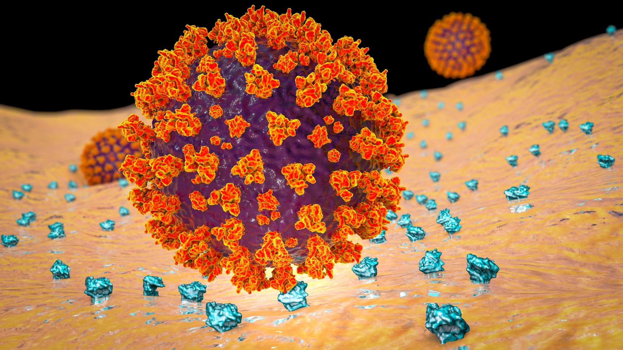
    Vidal, also an investigator in IU School of Medicine’s Stark Neurosciences Research Institute, said the research team used cryogenic electron microscopy to get a close-up, 3D view of the structure of Aβ and tau filaments in two individuals with both Down syndrome and Alzheimer’s disease. The study revealed two novel types of Aβ filaments in the vascular compartment with structures different from those previously reported in Alzheimer’s disease.

    Vidal said the study’s findings show it is important to include people with both Down syndrome and Alzheimer’s disease in clinical trials targeting the Aβ or tau filaments. He said there are similarities between the mechanisms at play in amyloid aggregation, but more research is needed to determine whether the differences observed in vascular Aβ deposition are unique to those with Down syndrome.

    “We are thrilled that our cryo-EM imaging and 3D modeling techniques have facilitated the determination of the atomic structures of amyloid beta and tau fibrils in individuals with Down syndrome, shedding light on the connection between Down syndrome and Alzheimer’s disease,” said Wen Jiang, PhD, professor of biology at Purdue University and co-corresponding author of the study. “We are fortunate to have the Purdue Cryo-EM Facility, which provides exceptional resources and services that have made this research possible. We are grateful to the patients who donated their brains to the research and thankful to the NIH for funding our work.”

    Other study authors include co-corresponding author Bernardino Ghetti, Anllely Fernandez, Grace Hallinan, Kathy Newell and Holly Garringer, all from the IU School of Medicine; and Rejaul Hoq, Daoyi Li, Sakshibeedu Bharath, Frank Vago, Xiaoqi Zhang and Kadir Ozcan, all from Purdue University.

    This research was funded by the National Institutes of Health and the IU School of Medicine Department of Pathology and Laboratory Medicine.

    Source:

    Journal reference:

    Fernandez, A., et al. (2024). Cryo-EM structures of amyloid-β and tau filaments in Down syndrome. Nature Structural & Molecular Biology. doi.org/10.1038/s41594-024-01252-3.

    [ad_2]

    Source link

  • Metformin boosts appetite-suppressing metabolite, new study finds

    Metformin boosts appetite-suppressing metabolite, new study finds

    [ad_1]

    A recent study published in the journal Nature Metabolism showed that metformin treatment significantly increases blood levels of N-lactoyl phenylalanine (Lac-Phe), an appetite-suppressing metabolite.

    Metformin, used for type 2 diabetes (T2D) treatment, reduces blood glucose and suppresses appetite. It is prescribed to more than 150 million individuals worldwide. However, the mechanisms of its therapeutic effects are not fully understood. It inhibits complex 1 of the electron transport chain at higher levels. Nevertheless, it is uncertain whether physiological levels of metformin are sufficient for complex 1 inhibition.

    Lac-Phe is a metabolite produced by carnosine dipeptidase 2 and has been reported as an appetite suppressant in obese mice. Lac-Phe correlates with weight loss in humans with regular exercise. It also increases in mitochondrial disease and phenylketonuria. Nonetheless, whether Lac-Phe has a role in the appetite-suppressing activity of metformin has not been explored.

    Study: Metformin and feeding increase levels of the appetite-suppressing metabolite Lac-Phe in humans. Image Credit: LuchschenF / ShutterstockStudy: Metformin and feeding increase levels of the appetite-suppressing metabolite Lac-Phe in humans. Image Credit: LuchschenF / Shutterstock

    The study and findings

    In the present study, researchers reported significant increases in Lac-Phe levels following metformin treatment. The study was conducted between August and December 2019 at Brigham and Women’s Hospital. Thirty-three volunteers who were 1) lean without T2D, 2) lean and prediabetic, 3) obese and prediabetic, 4) obese without T2D, or 5) obese with T2D were recruited.

    Diabetes was primarily managed with metformin, albeit some participants also received insulin. The team collected sera from participants and performed untargeted metabolomic profiling. This showed notable increases in all N-lactoyl amino acids in the obese T2D group relative to obese participants without T2D. These elevations were not related to body mass index (BMI) but to T2D.

    Moreover, Lac-Phe levels were 5.7 times higher in obese T2D subjects than in obese non-T2D volunteers. In addition, these increases were also significant compared to prediabetic individuals. Notably, Lac-Phe levels correlated with the concentrations of other N-lactoyl amino acids. Next, the team analyzed the metabolomic data from the TwinsUK cohort.

    Lac-Phe levels in this cohort were elevated in individuals with T2D. Further, there was a robust correlation between metformin and Lac-Phe levels among T2D patients in the Brigham cohort. Notably, while metformin use was a criterion for inclusion, one volunteer lacked detectable metformin levels and had the lowest Lac-Phe levels; the volunteer discontinued metformin.

    As such, the team speculated that metformin may elevate serum levels of Lac-Phe in individuals with T2D rather than T2D. This hypothesis was tested using the TwinsUK dataset; each participant had three samples collected during 1997-2012, a period when the role of metformin in T2D was growing considerably.

    This enabled analyses of the Lac-Phe trajectories in participants whose metformin and T2D status changed with time. The researchers noted that metformin treatment significantly increased Lac-Phe levels in individuals newly diagnosed with T2D. By contrast, for newly diagnosed diabetic individuals without metformin therapy, there were no significant changes in Lac-Phe levels.

    In participants who consistently had diabetes over successive sampling, metformin use caused substantial increases in Lac-Phe levels. On the other hand, those who did not receive metformin lacked significant changes in Lac-Phe levels. Further, the team analyzed two interventional studies from Denmark and Jordan to establish a causal link between metformin use and increases in Lac-Phe levels.

    In the Danish study, significant increases in N-lactoyl amino acids were evident following a 12-week metformin intervention in non-T2D and T2D groups. The Jordanian study reported rapid increases in Lac-Phe levels over 36 hours after a single metformin dose, and the peak Lac-Phe concentration corresponded to the maximum metformin concentration.

    Within the TwinsUK dataset, Lac-Phe levels correlated highly with the non-fasted state. As such, the researchers speculated whether metformin-related increases in Lac-Phe were influenced by feeding or fasting state. They observed a trend of higher Lac-Phe levels in the fed state. Moreover, while individuals not receiving metformin had lower Lac-Phe levels, Lac-Phe was significantly increased in fed subjects.

    Conclusions

    Taken together, the study highlighted that metformin elevates Lac-Phe levels. These increases were specific to metformin rather than T2D status and were evident in T2D subjects and healthy individuals. Further, Lac-Phe levels increase postprandially, paralleling other appetite suppressants’ patterns. Thus, pharmacological targeting of Lac-Phe could result in a more robust appetite-suppressing effect, leading to a new class of drugs for obesity.

    [ad_2]

    Source link

  • New calibration technique enhances accuracy of force measurements in microfluidic environments

    New calibration technique enhances accuracy of force measurements in microfluidic environments

    [ad_1]

    A study introduces a novel method for calibrating the spring constant of FluidFM micropipette cantilevers, crucial for the accurate measurement of forces in microfluidic environments. This method addresses the limitations of current calibration techniques, offering a significant advancement in the field of force microscopy.

    Fluidic force microscopy (FluidFM) combines the sensitivity of atomic force microscopy with microfluidics’ capabilities, necessitating precise calibration of its cantilevers for reliable data. Traditional methods, however, struggle with the unique internal structure of FluidFM cantilevers, leading to inaccuracies.

    A recent study (https://doi.org/10.1038/s41378-023-00629-6) published on February 18, 2024, in the journal Microsystems & Nanoengineering, researchers unveiled an innovative calibration technique for FluidFM micropipette cantilevers, pivotal for exact force measurements in microfluidic environments.

    The FluidFM is a tiny tool used in microscopic environments to measure forces with high precision. Unlike traditional methods that often fall short due to the complex inner structure of FluidFM cantilevers, this new approach leverages the cantilever’s resonance frequencies in both air and liquid environments. By focusing on these frequencies, the method circumvents the common pitfalls of the widely-used Sader method, which can introduce errors due to its reliance on geometric and fluidic assumptions that don’t hold up well for FluidFM’s unique cantilever designs. This innovative calibration technique was meticulously tested and validated on data obtained by the HUN-REN Nanobiosensorics Lab, Cytosurge, Nanosurf and Bruker, showing that it not only provides more accurate measurements but also simplifies the calibration process by reducing the effects of noise and eliminating the need for intricate experimental setups.

    Our method simplifies the calibration process, significantly reducing the influence of noise and eliminating the need for complex measurements, marking a significant step forward in the practical application of FluidFM technologies.”


    Dr. Attila Bonyár, study’s lead author

    The new calibration method promises enhanced accuracy in force measurements, with profound implications for biological, biophysical, and materials science research. It enables the precise manipulation of cells and nanoparticles, opening new avenues for investigation in these fields.

    Source:

    Journal reference:

    Bonyár, A., et al. (2024). Hydrodynamic function and spring constant calibration of FluidFM micropipette cantilevers. Microsystems & Nanoengineering. doi.org/10.1038/s41378-023-00629-6.

    [ad_2]

    Source link

  • WSU researchers find potential ‘trojan horse’ for treating difficult bacterial infections

    WSU researchers find potential ‘trojan horse’ for treating difficult bacterial infections

    [ad_1]

    Bacteria can be tricked into sending death signals to stop the growth of their slimy, protective homes that lead to deadly infections, a new study demonstrates.

    The discovery by Washington State University researchers could someday be harnessed as an alternative to antibiotics for treating difficult infections. Reporting in the journal, Biofilm, the researchers used the messengers, which they named death extracellular vesicles (D-EVs), to reduce growth of the bacterial communities by up to 99.99% in laboratory experiments.

    Adding the death extracellular vesicles to the bacterial environment, we are kind of cheating the bacteria cells. The cells don’t know which type of EVs they are, but they take them up because they are used to taking them from their environment, and with that, the physiological signals inside the cells change from growth to death.”


    Mawra Gamal Saad, first author on the paper and graduate student in WSU’s Gene and Linda Voiland School of Chemical Engineering and Bioengineering

    Bacterial resistance is a growing problem around the world. In the U.S., at least 2 million infections and 23,000 deaths are attributable to antibiotic-resistant bacteria each year, according to the U.S. Centers for Disease Control. When doctors use antibiotics to treat a bacterial infection, some of the bacteria can hide within their tough-to-penetrate, slimy home called a biofilm. These subpopulations of resistor cells can survive treatment and are able to grow and multiply, resulting in chronic infections.

    “They are resistant because they have a very advanced and well-organized adaptive system,” said Saad. “Once there is a change in the environment, they can adapt their intracellular pathways very quickly and change it to resist the antibiotics.” 

    In their new study, the researchers discovered that the extracellular vesicles are key to managing the growth of the protective biofilm. The vesicles, tiny bubbles from 30 to 50 nanometers or about 2,000 times smaller than a strand of hair, shuttle molecules from cells, entering and then re-programming neighboring cells and acting as a cell-to-cell communications system. 

    As part of this study, the researchers extracted the vesicles from one type of bacteria that causes pneumonia and other serious infections. They determined that the bacteria initially secrete vesicles, called growth EVs, with instructions to grow its biofilm, and then later, depending on available nutrients, oxygen availability and other factors, send EVs with new instructions to stop growing the biofilm. 

    The researchers were able to harness the vesicles with the instructions to stop growth and use them to fool the bacteria to kill off the biofilm at all stages of its growth. Even when the biofilms were healthy and rapidly growing, they followed the new instructions from the death EVs and died. The death EVs can easily penetrate the biofilm because they are natural products secreted by the bacteria, and they have the same cell wall structure, so the cells don’t recognize them as a foreign enemy.

    “By cheating the bacteria with these death EVs, we can control their behavior without giving them the chance to develop resistance,” said Saad. “The behavior of the biofilm just changed from growth to death.”

    WSU Professor and corresponding author Wen-Ji Dong, who has been studying the vesicles for several years initially thought that all of the bacterial-secreted vesicles would promote cell growth. The researchers were surprised when they found that older biofilms provided instructions on shutting themselves down. 

    “So now we’re paying attention to the extracellular vesicles secreted by older biofilms because they have therapeutic potential,” he said. 

    The researchers are applying for research funding from the National Institutes of Health to continue investigating exactly how the messengers work and how well the process works with other bacterial types or fungi. They are working with WSU’s Office of Commercialization and have applied for a provisional patent. 

    Source:

    Journal reference:

    Saad, M. G., et al. (2024). Dual roles of the conditional extracellular vesicles derived from Pseudomonas aeruginosa biofilms: Promoting and inhibiting bacterial biofilm growth. Biofilm. doi.org/10.1016/j.bioflm.2024.100183.

    [ad_2]

    Source link

  • Scientists uncover a new doorway for SARS-CoV-2 into human cells

    Scientists uncover a new doorway for SARS-CoV-2 into human cells

    [ad_1]

    In a recent study published in the journal Proceedings of the National Academy of Sciences, researchers demonstrated that human transferrin receptor (TfR) mediates severe acute respiratory syndrome coronavirus 2 (SARS-CoV-2) infection.

    Coronavirus disease 2019 (COVID-19), caused by SARS-CoV-2, presents influenza-like manifestations, including mild-to-severe pneumonia, acute respiratory distress syndrome, multiorgan failure, and fatal lung injury. Further, the etiology and pathogenesis of COVID-19 are not entirely understood and targeted therapies remain inadequate.

    The viral spike protein binds to the host receptor, angiotensin-converting enzyme 2 (ACE2), for cellular entry. Although SARS-CoV-2 preferentially infects cells in the respiratory tract, the virus has been detected in virtually all organs. Studies have revealed the presence of SARS-CoV-2 RNA in diverse cells lacking ACE2, suggesting that other receptors or co-receptors may mediate viral entry.

    Study: Human transferrin receptor can mediate SARS-CoV-2 infection. Image Credit: Kateryna Kon / ShutterstockStudy: Human transferrin receptor can mediate SARS-CoV-2 infection. Image Credit: Kateryna Kon / Shutterstock

    The study and findings

    In the present study, researchers identified TfR as an alternative receptor mediating the cellular entry of SARS-CoV-2. First, they used co-immunoprecipitation (Co-IP) to identify host proteins interacting with the viral spike in Calu-3 cells. This revealed 293 proteins, including 42 transmembrane proteins; two proteins were associated with entry (ACE2 and TfR). Next, the team evaluated TfR expression in the respiratory tract and liver in mice.

    TfR expression, both transcript and protein levels, was substantially higher in the lungs and trachea than in other tissues. Using immunohistochemical analysis, the researchers investigated the effects of SARS-CoV-2 on TfR expression in the lungs of humanized ACE2 (hACE2) mice and monkeys. This revealed a 1.5- and 1.8-fold increase in TfR expression in mice and monkeys, respectively.

    In addition, surface plasmon resonance revealed direct interactions between the viral spike and human TfR. Notably, the spike protein lacked interactions with Syrian hamster or mouse TfR. Docking analysis predicted two peptide sequences (QK8: QDSNWASK and SL8 SKVEKLTL) in TfR to be involved at the interface of TfR-spike interactions.

    Mutagenesis and Co-IP revealed that the A529 residue in TfR was essential for interactions with the spike. Further analysis indicated that physiological interactions between spike and TfR occurred at the cellular surface and during endocytosis. This was confirmed by electron microscopy using SARS-CoV-2 pseudoviral spike and HEK293/hACE2 and BHK-21/TfR cells.

    Next, the team evaluated the effects of soluble TfR, anti-TfR antibody, and SL8 and QK8 peptides on SARS-CoV-2 infection using reverse-transcription polymerase chain reaction (RT-PCR) and plaque assays. Results showed their inhibitory effects on SARS-CoV-2 in Vero E6 and Calu-3 cells. Cytotoxicity was not observed even at 1,000 nM.

    Confocal microscopy revealed that TfR was widespread on the surface of Calu-3 and Vero E6 cells, with the colocalization of TfR and SARS-CoV-2 at the surface and during endocytosis. Notably, treatment with the anti-TfR antibody inhibited the colocalization. Further, electron microscopy showed that viral particles were present in the cytosol and clathrin-coated pits in Vero E6 cells; likewise, treatment with anti-TfR antibody inhibited viral internalization.

    Next, ACE2 was knocked out (KO) from Calu-3 and Vero E6 cells and the cells were infected with SARS-CoV-2. This inhibited infection by 40% to 50%, suggesting that ACE2 might not be the only receptor mediating infection. In addition, TfR knockdown (KD) inhibited infection by 30%, whereas its overexpression (OE) promoted infection. TfR KO was not performed as it is lethal. TfR OE or KD did not impact ACE2 expression.

    Further, the team transfected C57 mice with adenovirus vector (Ad5) expressing hACE2 or humanized TfR (hTfR) and infected them with SARS-CoV-2. Viral load in the lungs in Ad5-hTfR and Ad5-hACE2 mice was significantly higher than in Ad5-empty mice. Finally, the researchers evaluated the effects of the anti-TfR antibody on infection in rhesus macaques. Anti-TfR antibody inhibited viral replication and reduced pneumonia.

    Viral load in the respiratory epithelium was also significantly lower between 3- and 7 days post-infection (dpi) compared to controls. Radiographs taken at 0 and 5 dpi revealed significantly less severe pulmonary infiltration in antibody-treated macaques relative to controls. Antibody-treated animals had no significant pulmonary lesions, while controls showed lung lesions of varying degrees.

    Conclusions

    Taken together, the study described the human TfR as a receptor for SARS-CoV-2. TfR can directly bind to the viral spike at an affinity comparable to that of ACE2. Notably, mouse TfR and the viral spike lacked interactions. Soluble TfR, SL8, and QK8 peptides and anti-TfR antibodies can inhibit infection. The team also illustrated the antiviral effects of the anti-TfR antibody in rhesus macaques. Overall, TfR could serve as an alternative infection pathway, facilitating viral entry through endocytosis.

    [ad_2]

    Source link

  • Red light therapy shown to significantly reduce blood sugar spikes, study finds

    Red light therapy shown to significantly reduce blood sugar spikes, study finds

    [ad_1]

    In a recent study published in the Journal of Biophotonics, scientists examined whether photobiomodulation of healthy subjects using red light of 670 nm wavelength impacted the circulating glucose levels in the plasma, using oral glucose tolerance tests.

    Study: Light stimulation of mitochondria reduces blood glucose levels. Image Credit: AlteredR/Shutterstock.comStudy: Light stimulation of mitochondria reduces blood glucose levels. Image Credit: AlteredR/Shutterstock.com

    Background

    Mitochondria are the organelles that carry out cellular respiration, using glucose and oxygen to produce adenosine triphosphate or ATP, the energy currency. The ability of the mitochondria to produce ATP reduces naturally with age and due to diseases.

    However, studies have found that the production of ATP can be increased through photobiomodulation using light in the visible and near-infrared ranges, between 650 nm and 900 nm.

    Photobiomodulation is also known to decrease reactive oxygen species levels, and this ability is believed to be conserved across species in the animal kingdom.

    Cytochrome C oxidase, which is part of the electron transport chain in the mitochondrial membrane, absorbs these longer wavelengths of light, increasing the membrane potential and production of ATP.

    Research has shown that photobiomodulation has brought about significant increases in regions of the body undergoing high levels of metabolic activity, such as the retina and the central nervous system.

    The increased ATP production could also increase the uptake of glucose, which might be evident in changes in the plasma glucose levels.

    About the study

    In the present study, the researchers used a standard glucose tolerance test to determine whether photobiomodulation using 670 nm light decreased blood glucose levels in healthy human subjects.

    The study included 30 healthy participants with no known medical conditions, half of whom underwent photobiomodulation with 670 nm light, and the other half were in the placebo group with no light.

    All the participants underwent an oral glucose tolerance test at the onset of the study, where they consumed 75 g of glucose dissolved in 150 mL of water, and finger prick blood samples were used to record the blood glucose levels.

    A second oral glucose tolerance test was administered after a week when the participants were administered the placebo or the intervention.

    About 45 minutes before the second oral glucose tolerance test was administered, the participants in the intervention group were exposed to 670 nm light for 15 minutes, while those in the placebo group were identically positioned but not exposed to the 670 nm light.

    The oral glucose tolerance tests were administered only after ensuring that the participants had fasted overnight.

    After consuming glucose dissolved in water, blood glucose concentrations and the end-tidal carbon dioxide (EtCO2) partial pressure were recorded every quarter of an hour for two hours when the participants were at rest.

    The 670 nm light exposure was directed at an 800 cm2 region in the upper back, using light-emitting diodes with a shield to prevent light leakage.

    The glucose tolerance test results were compared between the participants in the intervention and placebo groups.

    Additionally, participants in the intervention group were compared to each other, and similar comparisons were made within the placebo group for paired-participant analysis to account for individual variations.

    Results

    The results showed that exposure to 670 nm of light over 15 minutes resulted in a 27.7% decrease in glucose levels averaged over two hours.

    Additionally, a 7.5% decrease was also observed in maximum glucose spiking within the intervention group, and a 12.1% difference in peak glucose levels was seen between the placebo and intervention groups.

    The paired-participant analysis within the placebo group also showed no difference in the blood glucose levels between the two measurements.

    The impact of the light exposure was significant after approximately an hour and a half of local light exposure alone. The impact of this local light exposure on plasma glucose levels indicates an abscopal effect, where mitochondria in distal organs are also impacted.

    The researchers also discussed the potential mechanisms through which local light exposure could have such widespread impact, including the role of circulating cytokines and cell-free mitochondria in the blood that are competent to conduct cellular respiration.

    Conclusions

    To summarize, the findings showed that local exposure to 670 nm light for 15 minutes significantly reduced plasma and peak glucose levels.

    While these results have proven that longer wavelengths of light have a positive effect on mitochondrial function in healthy humans, the potential use of light exposure in helping regulate blood glucose levels in patients with diabetes needs to be explored.

    [ad_2]

    Source link

  • Maternal mRNA COVID-19 vaccination shields infants for six months

    Maternal mRNA COVID-19 vaccination shields infants for six months

    [ad_1]

    Women who receive an mRNA-based COVID-19 vaccination or booster during pregnancy can provide their infants with strong protection against symptomatic COVID-19 infection for at least six months after birth, according to a study from the National Institute of Allergy and Infectious Diseases (NIAID), part of the National Institutes of Health. These findings, published in Pediatrics, reinforce the importance of receiving both a COVID-19 vaccine and booster during pregnancy to ensure that infants are born with robust protection that lasts until they are old enough to be vaccinated.

    COVID-19 is especially dangerous for newborns and young infants, and even healthy infants are vulnerable to COVID-19 and are at risk for severe disease. No COVID-19 vaccines currently are available for infants under six months old. Earlier results from the Multisite Observational Maternal and Infant COVID-19 Vaccine (MOMI-Vax) study revealed that when pregnant volunteers received both doses of an mRNA COVID-19 vaccine, antibodies induced by the vaccine could be found in their newborns’ cord blood. This suggested that the infants likely had some protection against COVID-19 when they were still too young to receive a vaccine. However, researchers at the NIAID-funded Infectious Diseases Clinical Research Consortium (IDCRC), which conducted the study, did not know how long these antibody levels would last or how well the infants would actually be protected. The research team hoped to gather this information by following the infants through their first six months of life.

    In this portion of the study, researchers analyzed data from 475 infants born while their pregnant mothers were enrolled in the MOMI-Vax study. The study took place at nine sites across the United States. It included 271 infants whose mothers had received two doses of an mRNA COVID-19 vaccine during pregnancy. The remaining 204 infants in the study were born to mothers who had received both doses of an mRNA COVID-19 vaccine as well as a COVID-19 booster. To supplement data gathered during pregnancy and at birth, the infants were evaluated during at least one follow-up visit during their first six months after birth. Parents also reported whether their infants had become infected or had demonstrated COVID-19 symptoms.

    Based on blood samples from the infants, the researchers found that newborns with high antibody levels at birth also had greater protection from COVID-19 infection during their first six months. While infants of mothers who received two COVID-19 vaccine doses had a robust antibody response at birth, infants whose mothers had received an additional booster dose during pregnancy had both higher levels of antibodies at birth and greater protection from COVID-19 infection at their follow-up visits.

    While older children and adults should continue to follow guidance from the Centers for Disease Control and Prevention (CDC) to stay up-to-date on their COVID-19 vaccines and boosters, this study highlights how much maternal vaccination can benefit newborns too young to take advantage of the vaccine: During the course of this study, none of the infants examined required hospitalization for COVID-19. Researchers will continue to evaluate the data from the MOMI-Vax study for further insights concerning COVID-19 protection in infants.

    Source:

    Journal reference:

    Cardemil, C. V., et al. (2024). Maternal COVID-19 Vaccination and Prevention of Symptomatic Infection in Infants. Pediatrics. doi.org/10.1542/peds.2023-064252.

    [ad_2]

    Source link

  • Novel “Flash and Freeze-fracture” technique reveals neuronal communication secrets

    Novel “Flash and Freeze-fracture” technique reveals neuronal communication secrets

    [ad_1]

    Fear and addiction exert significant influence within society. Managing them is often challenging, as they are driven by intricate neuronal circuits in our brains. Understanding the underlying molecular mechanisms is crucial to intervene when these processes malfunction. Pioneered by scientists at the Institute of Science and Technology Austria (ISTA), the novel “Flash and Freeze-fracture” technique provides a unique glimpse into the respective brain region. The results were recently published in the journal PNAS.

    While looking for food, a bird encounters a fox. It gets away just in time, but the sight and the sound of the predator lingers. The negative experience will form a memory in its brain and will be associated with fear and stress from now on. Whenever it meets a fox again, the fear memory is revived. The bird’s attention spikes, its heart rate goes up, and it changes its behavior to reduce the risk of predation. Such memory is mediated by a specific brain region called the medial habenula, one of the epicenters for emotional processing.

    Peter Koppensteiner, together with Pradeep Bhandari, Cihan Önal and other members of Ryuichi Shigemoto’s research group at the Institute of Science and Technology Austria (ISTA) investigated this particular part of the brain to understand how its neurons (nerve cells) communicate with each other. Published in the journal PNAS, the results give an unprecedented look into this subject, utilizing a novel visualization technique called “Flash and Freeze-fracture”.

    Counter-intuitive brain cells

    Nerve cells in the medial habenula exhibit unusual behavior, contradicting the general understanding of how neurons transmit signals to each other. “Typically, communication between neurons is shut down, as soon as a specific molecule on the surface of the cells, known as the ‘GABAB’-receptor, is activated,” explains Peter Koppensteiner, previously a postdoc in the Shigemoto group and now a staff scientist at one of ISTA’s Scientific Service Units (SSUs). In neurons of the medial habenula, the exact opposite happens. “With the activation of GABAB, communication is elevated, to the extent that it shows the strongest synaptic facilitation throughout the entire brain,” he continues. The underlying mechanism, however, was still unknown.

    New method to uncover the inside of neurons

    Driven by curiosity, the ISTA scientists embarked on a journey to decipher this phenomenon. The goal was to thoroughly examine medial habenula neurons in mice after they had been activated with a light flash.

    It’s a very challenging task. The processes inside neurons occur in milliseconds, and classical electron microscope methods lack the temporal resolution to capture them.”


    Ryuichi Shigemoto

    A method formulated within the past decade, significantly influenced by Peter Jonas’ research group at ISTA called “Flash and Freeze”, proved to be a great starting point. It is a powerful tool, where neurons are frozen after being stimulated with light, to analyze the structure of neurons. The scientists now elevated it to the next level. Their new “Flash and Freeze-fracture” technique introduces the possibility of also depicting proteins and molecules. This advancement allows researchers to track their trajectories, i.e., where proteins are going after neuronal activation, and reveal why they occupy distinct positions.

    The latter is of particular importance. “The communication at the synapse varies depending on the localization of specific proteins. Our new method reveals that rapid position changes of some proteins strengthen the synapses,” explains Koppensteiner. Two proteins with previously unknown functions in particular, SPO and CAPS2, localize near the synapse, where CAPS2 anchors vesicles-;tiny bubbles carrying neurotransmitters-;to this region. A crucial event that enables a strong release of their messenger signals to the next nerve cell, thus facilitating communication between nerve cells.

    Understanding these details could potentially open new doors to actively strengthen synapses in neurodegenerative diseases, where they are not functioning properly anymore.

    Shigemoto adds, “I’m beyond excited about this remarkable publication that elucidates the mechanism of this peculiar phenomenon in the brain.”

    Source:

    Journal reference:

    Koppensteiner, P., et al. (2024). GABA B receptors induce phasic release from medial habenula terminals through activity-dependent recruitment of release-ready vesicles. PNAS. doi.org/10.1073/pnas.2301449121.

    [ad_2]

    Source link

  • Unraveling the architecture of poxvirus cores

    Unraveling the architecture of poxvirus cores

    [ad_1]

    A recent re-emergence and outbreak of Mpox brought poxviruses back as a public health threat, underlining an important knowledge gap at their core. Now, a team of researchers from the Institute of Science and Technology Austria (ISTA) lifted the mysteries of poxviral core architecture by combining various cryo-electron microscopy techniques with molecular modeling. The findings, published in Nature Structural & Molecular Biology, could facilitate future research on therapeutics targeting the poxvirus core.

    Variola virus, the most notorious poxvirus and one of the deadliest viruses to have afflicted humans, wreaked havoc by causing smallpox until it was eradicated in 1980. The eradication succeeded thanks to an extensive vaccination campaign using another poxvirus, the aptly named Vaccinia virus. The 2022-2023 re-emergence and outbreak of Mpox virus reminded us once more that viruses find ways to return to the forefront as public health threats. Importantly, this has highlighted the fundamental questions about poxviruses that have remained unanswered to this day.

    One such fundamental question lies, quite literally, at the core of the matter: “We know that for poxviruses to be infective, their viral core must be properly formed. But what is this poxviral core made of, and how do its individual components come together and function?” asks ISTA Assistant Professor Florian Schur, the corresponding author of the study.

    Schur and his team now put their finger on the missing link: a protein called A10. Interestingly, A10 is common to all clinically relevant poxviruses. In addition, the researchers found that A10 acts as one of the main building blocks of the poxviral core. This knowledge could be instrumental for future research on therapeutics targeting the poxviral core.

    “The most advanced cryo-EM techniques available today”

    The viral core is one of the factors common to all infectious poxvirus forms.

    Previous experiments in virology, biochemistry, and genetics suggested several core protein candidates for poxviruses, but there were no experimentally-derived structures available.”


    Julia Datler, ISTA PhD student, one of the co-first authors of the study

    Thus, the team started by computationally predicting models of the main core protein candidates, using the now-famous AI-based molecular modeling tool AlphaFold. In parallel, Datler was setting the project’s biochemical and structural foundations by drawing on her background in virology and the Schur group’s main expertise: cryogenic electron microscopy, or cryo-EM for short. “We integrated many of the most advanced cryo-EM techniques available today with AlphaFold molecular modeling. This gave us, for the first time, a detailed overall view of the poxviral core–the ‘safe’ or ‘bioreactor’ inside the virus that encloses the viral genome and releases it in infected cells,” says Schur. “It was a bit of a gamble, but we eventually managed to find the right mix of techniques to examine this complex question,” says postdoc Jesse Hansen, the study’s co-first author whose expertise in various structural biology techniques and image processing methods was pivotal for the project.

    A global 3D view of the poxvirus

    The ISTA researchers examined “live” Vaccinia virus mature virions and purified poxviral cores under every possible angle–quite literally. “We combined the ‘classic’ single-particle cryo-EM, cryo-electron tomography, subtomogram averaging, and AlphaFold analysis to gain an overall view of the poxviral core,” says Datler. With cryo-electron tomography, researchers can reconstitute 3D volumes of a biological sample as large as an entire virus by acquiring images while gradually tilting the sample. “It’s like doing a CT scan of the virus,” says Hansen. “Cryo-electron tomography, our lab’s ‘specialty,’ allowed us to gain nanometer-level resolutions of the whole virus, its core, and interior,” says Schur. In addition, the researchers could fit the AlphaFold models into the observed shapes like a puzzle and identify molecules that make up the poxviral core. Among these, the core protein candidate A10 stood out as one of the major components. “We found that A10 defines key structural elements of the core of poxviruses,” says Datler. Schur adds, “These findings are a great resource to interpret bits of structural and virological data generated over the last decades.”

    A rugged path to uncovering poxviral cores

    The path to these findings was all but straightforward. “We needed to find our own way from the start,” says Datler. Leveraging her expertise in biochemistry, virology, and structural biology, Datler isolated, propagated, and purified samples of Vaccinia virus and established the protocols to purify the complete viral core, all while optimizing these samples for structural studies. “Structurally, it was extremely hard to study these virus cores. But luckily, our perseverance and optimism paid off,” says Hansen.

    The ISTA researchers are convinced that their findings could provide a knowledge platform for future therapeutics that seek to target poxviral cores. “For example, one could think of drugs that prevent the core from assembling – or even disassembling and releasing the viral DNA during infection. Ultimately, fundamental virus research, as done here, allows us to be better prepared against possible future viral outbreaks,” concludes Schur.

    Source:

    Journal reference:

    Datler, J., et al. (2024). Multi-modal cryo-EM reveals trimers of protein A10 to form the palisade layer in poxvirus cores. Nature Structural & Molecular Biology. doi.org/10.1038/s41594-023-01201-6.

    [ad_2]

    Source link

  • MIT Chemists Unveil Proton Transfer Secrets

    MIT Chemists Unveil Proton Transfer Secrets

    [ad_1]

    Applying an Electric Potential Causes a Proton To Transfer From a Hydronium Ion (at Right) to an Electrode’s Surface

    Applying an electric potential causes a proton to transfer from a hydronium ion (at right) to an electrode’s surface. Using electrodes with molecularly defined proton binding sites, MIT researchers developed a general model for these interfacial proton-coupled electron transfer reactions. Credit: MIT

    A key chemical reaction — in which the movement of protons between the surface of an electrode and an electrolyte drives an electric current — is a critical step in many energy technologies, including fuel cells and the electrolyzers used to produce hydrogen gas.

    For the first time, MIT chemists have mapped out in detail how these proton-coupled electron transfers happen at an electrode surface. Their results could help researchers design more efficient fuel cells, batteries, or other energy technologies.

    “Our advance in this paper was studying and understanding the nature of how these electrons and protons couple at a surface site, which is relevant for catalytic reactions that are important in the context of energy conversion devices or catalytic reactions,” says Yogesh Surendranath, a professor of chemistry and chemical engineering at MIT and the senior author of the study.

    Among their findings, the researchers were able to trace exactly how changes in the pH of the electrolyte solution surrounding an electrode affect the rate of proton motion and electron flow within the electrode.

    MIT graduate student Noah Lewis is the lead author of the paper, which was recently published in Nature Chemistry. Ryan Bisbey, a former MIT postdoc; Karl Westendorff, an MIT graduate student; and Alexander Soudackov, a research scientist at Yale University, are also authors of the paper.

    Passing protons

    Proton-coupled electron transfer occurs when a molecule, often water or an acid, transfers a proton to another molecule or to an electrode surface, which stimulates the proton acceptor to also take up an electron. This kind of reaction has been harnessed for many energy applications.

    “These proton-coupled electron transfer reactions are ubiquitous. They are often key steps in catalytic mechanisms, and are particularly important for energy conversion processes such as hydrogen generation or fuel cell catalysis,” Surendranath says.

    In a hydrogen-generating electrolyzer, this approach is used to remove protons from water and add electrons to the protons to form hydrogen gas. In a fuel cell, electricity is generated when protons and electrons are removed from hydrogen gas and added to oxygen to form water.

    Proton-coupled electron transfer is common in many other types of chemical reactions, for example, carbon dioxide reduction (the conversion of carbon dioxide into chemical fuels by adding electrons and protons). Scientists have learned a great deal about how these reactions occur when the proton acceptors are molecules, because they can precisely control the structure of each molecule and observe how electrons and protons pass between them. However, when proton-coupled electron transfer occurs at the surface of an electrode, the process is much more difficult to study because electrode surfaces are usually very heterogeneous, with many different sites that a proton could potentially bind to.

    To overcome that obstacle, the MIT team developed a way to design electrode surfaces that gives them much more precise control over the composition of the electrode surface. Their electrodes consist of sheets of graphene with organic, ring-containing compounds attached to the surface. At the end of each of these organic molecules is a negatively charged oxygen ion that can accept protons from the surrounding solution, which causes an electron to flow from the circuit into the graphitic surface.

    “We can create an electrode that doesn’t consist of a wide diversity of sites but is a uniform array of a single type of very well-defined sites that can each bind a proton with the same affinity,” Surendranath says. “Since we have these very well-defined sites, what this allowed us to do was really unravel the kinetics of these processes.”

    Using this system, the researchers were able to measure the flow of electrical current to the electrodes, which allowed them to calculate the rate of proton transfer to the oxygen ion at the surface at equilibrium — the state when the rates of proton donation to the surface and proton transfer back to solution from the surface are equal. They found that the pH of the surrounding solution has a significant effect on this rate: The highest rates occurred at the extreme ends of the pH scale — pH 0, the most acidic, and pH 14, the most basic.

    To explain these results, researchers developed a model based on two possible reactions that can occur at the electrode. In the first, hydronium ions (H3O+), which are in high concentration in strongly acidic solutions, deliver protons to the surface oxygen ions, generating water. In the second, water delivers protons to the surface oxygen ions, generating hydroxide ions (OH), which are in high concentration in strongly basic solutions.

    However, the rate at pH 0 is about four times faster than the rate at pH 14, in part because hydronium gives up protons at a faster rate than water.

    A reaction to reconsider

    The researchers also discovered, to their surprise, that the two reactions have equal rates not at neutral pH 7, where hydronium and hydroxide concentrations are equal, but at pH 10, where the concentration of hydroxide ions is 1 million times that of hydronium. The model suggests this is because the forward reaction involving proton donation from hydronium or water contributes more to the overall rate than the backward reaction involving proton removal by water or hydroxide.

    Existing models of how these reactions occur at electrode surfaces assume that the forward and backward reactions contribute equally to the overall rate, so the new findings suggest that those models may need to be reconsidered, the researchers say.

    “That’s the default assumption, that the forward and reverse reactions contribute equally to the reaction rate,” Surendranath says. “Our finding is really eye-opening because it means that the assumption that people are using to analyze everything from fuel cell catalysis to hydrogen evolution may be something we need to revisit.”

    The researchers are now using their experimental setup to study how adding different types of ions to the electrolyte solution surrounding the electrode may speed up or slow down the rate of proton-coupled electron flow.

    “With our system, we know that our sites are constant and not affecting each other, so we can read out what the change in the solution is doing to the reaction at the surface,” Lewis says.

    Reference: “A molecular-level mechanistic framework for interfacial proton-coupled electron transfer kinetics” by Noah B. Lewis, Ryan P. Bisbey, Karl S. Westendorff, Alexander V. Soudackov and Yogesh Surendranath, 16 January 2024, Nature Chemistry.
    DOI: 10.1038/s41557-023-01400-0

    The study was funded by the U.S. Department of Energy Office of Basic Energy Sciences.


    [ad_2]

    Source link