Tag: Hepatitis C

  • Hepatitis C Fact sheet. World Health Organization http://www.who.int/mediacentre/factsheets/fs164/en/ (2024).

  • Torrents de la Pena, A. et al. Structure of the hepatitis C virus E1E2 glycoprotein complex. Science 378, 263–269 (2022).

    Article 
    ADS 
    CAS 
    PubMed 

    Google Scholar
     

  • Law, J. L. et al. A hepatitis C virus (HCV) vaccine comprising envelope glycoproteins gpE1/gpE2 derived from a single isolate elicits broad cross-genotype neutralizing antibodies in humans. PLoS ONE 8, e59776 (2013).

    Article 
    ADS 
    CAS 
    PubMed 
    PubMed Central 

    Google Scholar
     

  • Duncan, J. D., Urbanowicz, R. A., Tarr, A. W. & Ball, J. K. Hepatitis C virus vaccine: challenges and prospects. Vaccines 8, 90 (2020).

    Article 
    CAS 
    PubMed 
    PubMed Central 

    Google Scholar
     

  • Capella-Pujol, J. et al. Signatures of VH1–69-derived hepatitis C virus neutralizing antibody precursors defined by binding to envelope glycoproteins. Nat. Commun. 14, 4036 (2023).

    Article 
    ADS 
    CAS 
    PubMed 
    PubMed Central 

    Google Scholar
     

  • Freedman, H. et al. Computational prediction of the heterodimeric and higher-order structure of gpE1/gpE2 envelope glycoproteins encoded by hepatitis C virus. J. Virol. 91, e02309–e02316 (2017).

    Article 
    CAS 
    PubMed 
    PubMed Central 

    Google Scholar
     

  • Falson, P. et al. Hepatitis C virus envelope glycoprotein E1 forms trimers at the surface of the virion. J. Virol. 89, 10333–10346 (2015).

    Article 
    CAS 
    PubMed 
    PubMed Central 

    Google Scholar
     

  • Gerold, G., Moeller, R. & Pietschmann, T. Hepatitis C virus entry: protein interactions and fusion determinants governing productive hepatocyte invasion. Cold Spring Harb. Perspect. Med. 10, a036830 (2020).

    Article 
    CAS 
    PubMed 
    PubMed Central 

    Google Scholar
     

  • Augestad, E. H. et al. Global and local envelope protein dynamics of hepatitis C virus determine broad antibody sensitivity. Sci. Adv. 6, eabb5938 (2020).

    Article 
    ADS 
    CAS 
    PubMed 
    PubMed Central 

    Google Scholar
     

  • Prentoe, J., Velázquez-Moctezuma, R., Foung, S. K., Law, M. & Bukh, J. Hypervariable region 1 shielding of hepatitis C virus is a main contributor to genotypic differences in neutralization sensitivity. Hepatology 64, 1881–1892 (2016).

    Article 
    CAS 
    PubMed 

    Google Scholar
     

  • Tong, Y., Lavillette, D., Li, Q. & Zhong, J. Role of hepatitis C virus envelope glycoprotein E1 in virus entry and assembly. Front. Immunol. 9, 1411 (2018).

    Article 
    PubMed 
    PubMed Central 

    Google Scholar
     

  • Law, M. et al. Broadly neutralizing antibodies protect against hepatitis C virus quasispecies challenge. Nat. Med. 14, 25–27 (2008).

    Article 
    CAS 
    PubMed 

    Google Scholar
     

  • Giang, E. et al. Human broadly neutralizing antibodies to the envelope glycoprotein complex of hepatitis C virus. Proc. Natl Acad. Sci. USA 109, 6205–6210 (2012).

    Article 
    ADS 
    CAS 
    PubMed 
    PubMed Central 

    Google Scholar
     

  • Yanagi, M., Purcell, R. H., Emerson, S. U. & Bukh, J. Transcripts from a single full-length cDNA clone of hepatitis C virus are infectious when directly transfected into the liver of a chimpanzee. Proc. Natl Acad. Sci. USA 94, 8738–8743 (1997).

    Article 
    ADS 
    CAS 
    PubMed 
    PubMed Central 

    Google Scholar
     

  • Meunier, J. C. et al. Evidence for cross-genotype neutralization of hepatitis C virus pseudo-particles and enhancement of infectivity by apolipoprotein C1. Proc. Natl Acad. Sci. USA 102, 4560–4565 (2005).

    Article 
    ADS 
    CAS 
    PubMed 
    PubMed Central 

    Google Scholar
     

  • Khan, A. G. et al. Structure of the core ectodomain of the hepatitis C virus envelope glycoprotein 2. Nature 509, 381–384 (2014).

    Article 
    ADS 
    CAS 
    PubMed 
    PubMed Central 

    Google Scholar
     

  • Flyak, A. I. et al. HCV broadly neutralizing antibodies use a CDRH3 disulfide motif to recognize an E2 glycoprotein site that can be targeted for vaccine design. Cell Host Microbe 24, 703–716.e703 (2018).

    Article 
    CAS 
    PubMed 
    PubMed Central 

    Google Scholar
     

  • Tzarum, N. et al. An alternate conformation of HCV E2 neutralizing face as an additional vaccine target. Sci. Adv. 6, eabb5642 (2020).

    Article 
    ADS 
    CAS 
    PubMed 
    PubMed Central 

    Google Scholar
     

  • Tzarum, N. et al. Genetic and structural insights into broad neutralization of hepatitis C virus by human VH1-69 antibodies. Sci. Adv. 5, eaav1882 (2019).

    Article 
    ADS 
    PubMed 
    PubMed Central 

    Google Scholar
     

  • Kumar, A. et al. Structural insights into hepatitis C virus receptor binding and entry. Nature 598, 521–525 (2021).

    Article 
    ADS 
    CAS 
    PubMed 
    PubMed Central 

    Google Scholar
     

  • Kumar, A. et al. Regions of hepatitis C virus E2 required for membrane association. Nat. Commun. 14, 433 (2023).

    Article 
    ADS 
    CAS 
    PubMed 
    PubMed Central 

    Google Scholar
     

  • Kong, L. et al. Hepatitis C virus E2 envelope glycoprotein core structure. Science 342, 1090–1094 (2013).

    Article 
    ADS 
    CAS 
    PubMed 
    PubMed Central 

    Google Scholar
     

  • Pfaff-Kilgore, J. M. et al. Sites of vulnerability in HCV E1E2 identified by comprehensive functional screening. Cell Rep. 39, 110859 (2022).

    Article 
    CAS 
    PubMed 
    PubMed Central 

    Google Scholar
     

  • Olesen, C. H., Augestad, E. H., Troise, F., Bukh, J. & Prentoe, J. In vitro adaptation and characterization of attenuated hypervariable region 1 swap chimeras of hepatitis C virus. PLoS Pathog. 17, e1009720 (2021).

    Article 
    CAS 
    PubMed 
    PubMed Central 

    Google Scholar
     

  • Kong, L. et al. Structure of hepatitis C virus envelope glycoprotein E2 antigenic site 412 to 423 in complex with antibody AP33. J. Virol. 86, 13085–13088 (2012).

    Article 
    CAS 
    PubMed 
    PubMed Central 

    Google Scholar
     

  • Kong, L. et al. Structural basis of hepatitis C virus neutralization by broadly neutralizing antibody HCV1. Proc. Natl Acad. Sci. USA 109, 9499–9504 (2012).

    Article 
    ADS 
    CAS 
    PubMed 
    PubMed Central 

    Google Scholar
     

  • Kong, L. et al. Structural flexibility at a major conserved antibody target on hepatitis C virus E2 antigen. Proc. Natl. Acad Sci. USA 113, 12768–12773 (2016).

    Article 
    ADS 
    CAS 
    PubMed 
    PubMed Central 

    Google Scholar
     

  • Oliver, M. R. et al. Structures of the Hepaci-, Pegi-, and Pestiviruses envelope proteins suggest a novel membrane fusion mechanism. PLoS Biol. 21, e3002174 (2023).

    Article 
    CAS 
    PubMed 
    PubMed Central 

    Google Scholar
     

  • Perin, P. M. et al. Flunarizine prevents hepatitis C virus membrane fusion in a genotype-dependent manner by targeting the potential fusion peptide within E1. Hepatology 63, 49–62 (2016).

    Article 
    CAS 
    PubMed 

    Google Scholar
     

  • Lavillette, D. et al. Characterization of fusion determinants points to the involvement of three discrete regions of both E1 and E2 glycoproteins in the membrane fusion process of hepatitis C virus. J. Virol. 81, 8752–8765 (2007).

    Article 
    CAS 
    PubMed 
    PubMed Central 

    Google Scholar
     

  • Catanese, M. T. et al. Ultrastructural analysis of hepatitis C virus particles. Proc. Natl Acad. Sci. USA 110, 9505–9510 (2013).

    Article 
    ADS 
    CAS 
    PubMed 
    PubMed Central 

    Google Scholar
     

  • Rey, F. A. & Lok, S. M. Common features of enveloped viruses and implications for immunogen design for next-generation vaccines. Cell 172, 1319–1334 (2018).

    Article 
    CAS 
    PubMed 
    PubMed Central 

    Google Scholar
     

  • Augestad, E. H., Bukh, J. & Prentoe, J. Hepatitis C virus envelope protein dynamics and the link to hypervariable region 1. Curr. Opin. Virol. 50, 69–75 (2021).

    Article 
    CAS 
    PubMed 

    Google Scholar
     

  • Matsuura, Y. et al. Processing of E1 and E2 glycoproteins of hepatitis C virus expressed in mammalian and insect cells. Virology 205, 141–150 (1994).

    Article 
    CAS 
    PubMed 

    Google Scholar
     

  • Hu, T., Wu, Z., Wu, S., Chen, S. & Cheng, A. The key amino acids of E protein involved in early flavivirus infection: viral entry. Virol. J. 18, 136 (2021).

    Article 
    CAS 
    PubMed 
    PubMed Central 

    Google Scholar
     

  • Carlsen, T. H., Scheel, T. K., Ramirez, S., Foung, S. K. & Bukh, J. Characterization of hepatitis C virus recombinants with chimeric E1/E2 envelope proteins and identification of single amino acids in the E2 stem region important for entry. J. Virol. 87, 1385–1399 (2013).

    Article 
    CAS 
    PubMed 
    PubMed Central 

    Google Scholar
     

  • Serre, S. B., Krarup, H. B., Bukh, J. & Gottwein, J. M. Identification of alpha interferon-induced envelope mutations of hepatitis C virus in vitro associated with increased viral fitness and interferon resistance. J .Virol. 87, 12776–12793 (2013).

    Article 
    CAS 
    PubMed 
    PubMed Central 

    Google Scholar
     

  • Prentoe, J. et al. Hypervariable region 1 deletion and required adaptive envelope mutations confer decreased dependency on scavenger receptor class B type I and low-density lipoprotein receptor for hepatitis C virus. J. Virol. 88, 1725–1739 (2014).

    Article 
    PubMed 
    PubMed Central 

    Google Scholar
     

  • Dodev, T. S. et al. A tool kit for rapid cloning and expression of recombinant antibodies. Sci. Rep. 4, 5885 (2014).

    Article 
    PubMed 
    PubMed Central 

    Google Scholar
     

  • Jumper, J. et al. Highly accurate protein structure prediction with AlphaFold. Nature 596, 583–589 (2021).

    Article 
    ADS 
    CAS 
    PubMed 
    PubMed Central 

    Google Scholar
     

  • Emsley, P., Lohkamp, B., Scott, W. G. & Cowtan, K. Features and development of Coot. Acta Crystallogr. D 66, 486–501 (2010).

    Article 
    ADS 
    CAS 
    PubMed 
    PubMed Central 

    Google Scholar
     

  • Afonine, P. V. et al. Real-space refinement in PHENIX for cryo-EM and crystallography. Acta Crystallogr. D 74, 531–544 (2018).

    Article 
    ADS 
    CAS 

    Google Scholar
     

  • He, J., Li, T. & Huang, S.-Y. Improvement of cryo-EM maps by simultaneous local and non-local deep learning. Nat. Commun. 14, 3217 (2023).

    Article 
    ADS 
    CAS 
    PubMed 
    PubMed Central 

    Google Scholar
     

  • Williams, C. J. et al. MolProbity: more and better reference data for improved all‐atom structure validation. Protein Sci. 27, 293–315 (2018).

    Article 
    CAS 
    PubMed 

    Google Scholar
     

  • DeLano, W. L. Pymol: an open-source molecular graphics tool. CCP4 Newsl. Protein Crystallogr. 40, 82–92 (2002).


    Google Scholar
     

  • Pettersen, E. F. et al. UCSF Chimera—a visualization system for exploratory research and analysis. J. Comput. Chem. 25, 1605–1612 (2004).

    Article 
    CAS 
    PubMed 

    Google Scholar
     

  • Pettersen, E. F. et al. UCSF ChimeraX: Structure visualization for researchers, educators, and developers. Protein Sci. 30, 70–82 (2021).

    Article 
    CAS 
    PubMed 

    Google Scholar
     

  • Yariv, B. et al. Using evolutionary data to make sense of macromolecules with a “face-lifted” ConSurf. Protein Sci. 32, e4582 (2023).

    Article 
    CAS 
    PubMed 
    PubMed Central 

    Google Scholar
     

  • Rothwangl, K. B., Manicassamy, B., Uprichard, S. L. & Rong, L. Dissecting the role of putative CD81 binding regions of E2 in mediating HCV entry: putative CD81 binding region 1 is not involved in CD81 binding. Virol. J. 5, 46 (2008).

    Article 
    PubMed 
    PubMed Central 

    Google Scholar
     

  • Owsianka, A. M. et al. Identification of conserved residues in the E2 envelope glycoprotein of the hepatitis C virus that are critical for CD81 binding. J. Virol. 80, 8695–8704 (2006).

    Article 
    CAS 
    PubMed 
    PubMed Central 

    Google Scholar
     

[ad_2]

Source link

  • New drug candidate designed at the atomic level could help halt emerging SARS-CoV-2 variants

    New drug candidate designed at the atomic level could help halt emerging SARS-CoV-2 variants

    [ad_1]

    Although COVID-19 has faded from the headlines, SARS-CoV-2 – the coronavirus behind the pandemic – is still rampantly infecting people around the world. Public health officials fear as the virus continues to evolve, it will eventually hit upon a diabolical mutation that renders current treatments ineffective, triggering a new wave of severe infection and social disruption.

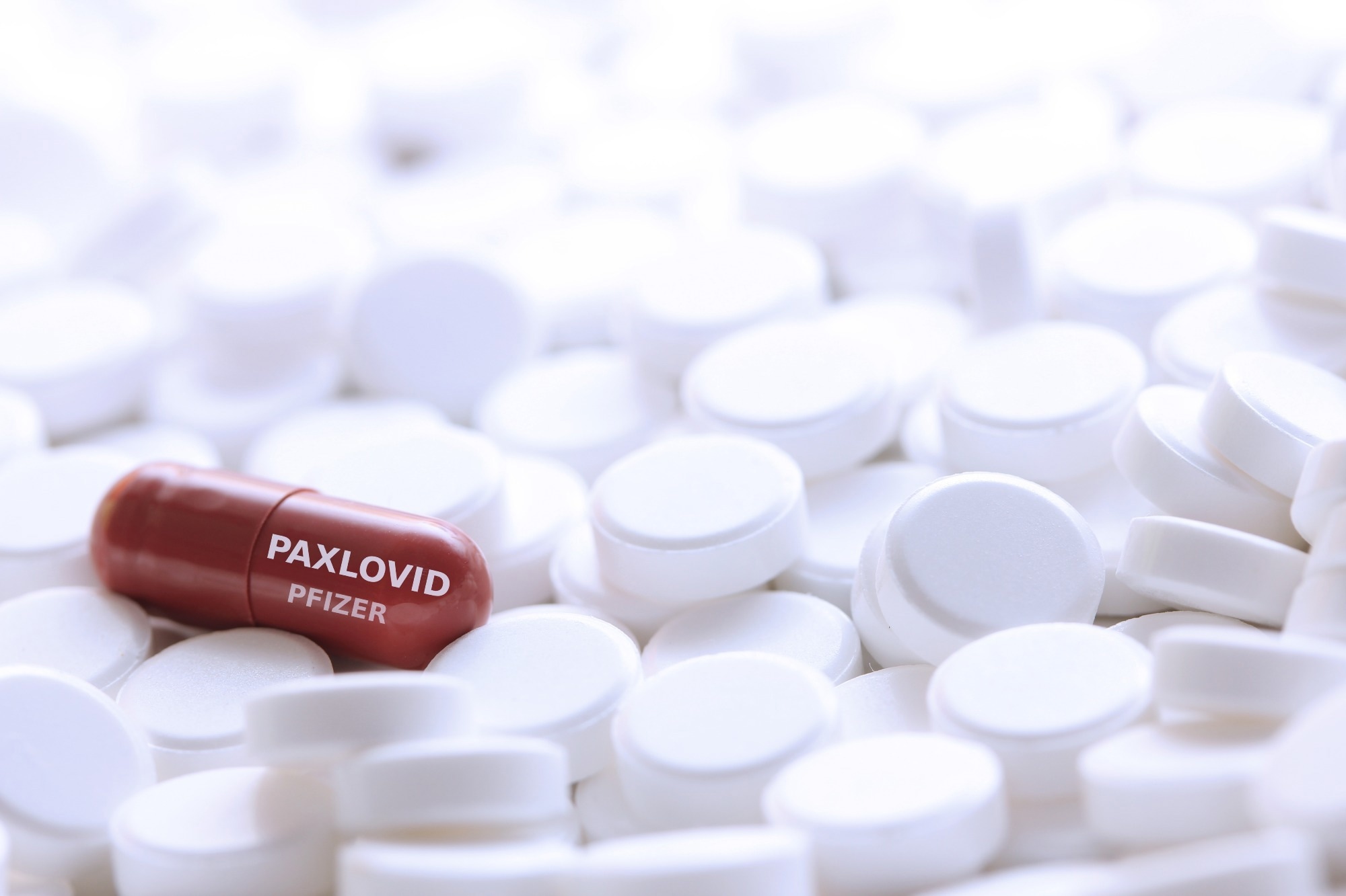
    In pursuit of new therapies to avoid this dark fate, researchers at Stanford have now unveiled a compound that measures up as a potentially powerful anti-coronavirus drug, detailed in a paper published March 13 in Science Translational Medicine. Dubbed ML2006a4, the compound works in the same way as Paxlovid – the most effective oral drug available to date – by binding to coronavirus particles and preventing the virus from making copies of itself. Compared to Paxlovid, though, ML2006a4 binds more tightly and durably, courtesy of the Stanford team custom-crafting the compound atom-by-atom.

    In preclinical experiments, the compound prevented deadly infections in mice at a superior rate compared to Paxlovid. In addition, the new compound is potent enough that it could likely be formulated without an additional component present in Paxlovid that poses severe drug interaction concerns. Importantly, ML2006a4 also performed well against coronavirus variants that have already evolved degrees of resistance to Paxlovid, suggesting the compound’s honed affinity makes it less vulnerable to mutant virus strains.

    At this point entering the fifth year of the pandemic, Paxlovid is our only really good drug against SARS-CoV-2, but it’s proven fairly easy for the virus to evolve resistance to it. As new waves of coronavirus keep crashing down, we need to have alternative drugs that are more tolerant of mutations and not as easy for the virus to defeat.”


    Michael Lin, senior author of the study, associate professor of neurobiology and of bioengineering in the schools of Medicine and Engineering and a member of Stanford Bio-X

    For the study, Lin worked closely with lead author Michael Westberg, now an assistant professor at Aarhus University in Denmark. From 2018 until 2022, Westberg worked in Lin’s lab as a visiting scholar at Stanford Bio-X, funded by the Novo Nordisk Foundation, through a joint program designed to strengthen international collaborations and the exchange of scientific expertise between Stanford and Denmark.

    Atomic-level precision

    Before the pandemic outbreak in 2020, Lin’s lab had already been investigating the broad class of drugs known as viral protease inhibitors. These drugs target protease enzymes that viruses need for disassembling bulky viral proteins as part of their replication cycle. Like a key fitting into a lock, protease inhibitors occupy the spaces, or active sites, where proteases normally link up with those bulky proteins, thus nipping replication in the bud.

    Specifically, the Stanford researchers had gained familiarity with hepatitis C virus protease, which has similarities to coronavirus versions. Although Westberg had come to Stanford to work on other projects, the global emergency prompted a pivot. “When the pandemic hit, we asked if we could put our expertise to good use,” said Lin.

    Their early research, posted online in September 2020, demonstrated that a hepatitis C drug, boceprevir, slotted reasonably well into the coronavirus protease site. Other scientists built off those findings, including at the pharmaceutical company Pfizer, which ultimately created Paxlovid and received regulatory approval for its use in December 2021. “We knew then that we were on the right track,” said Lin, “and we were motivated to keep going and make an even more effective drug.”

    The Lin lab pooled its collective chemical knowledge to design improvements to their iterative boceprevir-based compounds. Much of the work involved modifying the compound on the atomic scale in intricately detailed computer models to fit more snugly in the coronavirus protease active site.

    “Basically, you put your drug in the active site and you look for gaps where it doesn’t tightly fit. Then you fill those gaps,” said Lin.

    The Stanford researchers approached this challenge in a rational way by adding different configurations of atoms of carbon, nitrogen, and oxygen to the compounds as permitted by the laws of biochemistry.

    “There’s a lot of creativity and intuition involved because everyone is working with the same three atoms, but there are essentially infinite ways to arrange them,” said Lin. “Making these modifications, it’s like playing atomic Tetris.”

    The resulting compounds were then tested against actual coronavirus particles at the Stanford In Vitro Biosafety Level 3 Service Center. After multiple rounds of honing, Lin’s team arrived at the compound designated ML2006a4.

    A promising drug candidate

    In studies with SARS-CoV-2-infected mice, ML2006a4 worked as well as Paxlovid in promoting survival, while offering better protection of the rodents’ lungs and lowering overall virus load in the body.

    The researchers attribute this success to ML2006a4’s extremely refined fit inside coronavirus protease, where the compound boasted a 20-fold higher binding affinity than Paxlovid. That better fit equates to stronger chemical bonds, meaning the drug can stay bound to the protease for a longer time. In this temporal regard, ML2006a4 indeed proved quite sticky: The inhibitor remained attached for approximately 330 minutes, or greater than five hours, whereas the corresponding Paxlovid inhibitor typically fell off its target in just about two minutes.

    From a medication perspective, such staying power translates to spaced-out, smaller doses that can still prevent disease from worsening while giving the immune system a chance to kill off the invaders. “The long-lived drug-enzyme complex helps ensure that the virus doesn’t escape and replicate before your next medication dose,” said Lin.

    In this way, ML2006a4 offers other advantages compared to Paxlovid. Technically, Paxlovid is two drugs packaged together: nirmatrelvir, the actual protease inhibitor, and ritonavir, a drug that prevents the liver from quickly breaking down nirmatrelvir, boosting nirmatrelvir’s performance. Yet the slowing of the liver’s metabolism by ritonavir means that other drugs can toxically build up, forcing patients to take the risk of temporarily stopping their normal medications.

    According to Lin, an oral pill based on ML2006a4 might not require ritonavir to prop up drug levels enough between typical 12-hour administrations to effectively keep coronavirus in check, but “this would need to be tested to make sure,” said Lin. “We also continue to make improved versions of ML2006a4 with better potency and duration of activity,” he added.

    For the promising compounds to move forward, Lin and colleagues are seeking additional investment. So far, their funding has mostly consisted of small grants geared toward early-stage drug discovery. The group now feels their compounds are ready for expanded preclinical testing with an eye toward clinical trials in human patients.

    “We’re very excited how far we’ve come and how successful our drug discovery has been on a shoestring budget,” said Lin. “We hope to see this promising compound developed further to stay ready for what SARS-CoV-2 throws at us next.”

    Source:

    Journal reference:

    Park, T., et al. (2024) Single-mode squeezed-light generation and tomography with an integrated optical parametric oscillator. Science Advances. doi.org/10.1126/sciadv.adl1814.

    [ad_2]

    Source link

  • Nomogram could be a game-changer for predicting alcohol-related liver cancer risk

    Nomogram could be a game-changer for predicting alcohol-related liver cancer risk

    [ad_1]

    Liver cancer, unfortunately, is the sixth most common cancer and the third most frequent cause of cancer-related death globally. However, its distribution and causes vary greatly across different regions. While areas like Eastern Asia and sub-Saharan Africa see the most cases, the reasons behind them differ significantly.

    In high-income countries, liver cancer has been on the decline thanks to widespread newborn hepatitis B vaccination and antiviral drugs. Meanwhile, low-income countries witness a worrying rise, often linked to increased hepatitis B and C infections and injectable drug use.

    While viral hepatitis remains a major concern, another factor is gaining attention: alcohol consumption. Studies show that chronic alcohol consumption can directly cause about 10% of cancer cases in men and 3%, respectively, in women. In fact, a study at the Mayo Clinic revealed that alcoholic cirrhosis was the main culprit in 29% of patients with hepatocellular carcinoma (HCC), the most common liver cancer type.

    But the study doesn’t just sound the alarm; it also offers a potential solution. Researchers identified key risk factors for HCC in people with alcohol-related liver disease, including heavy drinking, age, diabetes, male sex, and liver cirrhosis. Based on these factors, they developed a novel tool called a nomogram that can predict HCC risk with high accuracy and ease of use.

    This nomogram could be a game-changer for doctors, allowing them to personalize treatment plans and identify individuals at highest risk for HCC. Early intervention could save lives and prevent unnecessary suffering.

    This study underscores the growing public health concern of alcohol-related liver cancer. The newly developed nomogram offers a valuable tool for doctors to identify high-risk individuals and personalize treatment plans, potentially saving lives and preventing unnecessary suffering.

    While the study provides valuable insights, it acknowledges limitations like its retrospective nature and the need for further validation in larger, prospective studies. Additionally, incorporating other factors like smoking, genetics, and dietary habits could further improve the prediction model.

    The researchers also highlight the need for future research on non-alcoholic fatty liver disease and its link to liver cancer, as this area remains under-investigated.

    Overall, this study shines a light on the rising threat of alcohol-related liver cancer and offers a promising tool for early detection and intervention. Further research and public health efforts are crucial to combat this growing health challenge.

    Source:

    Journal reference:

    Chang, B., et al. (2024). Prevalence and prediction of hepatocellular carcinoma in alcohol-associated liver disease: a retrospective study of 136 571 patients with chronic liver diseases. eGastroenterology. doi.org/10.1136/egastro-2023-100036.

    [ad_2]

    Source link

  • Paxlovid enhances treatment options for COVID-19 patients

    Paxlovid enhances treatment options for COVID-19 patients

    [ad_1]

    In a recent review published in the Pharmaceutics, a group of authors explored the design, synthesis, and mechanism of action of Paxlovid, a Protease inhibitor (PI) drug combination for treating coronavirus disease 2019 (COVID-19).

    Study: The Design, Synthesis and Mechanism of Action of Paxlovid, a Protease Inhibitor Drug Combination for the Treatment of COVID-19. Image Credit: Tobias Arhelger/Shutterstock.com

    Background 

    The COVID-19 pandemic, caused by the severe acute respiratory syndrome coronavirus 2 (SARS-CoV-2) virus, significantly challenged global healthcare systems and medical science.

    In response, researchers worldwide developed vaccines with innovative mechanisms and small-molecule antivirals targeting crucial viral proteins.

    Among these, PaxlovidTM, a blend of nirmatrelvir and ritonavir PIs, stands out for its effectiveness in treating COVID-19.

    Nirmatrelvir inhibits SARS-CoV-2’s main protease, vital for viral replication, while ritonavir boosts nirmatrelvir’s effectiveness by inhibiting Cytochrome P450 3A4 (CYP3A4), an enzyme that would otherwise degrade nirmatrelvir quickly.

    Further research is needed to develop alternative main protease (MPro) inhibitors despite the success of the nirmatrelvir-ritonavir combination, ensuring continued effectiveness against COVID-19.

    PIs as antivirals for Hepatitis C virus (HCV) and Human immunodeficiency virus (HIV) 

    PI Drugs for HCV and HIV Infections

    PIs are key in treating HCV and HIV infections. HCV, a small ribonucleic acid (RNA) virus causing hepatic diseases, is targeted by PIs like asunaporevir, telaprevir, and boceprevir, focusing on the nonstructural (NS)3/4A serine protease.

    These inhibitors are peptidomimetics, containing peptide bonds and a ‘warhead’ group that binds covalently but reversibly to the enzyme’s active site.

    HIV PIs target the virus’s aspartic acid protease, which is crucial for viral replication. They are used in antiretroviral therapy, transforming HIV from fatal to chronic.

    Development and mechanism of Nirmatrelvir

    Nirmatrelvir, developed from Pfizer’s earlier SARS-CoV-1 PI .. PF-00835231, faced challenges in oral absorption.

    Modifications like altering the warhead and substituting various molecular components enhanced its binding affinity and antiviral activity, eventually leading to nirmatrelvir with a nitrile warhead, improving solubility and synthesis.

    Despite different warheads, its structural similarity to boceprevir, and its role as a covalent inhibitor of SARS-CoV-2 Mpro makes it significant in COVID-19 treatment.

    Synthesis of nirmatrelvir

    Nirmatrelvir’s synthesis involves coupling the P1 building block and the P2-P3 dipeptide, with the final step being the formation of the nitrile warhead.

    The process starts with protected amino acid derivatives, proceeding through stages like Boc-deprotection, ester cleavage, and dipeptide formation.

    The synthesis yields nirmatrelvir with high efficiency and introduces a new approach involving a Ugi-type three-component reaction for higher diastereoselectivity.

    Synthesis and structure-activity relationship (SAR) study of nirmatrelvir analogs

    Research by Chia and co-workers led to the synthesizing nirmatrelvir analogs with different P1′ moieties, examining the role of the warhead in antiviral activity.

    These studies revealed varying levels of effectiveness in protease inhibition and antiviral activity, with some derivatives showing similar or superior effects to nirmatrelvir. However, challenges in cell penetration and specificity to SARS-CoV-2 limited the broader application of these analogs.

    Novel covalent and non-covalent inhibitors of SARS-CoV-2 Mpro

    Recent developments in SARS-CoV-2 Mpro inhibitors have introduced both peptidomimetic and non-peptidic inhibitors.

    These include warheads, such as epoxide rings and fluoromethyl groups, offering alternative mechanisms of covalent binding to the enzyme.

    Non-covalent inhibitors, like ensitrelvir, show lower reactivity but better selectivity due to their secondary interaction nature. These developments represent crucial steps in diversifying therapeutic options against COVID-19 and its evolving strains.

    Ritonavir as a pharmacokinetic enhancer

    Structure, activity, and interactions of ritonavir

    Originally an HIV protease inhibitor, Ritonavir is known for its efficacy at low doses (~100 mg) in inhibiting the CYP3A4 enzyme, a crucial element in drug metabolism.

    While high doses of Ritonavir are poorly tolerated, its low-dose effectiveness is leveraged in combination therapies with other HIV protease inhibitors, enhancing their half-lives and thus reducing required dosages.

    This unique use of Ritonavir has been explored even in early COVID-19 treatments. However, its use poses risks of significant drug–drug interactions, especially with medications metabolized by CYP3A4, potentially elevating their levels to toxic concentrations.

    Additionally, Ritonavir’s effect on other enzymes and transport proteins is noted, albeit of lesser importance in Paxlovid treatment.

    Synthesis of ritonavir

    developed at Abbott Laboratories, Ritonavir’s synthesis involves complex chemical processes, combining chiral amine and carboxylic acid building blocks.

    The synthesis starts with a cyclocondensation reaction involving thioformamide and ethyl 2-chloroacetate, followed by a series of steps leading to the formation of ritonavir.

    This intricate process involves various intermediate compounds and chemical reactions, including triethylamine and 4-dimethylaminopyridine, highlighting the sophistication required in pharmaceutical synthesis.

    The production of Ritonavir demonstrates the intricate chemical engineering necessary to develop effective pharmaceutical agents.

    Paxlovid—application and activity against mutant variants

    Paxlovid, combining nirmatrelvir and ritonavir, has shown significant efficacy in reducing COVID-19-related hospitalizations and mortality.

    While it has gained emergency use authorization in various regions, its effectiveness against emerging strains and mutant variants is under continuous scrutiny.

    The evolving landscape of SARS-CoV-2 mutations necessitates ongoing monitoring to ensure the sustained efficacy of treatments like Paxlovid.

    [ad_2]

    Source link