Tag: Hyperglycemia

  • Maternal diabetes linked to a slight increase in ADHD risk in children

    Maternal diabetes linked to a slight increase in ADHD risk in children

    [ad_1]

    In a recent study published in the journal Nature Medicine, researchers evaluated associations between maternal diabetes mellitus (MDM) and the risk of attention-deficit/hyperactivity disorder (ADHD) in the offspring.

    Sixteen percent of pregnant individuals experience hyperglycemia worldwide. The prevalence of MDM has increased globally, which is associated with the advancing maternal age, the growing obesity epidemic, and improved MDM diagnostic approaches. Animal studies have shown the adverse effects of hyperglycemia in pregnancy on intrauterine oxidative stress, inflammatory response, and epigenetic mechanisms, which might lead to poor neurodevelopment in the offspring.

    ADHD is a neurodevelopmental disorder characterized by impulsivity, hyperactivity, and inattentiveness. Globally, 2% to 7% of children are affected by ADHD. Besides, ADHD can have a substantial burden on families of affected individuals and society. Evidence suggests that gestational diabetes mellitus (GDM) and pre-GDM (PGDM) are associated with ADHD. A meta-analysis revealed that the offspring of diabetic mothers had a 40% increased risk of ADHD.

    Study: Maternal diabetes and risk of attention-deficit/hyperactivity disorder in offspring in a multinational cohort of 3.6 million mother–child pairs. Image Credit: Pixel-Shot / ShutterstockStudy: Maternal diabetes and risk of attention-deficit/hyperactivity disorder in offspring in a multinational cohort of 3.6 million mother–child pairs. Image Credit: Pixel-Shot / Shutterstock

    About the study

    In the present study, researchers assessed the association between MDM and ADHD risk in the offspring. This population-based cohort study used healthcare data from New Zealand, Hong Kong, Taiwan, and Nordic countries (Iceland, Sweden, Norway, and Finland). The study included children from live births within site-specific periods. Mother-child pairs were linked using exact deterministic linkage.

    Children without complete birth information, six years of follow-up, and valid linkage were excluded. Follow-up commenced at birth and continued until outcome occurrence, death, or end of study period. The primary exposure was MDM, including PGDM and GDM. MDM was stratified into (unmedicated and medicated) GDM and (types 1 and 2) PGDM.

    ADHD was defined using site-specific diagnosis and medication codes. The primary analyses compared ADHD status in children born to mothers with any type of diabetes with those born to non-diabetic mothers. Sibling-matched analyses compared the ADHD status in children of the same mother but with discordant GDM status.

    In secondary analyses, ADHD status was compared between children born to mothers with different subtypes of diabetes. The researchers computed hazard ratios of average treatment effect and 95% confidence intervals to examine associations between MDM status and ADHD using Cox proportional hazard regression models.

    Covariates included demographic factors, socioeconomic status, birth year, multifetal pregnancies, body mass index (BMI), use of psychotropic medication, alcohol and smoking status, neurologic and psychiatric conditions, and other chronic conditions. Several sensitivity analyses were also performed to evaluate the robustness and validity of the findings.

    Findings

    The study included more than 3.6 million mother-child pairs. About 6.6%, 8%, 4.1%, and 13.7% of children had mothers with diabetes in the Nordic countries, Hong Kong, New Zealand, and Taiwan, respectively. ADHD risk was higher among children whose mothers had any type of diabetes than in those born to non-diabetic mothers. ADHD risk was higher in children born to mothers with PGDM, type 1 PGDM, type 2 PGDM, or GDM compared to those born to non-diabetic mothers.

    Sibling-matched analyses did not find differential risks of ADHD. ADHD risk was similar among children whose mothers were diagnosed with GDM at different trimesters in Hong Kong. However, ADHD risk was the highest in children whose mothers had GDM diagnosis in the first trimester in New Zealand and Taiwan. Further, children whose mothers had GDM might have a lower ADHD risk than those born to mothers with PGDM.

    ADHD risks did not differ between children born to mothers with type 1 and type 2 PGDM. Children whose mothers had GDM and received anti-diabetic medications had a similar risk of ADHD compared to those born to mothers with GDM who were unmedicated. Sensitivity analyses produced similar results as primary analyses.

    Conclusions

    In sum, the findings revealed that MDM, PGDM, and GDM were associated with a small/moderate risk of ADHD in the offspring. ADHD risks did not differ between siblings with discordant GDM status in pregnancy, suggesting potential confounding by unmeasured, shared familial or genetic factors. Moreover, ADHD risk estimates were smaller compared to a previous meta-analysis. Future studies should reevaluate the specific roles of hyperglycemia and genetic factors in the relationship between MDM and ADHD.

    Journal reference:

    • Chan AYL, Gao L, Hsieh MHC, et al. Maternal diabetes and risk of attention-deficit/hyperactivity disorder in offspring in a multinational cohort of 3.6 million mother–child pairs. Nat Med, 2024, DOI: 10.1038/s41591-024-02917-8, https://www.nature.com/articles/s41591-024-02917-8

    [ad_2]

    Source link

  • Burdock roots outshine dandelion in antidiabetic potential study

    Burdock roots outshine dandelion in antidiabetic potential study

    [ad_1]

    In a recent study published in the journal Plants, researchers from Latvia analyzed and compared the chemical compounds in the roots of dandelion (Taraxacum officinale) and burdock (Arctium lappa) for their potential antidiabetic properties. They found that while burdock exhibited higher values for total phenolic content (TPC), tannin content, and α-amylase activity compared to dandelion, dandelion had higher total polysaccharide (TP) content. In vivo studies are warranted to confirm these findings and the antidiabetic potential of these plants.

    Study: Antidiabetic Properties of the Root Extracts of Dandelion (Taraxacum officinale) and Burdock (Arctium lappa). Image Credit: KatMoys / ShutterstockStudy: Antidiabetic Properties of the Root Extracts of Dandelion (Taraxacum officinale) and Burdock (Arctium lappa). Image Credit: KatMoys / Shutterstock

    Background

    Type 2 diabetes mellitus (T2DM) accounts for a majority of diabetes cases globally and is associated with various risk factors, including genetic predisposition, poor diet, and lack of physical activity, leading to insulin resistance and hyperglycemia-associated complications. Given the drawbacks and expenses of conventional hypoglycemic drugs, there is a growing interest in herbal medicine for diabetes management. Preclinical studies highlight the potential of edible plants for blood sugar control and offer promising alternatives with apparent efficacy and low toxicity.

    Dandelion and burdock, traditional medicinal plants belonging to the Asteraceae family, are rich in diverse phytochemicals with potential health benefits. They contain phenolic acids, coumarins, and polysaccharides, exhibiting various biological activities, suggesting their potential role in managing complex conditions like T2DM. The present study aimed to investigate the potential antidiabetic properties of chemical compounds in dandelion and burdock roots by assessing their effects on blood sugar levels and antioxidant capabilities.

    About the study

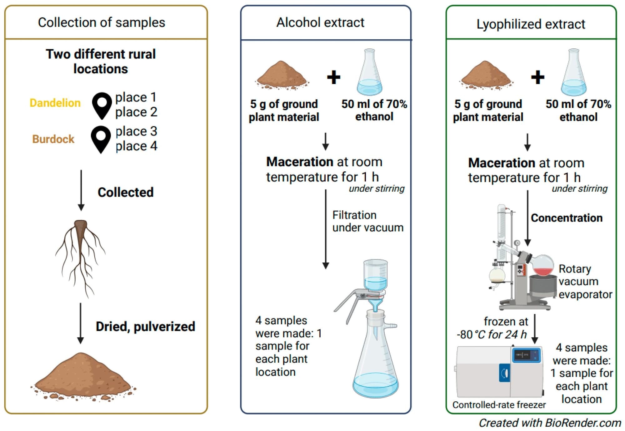
    Dandelion and burdock roots were collected from two distinct rural regions in Latvia and processed according to standardized methods. While dandelion roots were sourced from “Vecpiebalga” and “Kaļķis,” burdock roots were collected near “Viļani” and “Būdiņas.” The roots were washed, dried, and ground into a powder for extraction. Ethyl alcohol extracts (AE) and lyophilizate extracts (LE) were prepared from the powdered roots, and both extraction methods were analyzed comparatively. Analysis of the extracts included determination of inulin content, TPC, tannin level, and TP.

    Preparation of ethyl alcohol and lyophilizate extracts.Preparation of ethyl alcohol and lyophilizate extracts.

    Additionally, antioxidant activities were assessed using the DPPH (short for 2,2-diphenyl-1-picrylhydrazyl) assay, and hypoglycemic properties were assessed based on α-amylase activity. Trolox was used as a standard solution for constructing the standard curve in the antioxidant activity analysis. Half maximal inhibitory concentration (IC50) was determined for Trolox and compared with that of dandelion and burdock. Similarly, in the hypoglycemic activity analysis, acarbose was used as the standard solution.

    Liquid chromatography-mass spectrometry (LC-MS) was employed for qualitative analysis of the chemical components. Statistical analysis involved means and standard errors, analysis of variance, and the Mann–Whitney U test.

    Results and discussion

    The results of specific color-change-based chemical tests revealed the presence of inulin and the absence of starch in burdock and dandelion roots. Significant differences were observed in TPC between alcohol-based and lyophilizate extraction methods, with burdock showing higher TPC, particularly in LE. Dandelion roots showed negligible tannin content, while burdock roots exhibited low but detectable levels, with LE showing slightly higher values. However, no significant difference was found in terms of TPC and tannin obtained from samples of the two different Latvian rural regions in the study.

    Further, dandelion root extract showed higher values of TP compared to burdock root extract. No statistically significant differences were found in the TP between the two plants. Comparatively, LE exhibited significantly higher antioxidant activity compared to AE. Burdock LE outperformed Trolox, while dandelion AE showed the least favorable outcome.

    None of the plant extracts matched the IC50 of acarbose, with LE of burdock showing the most favorable outcomes and the AE of dandelion demonstrating the least favorable results. LE consistently showed significantly higher values compared to AE within the same plant samples.

    Diverse chemical compounds were found in root extracts, including amino acids, phenolic acids, and alkaloids, among others. Specific compounds like chlorogenic acid, phenylalanine, and valine were found in all the extracts, while others like caffeic acid and oleanolic acid were exclusive to burdock, and salicylic acid glucoside and protocatechuic acid were unique to dandelion. Burdock showed a wider array of unique compounds than dandelion, indicating its richer chemical profile.

    In the future, exploring additional compounds present in the roots and replicating tests with various solvents could provide further insights. Animal and human studies would be crucial to confirm these findings and explore the potential clinical applications of these compounds.

    Conclusion

    In conclusion, the present study found burdock root to be better than dandelion regarding its chemical composition and potential therapeutic activity. However, more research is needed to confirm the effectiveness of the two plants individually and in combination with other drugs for managing diabetes and other chronic ailments.

    Journal reference:

    • Antidiabetic Properties of the Root Extracts of Dandelion (Taraxacum officinale) and Burdock (Arctium lappa). Zolotova D. et al., Plants, 13(7):1021 (2024), DOI: 10.3390/plants13071021, https://www.mdpi.com/2223-7747/13/7/1021 

    [ad_2]

    Source link

  • Antioxidant-rich diets linked to lower type 2 diabetes risk, supplements less effective

    Antioxidant-rich diets linked to lower type 2 diabetes risk, supplements less effective

    [ad_1]

    Scientists at the Karolinska Institute in Sweden have conducted a systematic review and meta-analysis to determine the relationship between dietary antioxidant intake and type 2 diabetes risk.

    The study is published in the journal Advances in Nutrition

    Review: Vitamins C, E, and beta-carotene and risk of type 2 diabetes: a systematic review and meta-analysis. Image Credit: alicja neumiler / ShutterstockReview: Vitamins C, E, and beta-carotene and risk of type 2 diabetes: a systematic review and meta-analysis. Image Credit: alicja neumiler / Shutterstock

    Background

    Type 2 diabetes is a serious metabolic disease characterized by reduced secretion or impaired functioning of insulin and subsequent increase in blood glucose level (hyperglycemia). Insulin resistance and pancreatic beta-cell dysfunction are two major hallmarks of the disease.

    More than 10% of the global population is currently affected by type 2 diabetes. Its prevalence is sharply increasing worldwide mainly because of an increasing inclination towards unhealthy food habits and sedentary lifestyles.

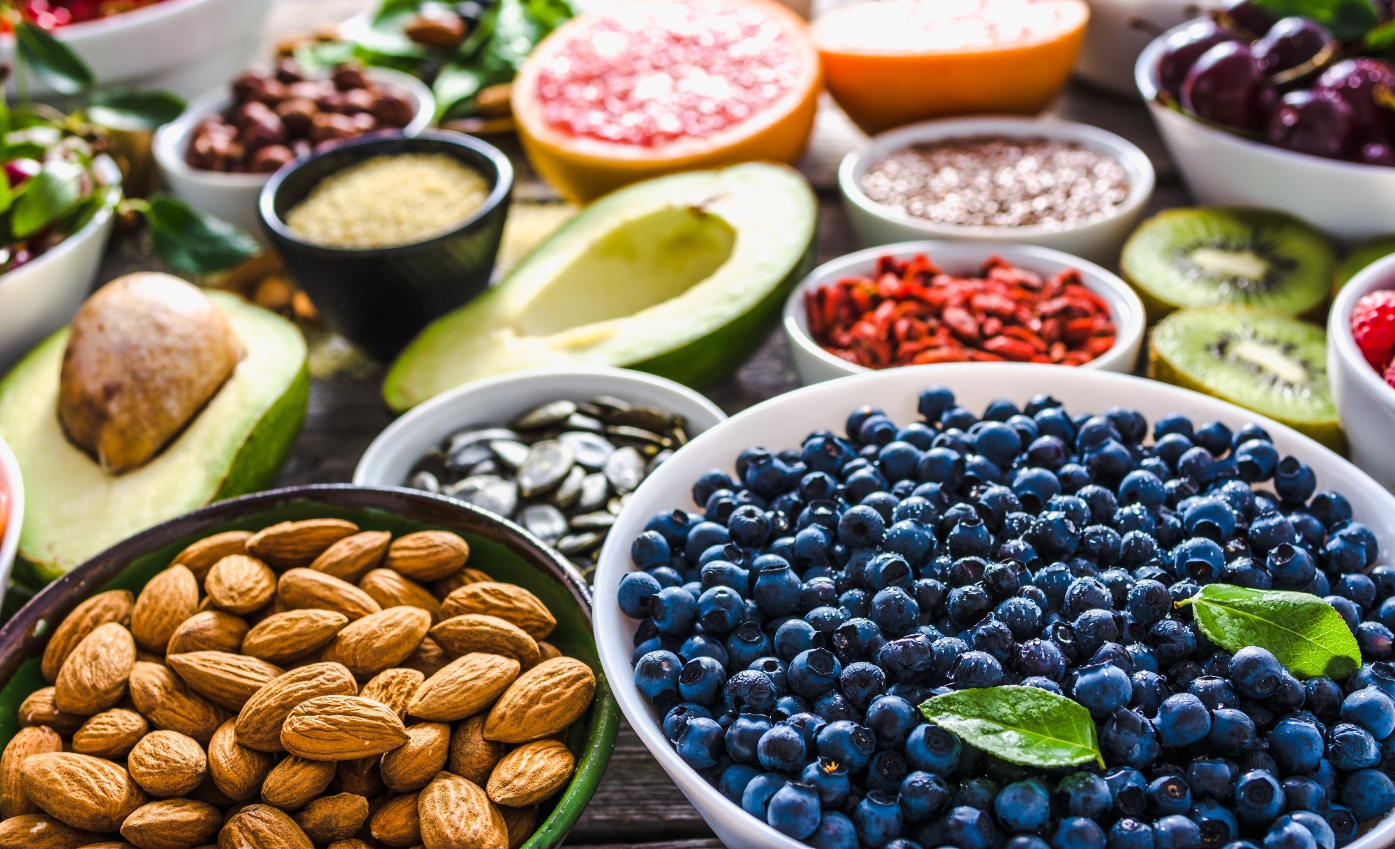
    Diet plays an important role in regulating the body’s metabolism, among various lifestyle factors. Evidence indicates that an increased adherence to healthy diets, such as the Mediterranean or DASH (Dietary Approach to Stop Hypertension), can significantly reduce the risk of type 2 diabetes. The fundamental characteristic of these diets is a higher intake of plant-based foods rich in antioxidants, including vitamins C, E, and beta-carotene.    

    In this systematic review and meta-analysis, scientists have assessed whether consumption of dietary vitamins C, E and beta-carotene can reduce the risk of type 2 diabetes.

    Vitamins C, E, and beta-carotene and risk of type 2 diabetes: a systematic review and meta-analysis

    Study design

    The scientists searched various electronic databases to identify studies investigating the association between dietary intakes, circulating levels, or supplementation of vitamin C, E, and beta-carotene and type 2 diabetes incidence or insulin resistance/sensitivity and beta cell function in non-diabetic individuals.

    The final screening led to the identification of 25 prospective observational studies and 15 randomized controlled trials. Moderate and serious risks of bias were observed in 21 and 4 observational studies, respectively. Among randomized controlled trials, 13 had a low risk of bias, and 2 had some concerns.

    Important observations

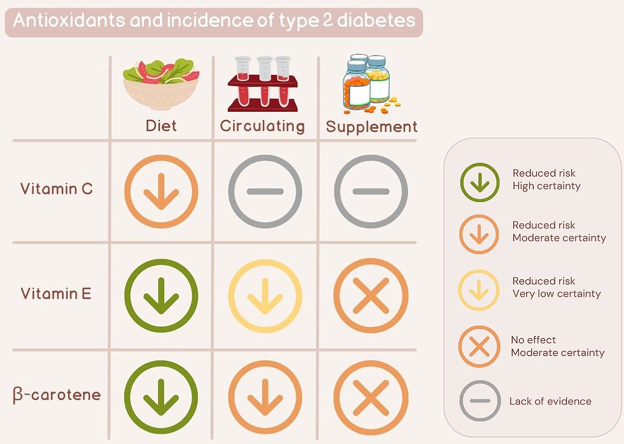
    The study found that moderate intakes of vitamins C, E, and beta-carotene can reduce the risk of type 2 diabetes.

    Vitamin C

    The meta-analysis of observational studies revealed that a vitamin C intake of up to 70 mg per day can reduce type 2 diabetes risk by 24%. However, no further risk reduction was observed for an intake higher than this level.

    An inverse association was observed between dietary intake of vitamin C and insulin resistance. Vitamin C intake also showed a positive impact on beta cell function.

    Vitamin E

    The meta-analysis of observational studies revealed that the vitamin E intake of up to 12 mg per day can reduce the risk of type 2 diabetes by 28%. Similar to vitamin C, no further risk reduction was observed for an intake higher than this level.

    The meta-analysis of randomized clinical trials revealed that vitamin E supplementation does not have any protective effect against type 2 diabetes development. However, vitamin E supplementation showed a positive impact on insulin sensitivity.

    Beta-carotene

    The meta-analysis of observational studies revealed that the beta-carotene intake of up to 4 mg per day can reduce the risk of type 2 diabetes by 22%. No further risk reduction was observed above this level.

    The meta-analysis of randomized controlled trials revealed that beta-carotene supplementation cannot reduce the risk of type 2 diabetes. It was also observed that circulating beta-carotene can reduce insulin resistance and increase insulin sensitivity.

    Study significance

    This systematic review and meta-analysis found an inverse association between dietary and circulating vitamins C, E, and beta-carotene and the risk of type 2 diabetes. However, no protective efficacy of supplementation with these antioxidants has been observed against type 2 diabetes.

    The robust antioxidant properties of these vitamins are mainly responsible for their anti-diabetic effects. Vitamin C is a water-soluble vitamin commonly found in fruits and vegetables. It can remove free radicals in the body’s hydrophilic compartments and regenerate vitamin E from its oxidized form.

    Vitamin E is a fat-soluble vitamin commonly found in nuts, seeds, and vegetable oils. It can prevent lipid peroxidation and protect lipid parts of the body, such as cell membranes. Beta-carotene is a fat-soluble provitamin A carotenoid commonly found in fruits and vegetables. Similar to vitamin E, beta-carotene can protect lipid parts of the body from free radical-mediated damage.

    Article Revisions

    • Mar 27 2024 – Addition of Graphical abstract illustration.

    [ad_2]

    Source link

  • Exploring the role of iodine in obesity, diabetes, and other metabolic conditions

    Exploring the role of iodine in obesity, diabetes, and other metabolic conditions

    [ad_1]

    In a recent study published in Frontiers in Nutrition, researchers reviewed recent data on the metabolic implications of iodine consumption and elucidated the underlying mechanisms.

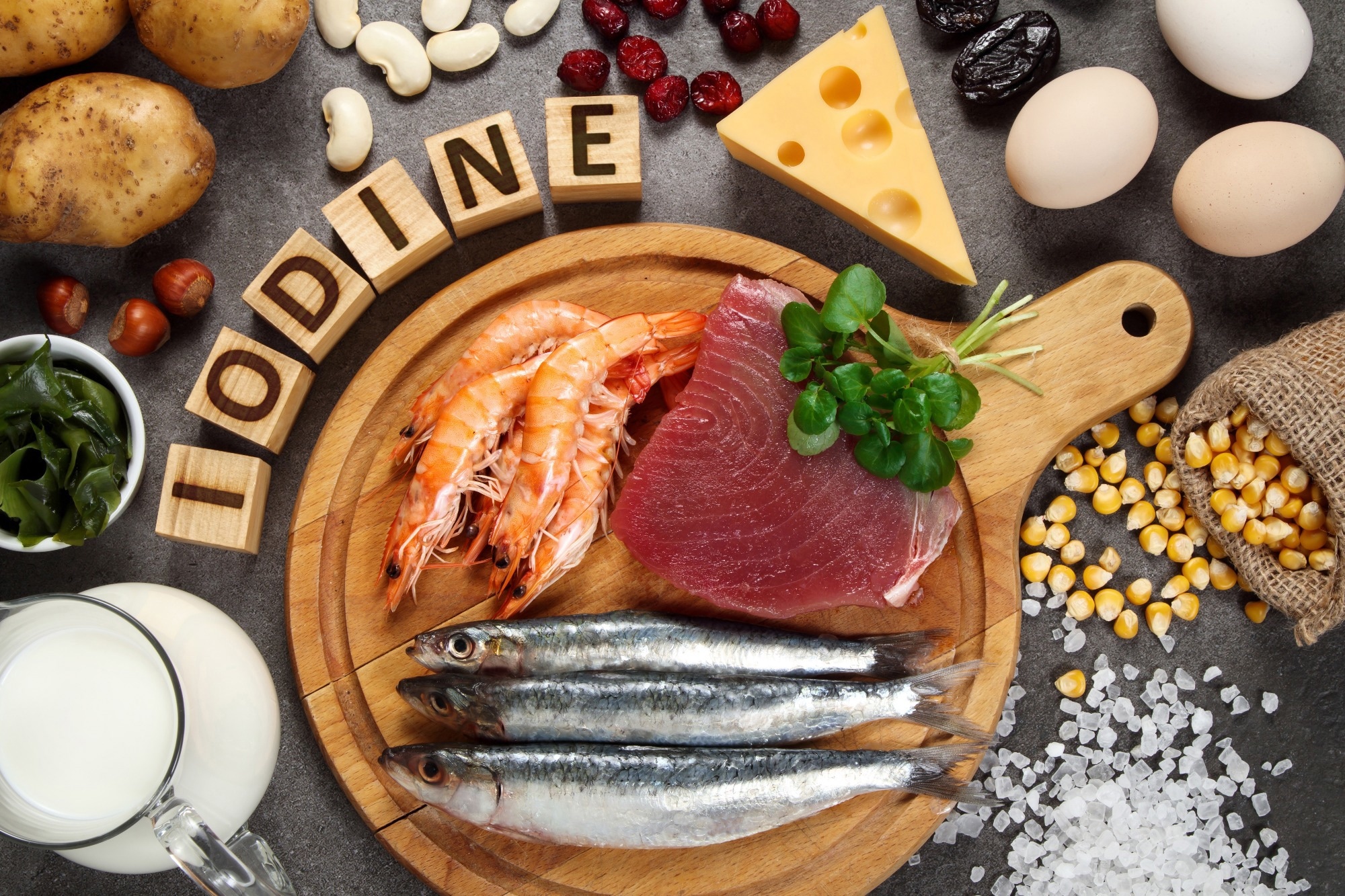
    Study: The correlation between iodine and metabolism: a review. Image Credit: Evan Lorne/Shutterstock.comStudy: The correlation between iodine and metabolism: a review. Image Credit: Evan Lorne/Shutterstock.com

    Background

    Iodine is an essential nutrient that aids in producing thyroid hormones and is associated with metabolic illnesses such as diabetes, obesity, dyslipidemia, and hypertension.

    However, the processes underlying these relationships are unknown. Iodine exerts immunomodulator, antioxidant, and differentiator effects in several tissues and organs, and alters the levels of thyroxine (T4) and tri-iodothyronine (T3), the primary regulators of energy metabolism.

    Metabolic syndrome (MetS), which includes hypertension, abdominal obesity, hyperlipidemia, and hyperglycemia, is common globally and can lead to cardiovascular disease, malignancies, and death. Oxidative stress, chronic inflammatory diseases, and dietary changes are all risk factors for MetS.

    The nutritional status of iodine may partly explain the incidence of metabolic syndrome. Further study on the relationship between iodine and metabolism will contribute to a better understanding of its role and promote an adequate and reliable iodine feeding standard.

    About the study

    In the present study, researchers explored the impact of iodine levels on metabolic health.

    Research on the effects of iodine on metabolism

    The recommended dietary allowance (RDA) of iodine ranges between 150 and 299 μg/day, with a moderately increased consumption potentially lowering the risk of prostate and breast cancer.

    Cross-sectional research indicates a U-shaped association between urinary iodine concentration (UIC) and metabolic syndrome prevalence, with a low point of 300 to 499 μg/L.

    In Korean postmenopausal women, consuming seaweeds and iodine showed inverse correlations with MetS incidence; however, excess seaweed intake demonstrated adverse effects among male MetS patients with TT and TG genotypes of the lipoprotein lipase gene (LPL). However, a study of school-aged children discovered associations between high UIC and MetS.

    Research in China indicated central adiposity decreased when UIC levels reached ≥300 μg/L. A randomized clinical trial found that individuals who received iodine-reduced kelp tablets had a significantly lower body fat percentage.

    A 28-day placebo-controlled trial discovered that fucoxanthin seaweed supplementation reduced waist circumference, fat mass, visceral fat, weight, and BMI among obese residents of Japan. However, among reproductive-age Colombian women, mUIC was shown to be positively linked with obesity.

    The TIDE study demonstrated a U-shape curve for the relationship between urinary iodine concentration and diabetes prevalence, with higher UIC levels increasing the likelihood of acquiring diabetes mellitus type 2 (T2DM). Patients with diabetes mellitus have lower UIC levels than healthy individuals.

    Increased iodine content in the placenta lowers gestational diabetes in pregnant women. The study also discovered a U-shaped curve in the correlation between UIC and hypertension prevalence, with individuals in iodine-excess (IE) and iodine-sufficient (IS) locations having higher blood pressure readings. Iodine deficiency is a risk factor for preeclampsia and pregnancy-related hypertension.

    Research has demonstrated an inverse relationship between UIC, hyperuricemia, and gout prevalence. Longitudinal data revealed higher death rates among patients with ID (UIC <100 μg/L).

    Iodine consumption can raise blood cholesterol levels in hens and cause hepatic steatosis in BaLB/c mice. In mice, higher iodine consumption enhanced lipid metabolism without affecting thyroid hormone levels or body weight.

    Mechanisms underlying the metabolic effects of iodine

    Iodine exerts antioxidative, antimicrobial, immunomodulatory, and molecular regulatory effects. Iodine alters the proportion of pathogenic and beneficial bacteria to restore the gut microbiome and reduce insulin resistance, obesity, and metabolic syndrome parameters.

    Iodine also reduces inflammation by lowering oxidative and endoplasmic reticulum stress caused by free radicals such as reactive oxygen species (ROS).

    Iodine acts on the Kelch-like ECH-associated protein 1-NF-E2-related factor 2 (KEAP1-NRF2) pathway to enhance the activities of antioxidant enzymes such as superoxide dismutase (SOD), catalase (Cat), and glutathione peroxidase (GSH-Px).

    In addition, iodine alters inducible nitric oxide synthase (iNOS) and cyclooxygenase-2 (COX2) levels, regulating mitogen-activated protein kinase (MAPK) and nuclear factor kappa B (NF-κB) pathways to reduce chronic inflammation and improve metabolic health.

    The mineral acts on type 2 deiodinase (D2) receptors that convert T4 to biologically active T3 to improve weight management and adaptive thermogenesis.

    Iodine also interacts with peroxisome proliferator-activated receptor-γ (PPARγ) receptors to enhance adipocyte differentiation, fatty acid uptake, and glucose metabolism by improving insulin sensitivity.

    Conclusions

    Overall, the review findings indicate that iodine impacts obesity, lipid metabolism, and glucose metabolism. Iodine’s antioxidant, immunomodulatory, gut-restoring, and antimicrobial effects explain the mineral’s effects.

    Iodine regulates the oxidative state related to variations in insulin sensitivity or metabolism. However, iodine shortages and persistent iodine excesses may increase the risk of thyroid diseases.

    Thus, it is critical to maintain iodine levels in an appropriate range at a population level. Future prospective studies and mechanism research must develop an evidence-backed and safe iodine nutrition standard.

    [ad_2]

    Source link

  • A call for targeted research and therapies

    A call for targeted research and therapies

    [ad_1]

    In a recent review published in the journal Cell Metabolism, researchers elucidated mechanisms and evaluated therapies for impaired skeletal muscle regeneration in diabetes, identifying research gaps and future directions.

    Study: Impaired skeletal muscle regeneration in diabetes: From cellular and molecular mechanisms to novel treatments. Image Credit: Crevis / ShutterstockStudy: Impaired skeletal muscle regeneration in diabetes: From cellular and molecular mechanisms to novel treatments. Image Credit: Crevis / Shutterstock

    Background 

    Diabetes, a growing public health issue, continues to surge despite extensive research and healthcare efforts. It leads to various forms of diabetic myopathy, irrespective of its type, causing a decline in skeletal muscle mass and function. This decline not only worsens obesity and hyperglycemia but also affects locomotion, energy metabolism, and glucose regulation, further deteriorating muscle structure and function. Additionally, diabetes impairs muscle regeneration, potentially worsening conditions like ischemia and foot ulcers by promoting fibrosis and hindering myofiber recovery. Further research is needed to better understand and develop targeted interventions for the complex mechanisms underlying impaired muscle regeneration in diabetes.

    Skeletal muscle abnormalities in diabetes

    Diabetes, alongside its comorbidities like obesity, hypertension, and dyslipidemia, significantly affects skeletal muscle structure, function, and metabolism. The complex nature of diabetes complicates the identification of effective therapeutic targets. Other contributing factors include aging, inactivity, and poor nutrition. Key observed abnormalities in diabetic patients include reduced muscle mass and strength, abnormal lipid deposition, fiber atrophy, and altered myokine secretion, contributing to decreased functional capacity and quality of life.

    Diabetes not only leads to muscle degeneration but also impairs the muscle’s ability to regenerate, complicating injuries such as ischemia and foot ulcers. The regeneration process, involving both muscle stem cells (MuSCs) and non-MuSCs, is hampered, as indicated by excessive fibrosis and delayed myofiber maturation.

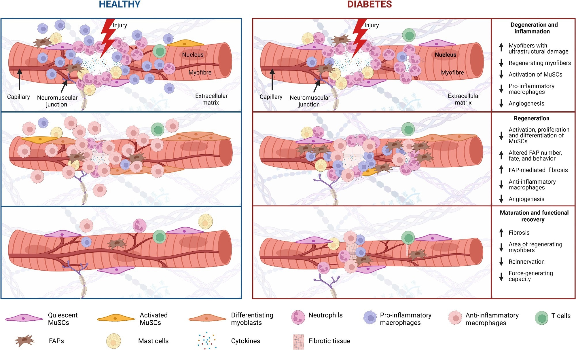
    Skeletal muscle regeneration in diabetes Diabetes and its associated complications, including obesity and hyperglycemia, impact multiple cell populations (MuSCs, neutrophils, macrophages, T cells, FAPs, and mast cells) that play a vital role in the process of muscle regeneration (i.e., degeneration and inflammation, regeneration, and maturation and functional recovery).

    Skeletal muscle regeneration in diabetes Diabetes and its associated complications, including obesity and hyperglycemia, impact multiple cell populations (MuSCs, neutrophils, macrophages, T cells, FAPs, and mast cells) that play a vital role in the process of muscle regeneration (i.e., degeneration and inflammation, regeneration, and maturation and functional recovery).

    Degeneration and inflammation

    Muscle injuries trigger necrosis and inflammation, marked by fiber breakdown and protein leakage into the serum. The process, essential for tissue repair, draws in immune cells like neutrophils and macrophages. Diabetes compounds this degeneration, amplifying damage, and hampering regeneration, highlighting the metabolic impact on muscle recovery.

    Regeneration process

    Diabetes negatively impacts the muscle regeneration process, notably affecting the activation, proliferation, and differentiation of MuSCs and the roles of fibro-adipogenic progenitors (FAPs). Treatments like metformin offer some hope by potentially modifying FAP activity. However, delayed regeneration in diabetic models underlines the urgent need for deeper insights into how diabetes disrupts muscle repair mechanisms.

    Challenges in muscle recovery

    Efficient muscle regeneration requires not only the formation of new myofibers but also the reconstitution of the extracellular matrix, vascular network, and innervation. Diabetes and obesity complicate this process, showing delayed functional recovery, increased collagen accumulation, and impaired neuromuscular junction adaptations.

    Diabetic impacts on muscle fiber and insulin signaling

    Diabetes shifts muscle fiber composition towards type II fibers, which are more prone to damage and impair regeneration. Insulin resistance disrupts muscle cell growth pathways, while hyperinsulinemia and lipotoxicity inhibit crucial recovery processes like autophagy and protein metabolism. These changes suggest that targeting fiber-type transitions and improving insulin signaling could enhance muscle regeneration in diabetes.

    Diabetic challenges in muscle regeneration signaling

    Diabetes triggers elevated pro-inflammatory cytokines and oxidative stress, disrupting muscle repair by inhibiting growth pathways and promoting protein breakdown. Concurrently, increased myostatin levels and NOTCH and WNT signaling alterations impair muscle cell proliferation and differentiation. Moreover, the compromised Adenosine Monophosphate-Activated Protein Kinase (AMPK) signaling pathway further hinders MuSC function and regeneration, highlighting complex challenges in diabetic muscle repair.

    Disentangling diabetes and comorbidity effects on muscle regeneration

    Diabetes significantly impairs muscle regeneration, but pinpointing whether diabetes itself or related comorbidities such as obesity and sarcopenia are responsible remains challenging. Muscle health is influenced by a number of factors, including genetics, diet, and physical activity, complicating the isolation of diabetes’ direct effects. Studies often struggle to establish control groups that adequately account for these variables, leading to ambiguity about the specific impacts of diabetes versus other conditions. For instance, research using obese diabetic mice versus lean controls has difficulty distinguishing whether observed effects are due to obesity or diabetes itself. 

    Challenges in research models and therapeutic approaches

    There is no definitive animal model for studying diabetes’ impact on muscle regeneration, complicating the translation of findings to humans. Treatments for muscle regeneration in diabetes are varied, spanning from exercise and dietary supplements to advanced cell therapies, yet their effectiveness often falls short in addressing muscle fibrosis. Despite the promise shown by certain therapies in improving muscle health in diabetes, rigorous clinical trials are needed to assess their true efficacy in muscle regeneration, specifically within diabetic populations.

    Future directions in muscle regeneration research

    Addressing these gaps requires a multifaceted approach. Research must refine its models and control groups to isolate the effects of diabetes from those of comorbidities and lifestyle factors. Advanced genetic and omics technologies offer new avenues to uncover the intricate mechanisms at play in diabetic muscle regeneration. Furthermore, integrating therapies such as exercise, dietary interventions, and possibly cell therapies may hold the key to enhancing muscle repair in diabetic patients. However, more research is essential to navigate the complexities of muscle regeneration in diabetes and develop effective treatments.

    [ad_2]

    Source link

  • Increased adherence to DASH diet related to decreased probabilities of metabolic disease conditions among adolescents, particularly overweight girls

    Increased adherence to DASH diet related to decreased probabilities of metabolic disease conditions among adolescents, particularly overweight girls

    [ad_1]

    In a recent study published in Scientific Reports, researchers evaluated the relationship between Dietary Approaches to Stop Hypertension (DASH) and metabolic health status among Iranian overweight and obese adolescents.

    Study: Association of priori-defined DASH dietary pattern with metabolic health status among Iranian adolescents with overweight and obesity. Image Credit: monticello/Shutterstock.com
    Study: Association of priori-defined DASH dietary pattern with metabolic health status among Iranian adolescents with overweight and obesity. Image Credit: monticello/Shutterstock.com

    Background

    Adolescent overweight and obesity are global health concerns associated with metabolic conditions such as hypertension, blood lipid abnormalities, impaired glucose metabolism, and insulin resistance. These diseases increase the risk of cardiovascular disease, type 2 diabetes, and early death—lifestyle variables such as food and physical exercise influence metabolic health.

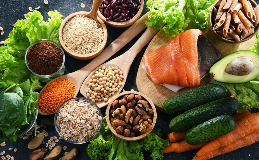
     Recent studies report favorable relationships between high vegetable and fruit intake and low sugary beverages and fats, implying a link between healthy diets and metabolically healthy overweight or obesity. The DASH diet, which contains more vegetables, fruits, whole grains, legumes, seeds, and low-fat-type dairy foods, has been researched in adolescents, but disputed findings call for more research.

    About the study

    In the present cross-sectional study, researchers explored the metabolic impact of DASH diets among overweight and obese adolescents.

    The team surveyed 203 adolescents aged 12 to 18 years with overweight or obesity status, as determined by body mass index (BMI) values using the Quetelet formula. Eligible students did not follow weight-loss diets, had no endocrinal or genetic disease, and did not use vitamin or mineral supplements or medications that could alter their metabolic profiles. 

    The researchers obtained dietary intake data using standardized food frequency questionnaires (FFQs). They also collected data on anthropometric parameters such as circulating insulin, blood pressure, lipid profile, and fasting blood sugar. The team characterized DASH scores based on eight components, i.e., higher intake of low-fat dairy foods, whole grains, seeds, nuts, vegetables, fruits, and legumes, and lower consumption of sodium, sweetened beverages, and processed and red meats.

    The team obtained blood samples from all participants for biochemical analysis. They measured insulin levels by enzyme-linked immunosorbent assays (ELISA) and ascertained metabolic health status based on insulin resistance, determined using the International Diabetes Federation (IDF) and Homeostatic Model Assessment for Insulin Resistance (HOMA-IR) criteria.

    The researchers assessed physical activity levels using the Physical Activity Questionnaire for Adolescents (PAQ-A) and total calorie intake using the United States Department of Agriculture (USDA) food composition database. They used multivariate logistic regression to calculate the odds ratios (ORs) for the association between DASH diets and metabolic health, adjusting for age, gender, physical activity, socioeconomic status, and total calorie intake.

    Results

    The mean values for age and BMI of the study participants were 14 years and 27 kg/m2, respectively. Among participants, 79 (42 girls and 37 boys, 39%) suffered from metabolically unhealthy overweight or obesity (MUO) by the IDF definition, and 62 (32 girls and 35 boys, 33%) were MUO following the IDF and HOMA-IR guidelines. Using the IDF definition, MUO prevalence in the DASH diet’s highest tertile was lower compared to the lowest statistical tertile (10 vs. 67%). Using HOMA-IR guidelines yielded similar findings (10 vs. 61%).

    Individuals in the uppermost tertile of the DASH diet were more physically active, with higher HDL-c levels and lower blood pressure, fasting blood sugar, insulin, triglyceride, and HOMA-IR levels, compared to those in the lowest tertile. Confounder adjustment showed that individuals in the highest vs. lowest DASH tertile had 91% and 92% lower MUO odds using the IDF/IR (OR, 0.09) and IDF definition (OR, 0.08), respectively.

    The highest vs. lowest DASH adherence was associated with decreased odds of hyperglycemia, hypertriglyceridemia, insulin resistance, and low HDL cholesterolemia, with odds ratios of 0.1, 0.3, 0.1, and 0.3, respectively. Subgroup analysis by BMI and sex indicated that the association was more robust among females (OR, 0.02) than males (OR, 0.09). The DASH diet likely improves metabolic health by lowering inflammation because of its high fiber, antioxidants, potassium, magnesium, and low salt content.

    Conclusions

    Overall, the study findings showed that increased adherence to the DASH diet was related to decreased probabilities of metabolic disease conditions among Iranian adolescents, particularly overweight girls. The study also found that adhering to the DASH diet reduced the risk of hypertriglyceridemia, hyperglycemia, insulin resistance, and low HDL cholesterol. Further research, including prospective surveys, could validate the study findings.

    The inverse association observed between the DASH dietary pattern and the MUO category in the current study indicates that adolescents must increase the intake of health-associated DASH components, including fruits, vegetables, legumes, low-fat-type dairy foods, whole grains, seeds, and nuts while limiting the consumption of unhealthy foods such as processed and red meats, sweetened beverages, and salt to improve dietary quality and decrease the metabolic disease burden.

    [ad_2]

    Source link

  • Spirulina shows promise in battling heart disease and diabetes

    Spirulina shows promise in battling heart disease and diabetes

    [ad_1]

    In a recent study published in the journal Nutrients, a team of Italian researchers reviewed clinical and experimental findings from recent studies to understand the therapeutic contributions of Spirulina, also called blue-green cyanobacteria, in managing cardiovascular disease and its risk factors.

    Study: Beneficial Effects of Spirulina Supplementation in the Management of Cardiovascular Diseases. Image Credit: baibaz / ShutterstockStudy: Beneficial Effects of Spirulina Supplementation in the Management of Cardiovascular Diseases. Image Credit: baibaz / Shutterstock

    Background

    Although Spirulina has recently gained popularity as a ‘superfood’ because of its high nutritional content, the use of microalga in diet dates back to the ancient times of the Aztecs in Mexico. Spirulina is also known as blue-green cyanobacteria and are microscopic, photosynthesizing, filamentous microalgae of the genus Arthrospira, with A. plantensis and A. maxima being the two species most commonly used for their therapeutic and nutritional value.

    They grow in the tropics, in alkaline lakes with high bicarbonate and carbonate salt concentrations, although they have been known to survive in extremely cold temperatures. Spirulina is considered a ‘superfood’ because 60% to 70% of its dry weight is composed of protein, while it is also abundant in minerals, vitamins, carbohydrates, phycocyanin, carotenes, and fatty acids. As a nutraceutical, it has been added to various types of foods, including sports supplements and baby foods, while the pharmaceutical industry has popularized it in the form of capsules, dehydrated powders, and tablets.

    Therapeutic effects of Spirulina

    Research indicates that Spirulina exhibits a wide range of therapeutic effects such as anti-inflammatory, antidiabetic, antioxidant, hypolipidemic, and neuroprotective properties. The antioxidant properties are attributed mainly to the pigments phycocyanin, β-carotene, diatoxanthin, and diadinoxanthin found in Spirulina.

    Given its hypolipidemic and antioxidant properties, supplementation with Spirulina could be beneficial in lowering the risk of cardiovascular disease. Furthermore, diabetes, along with dyslipidemia and hypertension, is one of the risk factors for cardiovascular disease. Therefore, the present review examined how the cumulative health benefits of Spirulina could lower the overall risk of cardiovascular disease, which continues to be one of the major causes of mortality across the globe.

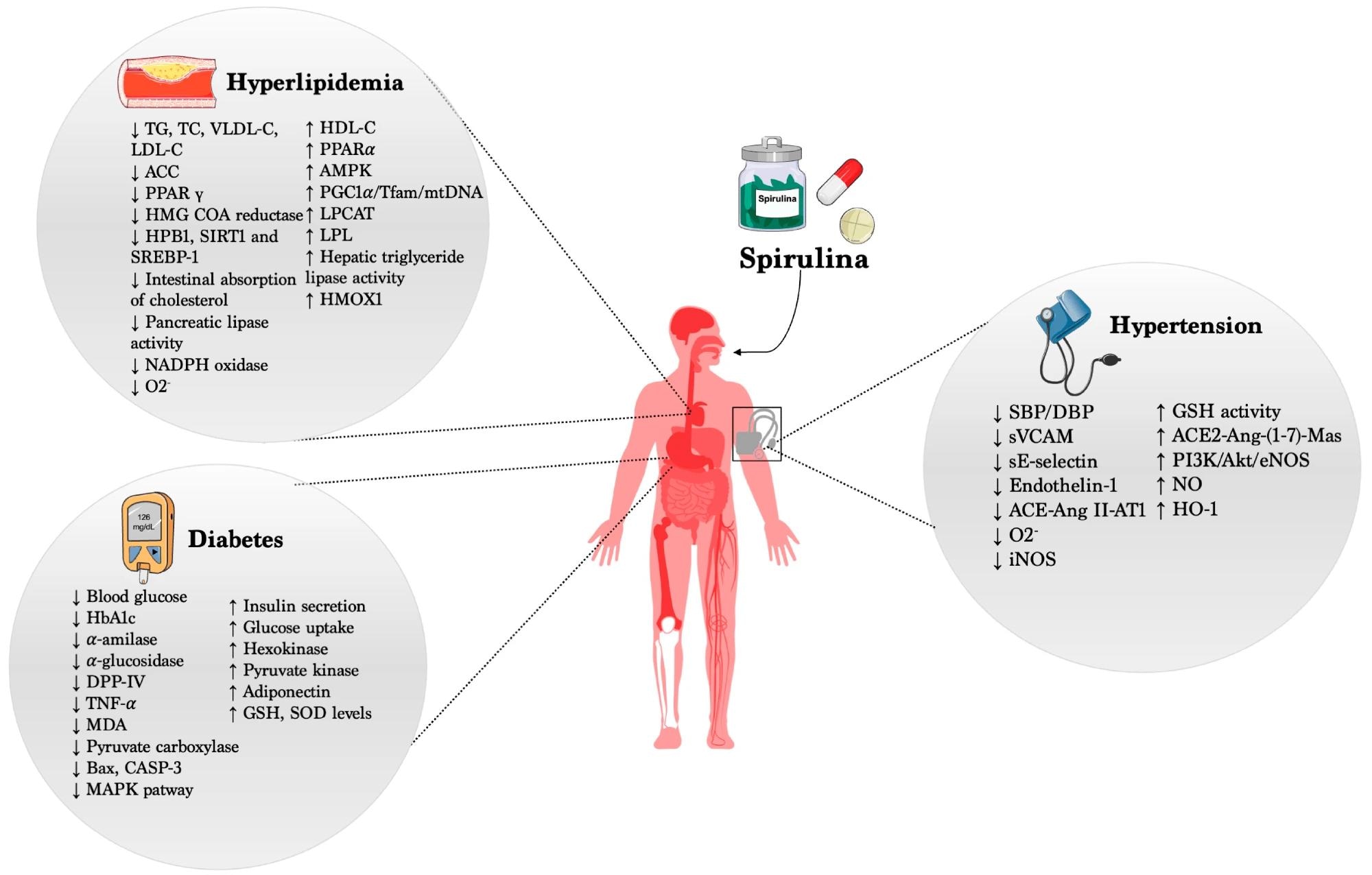
    Beneficial effects of Spirulina in CVDs.Beneficial effects of Spirulina in CVDs.

    Spirulina and hypertension

    The impact of Spirulina in lowering the risk of hypertension and stroke has been studied extensively in clinical trials, and the findings from these studies have shown that daily consumption of Spirulina, even added to foods such as salad dressing, significantly reduced the diastolic and systolic blood pressure.

    Consumption of Spirulina in the form of nutraceutical tablets also showed similar hypotensive results. Furthermore, animal studies using hypertensive rat models have shown that the high silicon content of Spirulina could be responsible for improving the elasticity of the arterial walls, along with angiotensin-converting enzyme-inhibiting properties that result in hypotensive effects.

    Antidiabetic effects of Spirulina

    Diabetes mellitus increases the risk of cardiovascular events such as heart failure, myocardial infarction, stroke, and peripheral vascular disease due to the micro- and macrovascular consequences of hyperglycemia. Cellular membrane integrity is also impacted by hyperglycemia, causing the peripheral tissues and liver to become insulin-resistant, increasing the generation of reactive oxygen species.

    In comparison to metformin, which is the standard treatment for hyperglycemia during diabetes, supplementation with Spirulina is believed to not only lower the levels of circulating glucose but also have a positive impact on lipid metabolism, which is linked to diabetes. The hypoglycemic and hypolipidemic properties of Spirulina are believed to have a cumulative effect in decreasing the risk of cardiovascular disease.

    The review discussed various clinical trials and studies using animal models of diabetes mellitus that have investigated the hypoglycemic properties of Spirulina and compared its efficacy in lowering blood sugar levels with that of metformin.

    While the mechanism through which Spirulina impacts blood glucose levels is not yet fully understood, the researchers believe that it could be influencing the secretion of insulin from the β-cells in the islets of Langerhans in the pancreas or further downstream, facilitating the transport of glucose from blood to all the peripheral tissue.

    Hyperlipidemia and Spirulina

    Spirulina has also demonstrated hypolipidemic properties by lowering the concentrations of low-density-lipoprotein cholesterol and triglycerides in the plasma while increasing the levels of high-density lipoprotein cholesterol, with the beneficial effects not being dose-dependent or toxic at high concentrations.

    Studies in animal models and overweight or obese human participants have reported significant benefits of Spirulina supplementation in lowering triglyceride levels, either as food additives or as nutraceutical pills or tablets. Spirulina was also found to be beneficial as an adjunct therapy to metformin in overweight diabetes patients.

    Conclusions

    Overall, this comprehensive review reported that consumption of Spirulina, either as an additive to regular foods or as a nutraceutical supplement, had numerous potential benefits, such as hypoglycemic, antioxidant, and hypolipidemic effects. However, the dosage and timing of Spirulina supplementation need to be standardized for optimal benefits in lowering the risk of cardiovascular disease.

    In conclusion, based on these data, more rigorous studies should be planned in the future aiming to address these critical questions, putting the foundations for developing a common guideline on “how and when” to use Spirulina.

    Journal reference:

    • Prete, V., Abate, A. C., Pietro, D., Lucia, D., Vecchione, C., & Carrizzo, A. (2024). Beneficial Effects of Spirulina Supplementation in the Management of Cardiovascular Diseases. Nutrients, 16(5). DOI: 10.3390/nu16050642, https://www.mdpi.com/2072-6643/16/5/642

    [ad_2]

    Source link