Tag: Male Infertility

  • The complex relationship between obesity and male reproductive function

    The complex relationship between obesity and male reproductive function

    [ad_1]

    In a review article published in the journal Molecular Aspects of Medicine, authors have analyzed current evidence on the impact of obesity on the male reproduction system.

    They have thoroughly discussed molecular mechanisms responsible for male infertility in obese or overweight individuals.

    Study: Obesity and male fertility disorders. Image Credit: Shidlovski/Shutterstock.comStudy: Obesity and male fertility disorders. Image Credit: Shidlovski/Shutterstock.com

    Background

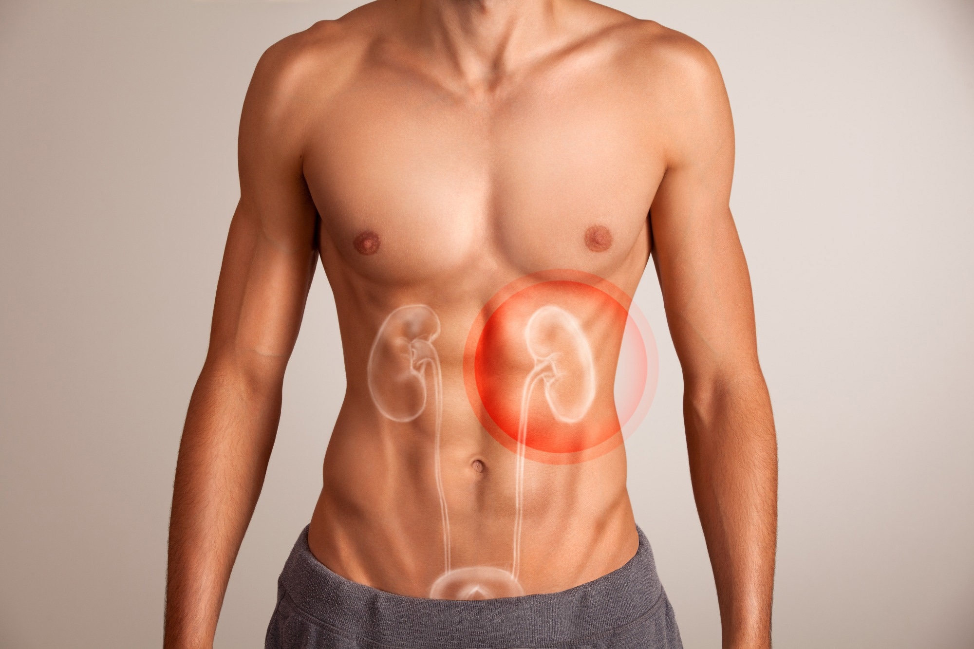
    Obesity is considered to be one of the major causes of male infertility globally. An increased body weight is known to impair testicular development and function starting from prenatal age. Moreover, recent evidence shows that obesity can significantly reduce sperm parameters in adults.

    According to the World Health Organization, more than one billion people are living with obesity worldwide.

    With an ever-increasing prevalence of obesity in the global population, it has become necessary to precisely understand the relationship between obesity and male reproductive dysfunctions.

    Impact of obesity on male infertility

    A body mass index (BMI) of 30kg/m2 or more is defined as obesity. The body fat percentages of more than 25% in men and 30% in women are also described as obesity, which are often poorly correlated with BMI in the context of obesity diagnosis.

    Studies conducted on couples with an obese male partner have shown that male obesity can significantly increase the risk of infertility. However, studies investigating the direct effect of obesity on conventional sperm parameters have produced mixed or conflicting results.

    Studies involving couples undergoing fertility-related treatments have shown that obesity does not have any significant impact of obesity on sperm count, morphology, and motility.

    In contrast, findings of meta-analyses have indicated that obesity can reduce total sperm count, sperm concentration, semen volume, sperm vitality, and total sperm motility.

    One most recent meta-analysis, including studies following the 2010 WHO manual for sperm parameter analysis, has shown that obesity can significantly reduce total sperm count, sperm concentration, and sperm progressive, and total motility.

    This study has also shown that obesity affects overall sperm quality through the induction of hypogonadism (reduced production of male sex hormones).

    Regarding sperm bio-functional parameters, evidence indicates that obesity can lead to sperm DNA fragmentation and reduced mitochondrial membrane potential. These parameters might be associated with reduced sperm quality and motility.

    Regarding serum hormone levels, evidence indicates that obesity can reduce testosterone and sex hormone-binding globulin levels and increase estrogen levels.

    Mechanisms involved in obesity-related male infertility

    One of the potential factors responsible for hypogonadism is excess visceral fat deposition. Hypogonadism is associated with excessive conversion of testosterone into 17ß-estradiol by adipocytes, which further promotes the secretion of sex hormone-binding globulin by the liver.

    This protein can bind to testosterone and inhibit its biological functions. Furthermore, low blood levels of testosterone due to hypogonadism can trigger fat accumulation in the body.

    A reduced testosterone can lead to impaired proliferation and differentiation of Sertoli cell (somatic cells of the testis) and spermatogonial stem cells, negatively affecting spermatogenesis or sperm cell production.

    A high blood estrogen level due to hypogonadism can also negatively affect male reproductive system by inhibiting the release of lactate (an essential substrate) to germ cells, as well as by impairing the integrity of blood-testis barrier.

    Increased visceral fat can induce insulin resistance, reducing sex hormone-binding globulin secretion and subsequent induction in free estrogen levels. Free estrogen and inflammatory mediators produced due to insulin resistance can negatively affect the hypothalamic-pituitary-gonadal axis.

    Insulin resistance can also interfere with follicle-stimulating hormone signaling pathways at the testicular level, leading to impaired spermatogenesis.

    An increased insulin level in the blood can impair the growth, proliferation, metabolism, and survival of testicular cells, which in turn can impair male reproductive functions.

    Obesity-related low-grade chronic inflammation can influence male reductive functions in many ways. Increased production of pro-inflammatory cytokines can regulate Leydig cell function and subsequently reduce testosterone production.

    Obesity-related chronic inflammation can also increase the production of free radicals, leading to sperm DNA damage and reduced sperm quality.

    Obesity can affect the levels of adipokines produced by fat cells. These adipokines, including adiponectin, chemerin, leptin, resistin, and visfatin, play vital roles in modulating the immune, metabolism, and reproductive systems.

    Leptin is the most studied adipokine that regulates food intake, reproductive functions, and proinflammatory immune responses. A high-fat diet is known to induce leptin resistance in obese people. Highly increased blood levels of leptin characterize this condition.

    An increased leptin level can reduce lactate dehydrogenase activity and activate the PI3K/AKT/mTOR signaling pathway, leading to reduced lactate production by Sertoli cells and impaired nutritional support to germ cells.

    Sirtuins are NAD+-dependent deacetylases that play a role in modulating spermatogenesis. Sirtuin 1-knock-out mice have been found to have reduced sperm count and increased sperm DNA fragmentation.

    Gut hormones, such as ghrelin, Glucagon-like peptide-1, and glucose-dependent insulinotropic polypeptide, secreted by gastrointestinal tract cells, play important roles in regulating lipid and glucose metabolism. An increased secretion of these hormones can lead to impaired functioning of Sertoli cells and Leydig cells.

    The gut microbiota provides essential nutrients and factors required for testicular function. Any alteration in gut microbiota composition and function can lead to local inflammation, which in turn can cause Leydig cell death, disrupted blood-testicular-barrier, and abnormal spermatogenesis.

    Sperm RNAs, including messenger RNAs (mRNAs), long non-coding RNAs (lncRNAs), micro RNAs (miRNAs), Piwi-interacting RNAs (piRNAs), and transfer of RNA-derived small RNAs (tsRNAs), play vital roles in spermatogenesis, fertilization, and embryo development.

    Certain sperm miRNAs have been found to induce inflammatory responses and impair iron homeostasis, glucose metabolism, meiotic recombination, fertilization, and sperm maturation and motility.

    [ad_2]

    Source link

  • Infertile men more likely to have impaired kidney function, study says

    Infertile men more likely to have impaired kidney function, study says

    [ad_1]

    In a recent study published in the journal Scientific Reports, researchers investigated the association between male infertility and renal function. They used a large cohort comprising 11,602 participants (5,494 childless men and 6,108 fathers). They found that childless men were likelier to have a low estimated glomerular filtration rate (< 60 ml/min/1.73m2) and dipstick proteinuria independent of age, socioeconomic status, or traditional renal risk factors such as hypertension, diabetes, and metabolic function.

    These findings contribute novel evidence to a growing body of literature highlighting the role of male fertility in altering the risk or pathology of several non-communicable chronic diseases. Study outcomes suggest that men with reduced fertility may represent a population in need of routine kidney function evaluation.

    Study: Prevalence of impaired renal function among childless men as compared to fathers: a population-based studyImage Credit: BigBlueStudio / ShutterstockStudy: Prevalence of impaired renal function among childless men as compared to fathers: a population-based study. Image Credit: BigBlueStudio / Shutterstock

    The non-reproductive impacts of male infertility

    Infertility has long been down upon in cultures across the world, with many traditions considering a person’s inability to reproduce an ill omen, curse, or divine punishment. Recent research suggests that while male infertility-associated ill omens are yet to be discovered, the dangers of the condition may extend far beyond just reproduction or the reproductive system. Previous studies have identified infertile men at heightened risk of ischemic disease and diabetes.

    Male childlessness, a commonly invoked proxy for the much-harder-to-elucidate male infertility, has been associated with cardiovascular risk factors, including hyperlipidemia, hyperglycemia, and hypertension, with these individuals being much more likely than fertile men to consume prescription metabolic syndrome and hypertension medication. Unfortunately, research extending these investigations into renal evaluations remains lacking. The current study aims to add to the work of Eisenberg et al., hitherto the sole publication exploring the association between male infertility and renal disease.

    About the study

    The present study aims to evaluate if male childlessness (herein a proxy for male in- or subfertility) is associated with impaired renal function (eGFR < 60 mL/min/1.73 m2) or dipstick proteinuria. The study sample cohort was derived from the Malmö Preventive Project (MPP), a long-term, longitudinal, population-based sample group established in the 1970s, with detailed information on creatinine levels and urine dipstick results essential from the measurement of glomerular filtration rate (eGFR) and dipstick test for protein in the urine. MPP also records and maintains participants’ fatherhood status, further meeting the current study’s requirements.

    MMP data revealed 22,444 men between the ages of 25 and 63 enrolled between 1974 and 1994. Data collection included socioeconomic, demographic, lifestyle, and medical history records obtained from participants via a generalized questionnaire. Experimental assays and characterizations were carried out using participant-submitted urine samples and author-recorded physical examinations. The Swedish Tax Agency Statistics (STAS) provided data on the number of children per participant at baseline, with each record being associated with a unique personal identification number.

    Jaffe’s alkaline picrate assay and the CKD-EPI creatinine formula (2021) were used to quantify the concentrations of serum creatine and eGFR, respectively. Proteinuria was investigated using a semi-quantitative urine dipstick test. Finally, two logistic regression models were used to elucidate any statistical associations (expressed as crude odds ratios [ORs]) between male childlessness and eGFR and/or dipstick proteinuria. The first model took into consideration men’s marital, socioeconomic, and occupational status, while the second adjusted for previously reported renal risk factors such as age, marital status, smoking status, and CKD-associated comorbidities.

    Study findings and conclusions

    “In this population-based study we found that childless men, as compared to fathers, are more prone to show signs of renal disease as decreased eGFR and dipstick proteinuria. The likelihood of dipstick proteinuria, with or without concomitant decrease in eGFR, remained statistically significant even after adjustment for comorbidities and traits known to be linked to impaired renal function.”

    Of the 22,444 participants initially scouted from the MPP cohort, the exclusion of individuals with incomplete data and those above the age of 45 years resulted in a final cohort size of 11,602 individuals – 47.3% (n = 5494) were childless. eGFR evaluations revealed that childless men (3.1%) were more likely to present with an eGFR < 60 mL/min/1.73 m2 than their fertile counterparts (2.3%). These findings were consistent with those found during dipstick proteinuria evaluations (7.1% in childless men and 4.9% in fathers). Surprisingly, these associations remained significant in all but one, including the logistic regression model.

    These findings highlight the heightened renal disease risk of childless (in- or sub-fertile) men when compared to Fathers, suggesting that the former is a target population from frequent renal monitoring, potentially presenting a novel tool in the clinicians’ future arsenal against renal diseases.

    [ad_2]

    Source link

  • Research identifies cancer risk patterns in families of men with fertility problems

    Research identifies cancer risk patterns in families of men with fertility problems

    [ad_1]

    For the first time researchers have identified patterns of risk for several different types of cancer in men with fertility problems and their families.

    The study, which is published today (Thursday) in Human Reproduction, one of the world’s leading reproductive medicine journals, found that families of men who have very few or no sperm in their semen have a higher risk of developing cancer, including developing cancer at younger ages, compared to families of fertile men.

    The risk and the type of cancer varied greatly depending on whether the men had low numbers of sperm (oligozoospermic) or none (azoospermic), with several cancers identified in distinct clusters of families.

    The researchers, led by Dr Joemy Ramsay, assistant professor at the University of Utah, Salt Lake City, USA, hope their findings will improve their understanding of the biological mechanisms involved in both cancer and infertility. This would enable doctors to make more accurate predictions of risk of cancer for men with fertility problems and their families, and to improve the counselling that could be offered to them.

    Previous research has shown that male infertility is linked to an increased risk of cancer in the men and their families, but the results have been inconsistent. Increased risks and the types of cancer varied considerably between family groups and depending on whether the men were oligozoospermic or azoospermic.

    In this study, we wanted to describe the extent to which patterns of cancer risk vary between families of subfertile men, and whether this risk is seen in all families or is driven by a small subset of families, akin to the way mutations in the BRCA gene increase the risk of breast cancer in families that carry this mutation. By identifying families with similar patterns of cancer, we may be able to discover factors that are involved in both infertility and cancer.”


     Dr Joemy Ramsay, Assistant Professor, University of Utah, Salt Lake City

    Dr Ramsay and colleagues took results from semen analyses carried out between 1996 and 2017 from 786 men attending fertility clinics in Utah, and they matched them with information from 5674 fertile men in the general population who had at least one child to ensure they were fertile. Among the men with fertility problems, 426 were azoospermic and 360 were severely oligozoospermic (with less than 1.5 million sperm per millilitre of semen).

    The researchers collected information on first, second and third degree relatives using the Utah Population Database. Cancer diagnoses were identified from the Utah Cancer Registry.

    “We simultaneously assessed the risk for multiple types of cancer within each family and then we performed a cluster analysis to find groups of families with similar patterns of risk for multiple cancers,” said Dr Ramsay. “This is the first study to describe these multicancer patterns in families of subfertile men.”

    When the researchers looked at all families of azoospermic men, they saw a significantly increased risk of five cancers: bone and joint cancer (156% increased risk), soft tissue cancers such as sarcomas (56% increased risk), cancers of the womb (27% increased risk), Hodgkin Lymphomas (60% increased risk), and thyroid cancers (54% increased risk).

    Families of the severely oligozoospermic men had a significantly increased risk of three cancers: colon cancer (16% increased risk), bone and joint (143% increased risk), and testicular cancer (134% increased risk). The researchers also found a 61% decreased risk of oesophageal cancer (cancer of the gullet).

    The researchers found the risk of cancer and the types of cancer varied greatly among the families of men with fertility problems, both by type of subfertility and also within subfertility type. This could explain the inconsistent associations between subfertility and cancer in previous studies. For example, the study found an increased risk of testicular cancer in only a third of the clusters of families of oligozoospermic men, but the increased risk ranged from four- to 24-fold depending on family cluster.

    Among the families of azoospermic men, the researchers identified 13 clusters of families. One cluster, which included the majority of the families, had a risk of cancer that was similar to that in the general population. However, the remaining 12 clusters all had increased risks of developing at least one type of cancer. Among the families of oligozoospermic men, there were 12 distinct clusters and all of them had an increased risk for at least one type of cancer.

    “Our study identified several unique patterns of cancer risk in families of men with poor fertility. When family members share cancer risk patterns, it suggests that they have genetic, environmental, or health behaviours in common. Genetic and environmental exposures can also act together to increase cancer risk. By identifying which groups of families have similar cancer risk patterns we can improve our understanding of the biological mechanisms of both cancer and infertility,” said Dr Ramsay. “It will help us to assess the risk of cancer for families and provide improved patient counselling.”

    The researchers have carried out genetic sequencing studies to look for specific genetic mutations that may be driving the associations between subfertility and cancer seen in this study.

    Strengths of the study include the use of data from population registries for family structure, cancer diagnosis and subfertility. Limitations include lack of semen measures for the fertile men, lack of information on other health conditions, lifestyle risk factors, such as smoking and body mass index, and exposure to environmental risk factors among the subfertile men; and, finally, that the men with fertility problems in this study were all seen at a fertility clinic and, therefore, represent a subset of the overall population of subfertile men who had the socioeconomic means to be evaluated by a doctor.

    Source:

    Journal reference:

    Ramsay, J. M., et al. (2024) Describing patterns of familial cancer risk in subfertile men using population pedigree data. ​​ Human Reproduction. doi.org/10.1093/humrep/dead270.

    [ad_2]

    Source link

  • “Laboratory testicles” could solve male infertility mysteries

    “Laboratory testicles” could solve male infertility mysteries

    [ad_1]

    The testis is responsible for sperm production and testosterone synthesis. Abnormalities in testis development and function lead to disorders of sex development (DSD) and male infertility. Currently, no in vitro system exists for modeling the testis.

    Dr. Nitzan Gonen, a researcher specializing in the process of fetal sex determination, together with research students Aviya Stopel, Cheli Lev and Stav Dahari, has succeeded in creating “laboratory testicles” that may significantly advance understanding of the mechanisms involved in sex determination and provide solutions for male infertility, which affects one in 12 men worldwide.

    The artificial testicles produced in Dr. Gonen’s lab at the Goodman Faculty of Life Sciences and the Institute of Nanotechnology and Advanced Materials at Bar-Ilan University, are testis organoids – tiny, artificial organs produced from real mouse testis. Development of organoids has greatly advanced in the last decade with the realization that two-dimensional cellular sample in vitro cannot mimic the behavior of an entire organ. Today, organoids of the brain, kidneys, intestines, and other organs have already been produced. The testicular organoids created by Gonen’s group closely simulate a natural testicle.

    The artificial testicles were cultured from immature testicular cells sampled from neonatal mice. The research team realized the procedure was a success when they identified tubule-like structures and cellular organization highly resembling that of the in vivo testis. These tubular structures parallel the multiple seminiferous tubules present in the natural testicle, where the sperm is produced.

    The organoids were successfully cultured in vitro for nine weeks. This is considered a long period of time and can, theoretically, be enough time to complete the process of sperm production and hormone secretion. In mice this takes 34 days, so the relatively long lifespan of the organoids may allow these processes to occur in vitro. Dr. Gonen doesn’t yet know if the existing model will actually produce sperm cells, but the laboratory team has already noticed signs of the beginning of meiosis, a process in which gametes are produced. Gametes are reproductive cells, in this case sperm cells with half the number of chromosomes as in a normal cell, that “await” for the completion of the other half from another gamete, in this case an egg, upon fertilization.

    Organoids usually resemble organs in the embryonic stage. In this case the researchers created conditions that allowed the organoid to mature in the laboratory and showed that even testicles grown from embryonic cells can develop and grow clear sperm tubes. The team was unsuccessful in its attempt to grow organoids from adult mice testis.

    Artificial testicles are a promising model for basic research on testicle development and function, which can be translated into therapeutic applications for disorders of sexual development and infertility.”


    Dr. Nitzan Gonen, Researcher

    In the future she plans to produce organoids using human samples. A testis produced from human cells, for example, could help children being treated for cancer, which may impair their ability to produce functional sperm. As children are too young to produce their own sperm, these samples can be frozen and used in the future to have children. Gonen’s vision is to grow testes organoids from biopsies of children with cancer and hopefully grow fertile sperm in vitro.

    The finding of this study were recently published in the International Journal of Biological Sciences.

    Source:

    Journal reference:

    Stopel, A., et al. (2024). Towards a “Testis in a Dish”: Generation of Mouse Testicular Organoids that Recapitulate Testis Structure and Expression Profiles. International Journal of Biological Sciences. doi.org/10.7150/ijbs.89480.

    [ad_2]

    Source link

  • Mediterranean diet linked to better semen quality, study finds

    Mediterranean diet linked to better semen quality, study finds

    [ad_1]

    In a recent systematic review published in the journal Frontiers in Nutrition, researchers from Spain investigated the effect of the Mediterranean diet on the quality of semen in men of reproductive age. They found that adherence to the Mediterranean diet could improve male reproductive health by positively influencing semen quality.

    Study: Influence of the Mediterranean diet on seminal quality—a systematic review. Image Credit: leonori / ShutterstockStudy: Influence of the Mediterranean diet on seminal quality—a systematic review. Image Credit: leonori / Shutterstock

    Background

    Fertility research has surged as about 15% of the global population, comprising 70 million reproductive-age couples, suffer from infertility across geographies and income groups. The World Health Organization investigated infertility across 25 nations and found that male factors contributed to about half of the couples, challenging the older notion that infertility is solely a female concern. The major causes of male infertility are impaired spermatogenesis, idiopathic causes, endocrine disorders, and altered sperm motility. Clinically investigating the cause of male infertility warrants thorough history, physical examination, and semen analysis. Additionally, non-modifiable factors such as genetics and age, alongside modifiable factors such as diet and lifestyle, are also known to influence male fertility.

    Popular for its potential health benefits, the Mediterranean diet demonstrates positive associations with male reproductive health, including improved semen quality attributed to its anti-inflammatory and antioxidant properties. Evidence suggests that optimal adherence to this diet may lower the risk of various chronic diseases and potentially enhance male fertility by addressing metabolic factors affecting sperm function. Therefore, researchers in the present study conducted a systematic review to understand the effect of the Mediterranean diet on the quality of semen in men of reproductive age.

    About the study

    Data for this review were gathered through electronic searches in PubMed, the Cochrane Library, Scopus, and Web of Science, as well as reference lists. Full-text articles were screened based on criteria evaluated independently by two authors. The quality of studies was assessed using appropriate tools such as the Crombie criteria, the Newcastle-Ottawa Scale, and the PEDro scale. Interrater reliability was evaluated using Cohen’s kappa statistic.

    Inclusion criteria were open-access articles published in English or Spanish between 2012 and 2022, focusing on men aged 18 to 55. The exclusion criteria excluded unrelated articles, systematic reviews, meta-analyses, conference proceedings, and studies of specific medical conditions. A total of 10 studies met the inclusion criteria, including a total of 2,032 participants across various countries, primarily Spain. Study designs included cross-sectional, cohort, case-control, and randomized controlled trials. Data extraction focused on variables such as nutrition status, diet, and semen quality assessment techniques. After extraction, data were grouped based on assessment techniques and associations between semen quality and the Mediterranean diet.

    The quality of included studies was evaluated using the Cochrane Collaboration Risk of Bias tool, evaluating domains such as randomization, intervention deviations, missing outcome data, outcome measurement, selection of reported results, and other biases, classifying each item as having a low, high, or unclear risk of bias.

    Nutrition status was evaluated using weight, height, body mass index (BMI), and waist circumference, while adherence to the Mediterranean diet was assessed using validated food frequency questionnaires or specific scores. Semen quality was primarily assessed through sperm concentration, motility, morphology, volume, total antioxidant capacity, and hormone levels. Some studies also explored chromosome stability, DNA (short for deoxyribonucleic acid) fragmentation, global sperm DNA methylation, microRNA (short for micro ribonucleic acid) expression, and reactive oxygen species.

    Results and discussion

    Six out of 10 studies demonstrated a positive association between semen quality and following the Mediterranean diet, particularly in parameters like sperm concentration, motility, and total sperm count. In three of them, men with higher adherence levels showed significantly higher semen quality. However, two studies found no significant association between Mediterranean diet adherence and semen quality.

    The present study is the first to comprehensively investigate the positive link between the Mediterranean diet and semen quality. However, the study is limited by its small sample size, observational design, and low generalizability, with evidence quality rated as very low to moderate using the GRADE (short for Grading of Recommendations, Assessment, Development, and Evaluation) system. Risk of bias assessment for randomized controlled trials indicated one trial with low risk and another with bias concerns related to intervention deviations and result evaluation. Further research with larger sample sizes and randomized controlled trials is warranted to confirm these findings.

    Conclusion

    In conclusion, healthy dietary habits, particularly adherence to the Mediterranean diet, are found to be associated with improved semen quality in men of reproductive age. This diet, rich in polyunsaturated fatty acids, antioxidants, and anti-inflammatory substances, may reduce oxidative stress and protect against its adverse effects on sperm. These findings highlight the importance of dietary counseling for couples planning a pregnancy or undergoing assisted reproductive technology. However, further research is necessary to explore the relationship between diet and semen quality, informing strategies for improved fertility and health outcomes.

    [ad_2]

    Source link