Tag: Medical research

  • The future of precision cancer therapy might be to try everything

    The future of precision cancer therapy might be to try everything

    [ad_1]

    The blood cancer had returned, and Kevin Sander was running out of treatment options. A stem-cell transplant would offer the best chance for long-term survival, but to qualify for the procedure he would first need to reduce the extent of his tumour — a seemingly insurmountable goal, because successive treatments had all failed to keep the disease in check.

    As a last throw of the dice, he joined a landmark clinical trial. Led by haematologist Philipp Staber at the Medical University of Vienna, the study is exploring an innovative treatment strategy in which drugs are tested on the patient’s own cancer cells, cultured outside the body.

    In February 2022, researchers tried 130 compounds on cells grown from Sander’s cancer — essentially trying everything at their disposal to see what might work.

    One option looked promising. It was a type of kinase inhibitor that is approved to treat thyroid cancer, but it is seldom, if ever, used for the rare subtype of lymphoma that Sander had. Physicians prescribed him a treatment regimen that included the drug, and it worked. The cancer receded, enabling him to undergo the stem-cell transplant. He has been in remission ever since. “I’m a bit more free now,” says Sander, a 38-year-old procurement manager living in Podersdorf am See, Austria. ”I do not fear death any more,” he adds. “I try to enjoy my life.”

    His story is a testament to this kind of intensive and highly personalized drug-screening method, referred to as functional precision medicine. Like all precision medicine, it aims to match treatments to patients, but it differs from the genomics-guided paradigm that has come to dominate the field. Instead of relying on genetic data and the best available understanding of tumour biology to select a treatment, clinicians throw everything they’ve got at cancer cells in the laboratory and see what sticks.

    But what it sometimes lacks in elegance, it could make up for in results: in pilot studies, Staber and his colleagues found that more than half of people with blood cancer whose treatment was guided by functional drug testing enjoyed longer periods of remission compared with their experiences of standard treatments1,2. Large-scale testing of genome-directed approaches suggests that the techniques are very effective against some cancers, yet they benefit, at most, only around 10% of patients overall3. Staber and his group’s latest trial is the first to compare functional- and genome-guided approaches head-to-head alongside treatments directed by standard pathology and physician intuition.

    “That’ll be a very powerful study, and it will probably vindicate the utility of these functional assays,” says Anthony Letai, a haematologist at the Dana-Farber Cancer Institute in Boston, Massachusetts, and president of the Society for Functional Precision Medicine, a professional organization founded in 2017 to advance the field. And, if anecdotal reports serve as any indication, the try-everything tactic seems to bring about meaningful improvements, even when the genetic sequence of a tumour provides no actionable information, as was the case for Sander.

    Companies around the world are already offering these kinds of personalized drug testing service. But proponents of the strategy still have much to prove. Although the concept of screening a bunch of drugs seems simple, the methods used to culture cancer cells outside the body can be technically demanding, time-consuming and costly.

    The challenges are particularly acute for solid tumours, which live in complex environments inside the body; replicating those conditions is no easy feat. Researchers are trying wildly differing methods that range from growing tumour samples in mice and chicken embryos to cultivating carefully engineered organoids, and even the delivering infinitesimal amounts of various medicines to a tumour while it’s still in a patient.

    Figuring out what works and what is practical, with regard to cost and scale, won’t be easy. But momentum is growing, says Christopher Kemp, a cancer biologist at the Fred Hutchinson Cancer Center in Seattle, Washington. “This is a revolution. Patients are demanding this approach.”

    Behind the screen

    Down a long corridor, beyond a set of tangerine-coloured doors, lies the Vivi-Bank at the Medical University of Vienna. Short for ‘Viable Biobank’, the room is brimming with liquid-nitrogen dewars, each containing frozen lymphoma samples.

    When surgeons extract biopsies from cancerous lymph nodes, they usually immerse the tissue in formaldehyde to prepare for standard pathology analyses. That kills the cells, however, rendering them useless for functional testing. So, to enable drug screens, Staber and haematopathologist Ingrid Simonitsch-Klupp, who jointly oversee the Vivi-Bank, had to convince their surgical colleagues to change their ways, keeping the tissue alive and sending it quickly for processing and storage. “Fresh tissue is the most important thing,” Simonitsch-Klupp says.

    Some of that tissue arrives in Staber’s lab, where researchers break up the cells using a knife, forceps and a nylon strainer, creating a slurry to distribute across a 386-well plate. In each well, they test a different drug compound — chemotherapy agents, enzyme-targeted drugs, immune-modulating therapies and more. After a night of incubation, lab testing reveals which drugs are active against the cancer and which ones are not.

    A team of clinicians, known as a molecular tumour board, then uses this information to determine the most appropriate course of treatment for each patient.

    Several groups have reported success with this general approach. In a trial from the University of Helsinki, for example, researchers found that individualized drug screening of leukaemia cells provided informative results substantially faster than did genomic profiling, yielding impressive clinical responses as well4. Of 29 people with treatment-resistant acute myeloid leukaemia (AML), 17 responded to drug-screening-informed therapies and entered remission.

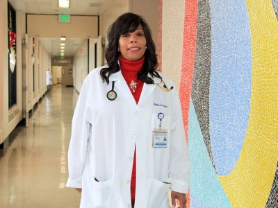
    Likewise, Candace Howard, a radiologist at the University of Mississippi Medical Center in Jackson, and her colleagues published a study last year showing that people with aggressive brain tumours live longer when their chemotherapy regimens are guided by lab testing than when their treatment is directed by a physician’s intuition alone5 — with lower annual health-care costs to boot6.

    A plate containing different cancer drugs and a graphical drug sensitivity testing result output

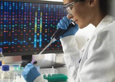
    Multi-well plates can be used to test the effectiveness of many cancer drugs at once.Credit: FIMM, University of Helsinki

    “It’s cheaper and it’s more effective,” says Jagan Valluri, a cell biologist at Marshall University in Huntington, West Virginia, who co-founded a company called Cordgenics, also based in Huntington, to commercialize the assay used in Howard’s trial.

    Functional drug testing is not a new idea. It was embraced by cancer researchers in the late twentieth century, but soon fell out of favour — largely owing to the limitations of assays at the time and a restricted repertoire of anti-cancer drugs. Technological improvements and an expanded pharmacopoeia have changed the picture. Yet, as with most lab-based testing systems, the necessary equipment can be expensive and requires trained personnel to operate it.

    That’s a big limitation according to Joan Montero, a biochemist at the University of Barcelona in Spain, because it hinders the broad implementation of functional precision drug testing, especially in low-resource settings. To address these challenges, Montero and his colleagues have been developing inexpensive and portable microfluidic devices for rapid, on-site testing of cancer cells7.

    Their microfluidic platform remains years away from practical use, however. And it might guide treatment only for certain types of cancer. That’s because protocols developed for tailoring therapies against blood cancers do not always work in solid tumours of the breast, lung, liver and other organ systems.

    Biopsies from solid tumours often yield lower cell counts, requiring extra steps to culture the cells before drug screening. Moreover, solid tumours have complex interactions with healthy cells in their surroundings, meaning that models should be more sophisticated.

    Growing pains

    The first challenge remains growing enough tumour tissue to test. David Ziegler, a paediatric neuro-oncologist at Sydney Children’s Hospital in Australia, had set out to perform individualized drug screens for around 1,000 children with high-risk cancers as part of the country’s Zero Childhood Cancer Program. But in pilot testing, he and his team discovered that, after several days under lab conditions, up to one-fifth of the patient samples either contained no cancer cells at all, or the cancer cells were being outcompeted by normal, healthy cells8. The researchers quickly learnt to check cultures for tumour cells — through imaging, cellular analysis or genetic profiling — before testing them against drugs.

    Cell cultures from solid tumours can, in principle, be subjected to the same kind of testing used for blood cancers. But an increasing number of research teams are crafting elaborate structures, known as organoids, to test. These patient-derived 3D tissue models — made by growing tumour samples in specialized scaffolds over the course of several weeks — are designed to replicate the intricate tissue architecture of a tumour, thereby offering a more accurate representation of the cancer that physicians are looking to treat.

    “We want to put the tumour cells in an environment that’s as close [as possible] to how they were growing in the body,” says Alice Soragni, a cancer biologist at the University of California, Los Angeles.

    The process can add weeks to the timeline for obtaining drug sensitivity data. But the extra effort and time investment is worth it, says Carla Grandori, co-founder and chief executive of SEngine Precision Medicine in Bothell, Washington.

    In clinical validation studies, Grandori and her SEngine colleagues found that the drug-screening results using organoids aligned with patient outcomes with around 80% accuracy. Those findings are not yet published, but the company — which counts Kemp among its founders — has put out case reports over the past year describing people with difficult-to-treat cancers who, after seemingly running out of treatment options, found unexpectedly effective remedies through organoid drug testing9,10.

    Heidi Gray, a gynaecological oncologist at the University of Washington Medical Center in Seattle, treated one of these patients, a woman with ovarian cancer. “Her response was definitely one of the best I’ve seen,” she says. The drug they tried is generally used to treat leukaemia, but it helped to beat back the woman’s ovarian tumour for more than a year, allowing her to travel and enjoy precious time with loved ones before ultimately succumbing to the disease. “We profoundly improved her quality of life,” Gray says, “and that would not have happened without the knowledge provided by this test.”

    Model of efficiency

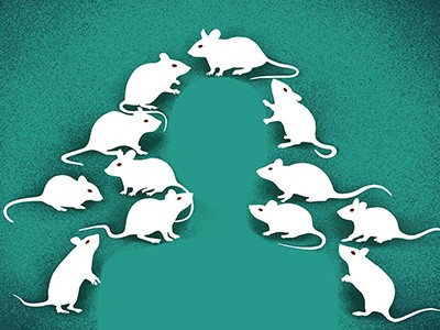
    In the hope of testing drugs against even more realistic cancer systems, some researchers have opted to study mice implanted with fresh tumour specimens, a model system known as a patient-derived xenograft.

    These personalized ‘avatars’ were once heralded as the next big thing in cancer care. But it soon became evident that many tumours do not grow in mice, that drug screening in xenografts takes too long to provide timely recommendations and that the cost of the approach — often exceeding US$50,000 — is more than most patients and health-care systems can bear.

    “It was too slow, too expensive and not robust enough,” says David Sidransky, an oncologist at Johns Hopkins University School of Medicine in Baltimore, Maryland, and a co-founder of Champions Oncology, a leading developer of xenograft models, based in Hackensack, New Jersey.

    Albert Manzano and Joan Montero work at a fume hood on a microfluidic device used to predict cancer therapy response

    Joan Montero (standing) and his colleagues are developing a low-cost microfluidic device.Credit: University of Barcelona

    Although some drug companies continue to use xenografts for research, and some oncologists think that there are certain situations in which they can inform patient care, for the most part, researchers have moved away from mice for functional testing in the clinic. Some have moved on to other living systems.

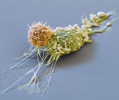
    One such alternative comes from cancer biologist Hon Leong and his colleagues at Sunnybrook Hospital in Toronto, Canada, who devised a system for screening drugs on tumour biopsy samples cultivated on developing chicken embryos. The approach is both rapid and inexpensive, says Leong, allowing researchers to assess different drug options in a matter of weeks rather than the months required for mice.

    In ongoing trials focused on advanced breast and kidney cancers that have spread to other parts of the body, Leong and his team have successfully used the chicken-embryo system to identify individuals who would benefit from immune therapies. These are among the most effective cancer treatments today, and a drug class that few other avatar systems can accurately assess, says Leong.

    Another approach comes from Ross Cagan, a developmental biologist at the University of Glasgow, UK, who uses genomic sequencing and genetic engineering to recreate the unique characteristics of a patient’s tumour in a custom-made fruit fly. This involves introducing mutated forms of cancer-promoting genes or incorporating sequences that restrict cancer-suppressing genes — generally between 5 and 16 alterations in total. Feeding the flies with food containing various medications can then reveal therapeutic regimens that suppress cancer growth, either by acting directly on tumour cells or by influencing the animal’s biology in ways that indirectly impede cancer progression.

    This is how Cagan and his colleagues identified a new three-drug cocktail — consisting of a lymphoma treatment, a blood-pressure medicine and an arthritis therapy — that, when administered to a man with a rare tumour of the salivary glands, helped to stabilize the cancer for a year11. In another case, involving a man with an aggressive form of colon cancer, the use of fly avatars guided the team to administer a melanoma drug alongside a bone-strengthening agent, resulting in notable tumour shrinkage and a clinical response that lasted for nearly a year12. A biotech start-up in London called Vivan Therapeutics now offers this bespoke fly-making and drug-screening service for $15,000 per patient.

    Close up of chicken embryos in blue plastic trays which are prepared for growing cancer tumors

    A researcher prepares developing chicken embryos to grow model tumours.Credit: Hon Sing Leong

    Any model invariably has biological limitations, however, and so some researchers have elected to do away with animal stand-ins or cellular replicas entirely. Instead, they have developed implantable devices that allow clinicians to test drugs directly on patient tumours — and to do so while the cancer is still inside the body.

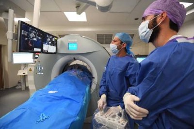
    Last year, bioengineer Oliver Jonas at Brigham and Women’s Hospital in Boston, and his colleagues demonstrated the feasibility of this strategy in people with lung13 and brain14 cancers. In small trials, surgeons inserted tiny drug-releasing devices, each loaded with nanodoses of up to 12 drugs, into tumours as people underwent cancer-removal surgery. Over the course of the operation, drugs flowed into the surrounding tissue from separate reservoirs in a device the size of a grain of rice.

    Those tissues, along with the device itself, were then removed at the end of the procedure, and subsequently inspected for molecular indicators of drug action. So far, the data collected haven’t been used to guide treatments, but retrospective analyses hinted at potential benefits if they had. Two companies — Boston-based Kibur Medical, co-founded by Jonas, and Presage Biosciences, headquartered in Seattle — are now developing these kinds of in situ drug-testing platform.

    A choice opportunity

    An assay’s treatment predictions are only as good as a patient’s ability to access the recommended drugs — and, when those are expensive cancer agents that have not been approved for the desired use, costs and insurance reimbursement can be impediments.

    Pamela Becker, a haematologist at City of Hope cancer centre in Duarte, California, has encountered some of these problems when trying to prescribe drugs that were identified during assay-guided treatment trials for people with multiple myeloma and other blood cancers. “I couldn’t get my top choice,” she says. Becker had to go down the list of recommendations, eventually finding drugs that would be covered by insurance.

    Another financial obstacle remains reimbursement for the functional tests themselves. In the United States, an official policy enacted in 1996 classifies drug-sensitivity assays as ‘experimental’, making them ineligible for coverage under Medicare, the federal government’s giant health insurance programme for older people. Changing reimbursement rules will thus require reversing that decades-old decision, says Bruce Yeager, an independent consultant in functional precision diagnostics based in Johns Creek, Georgia — an extra hurdle that means “we’re not starting from a point of neutrality”, he says. “We’re starting from negativity.”

    Combating such policies and entrenched practices hinges on the availability of compelling clinical data. But accumulating such data can be challenging when the medical establishment is not geared towards enabling functional drug testing. It’s something of a catch-22, says Letai. “But that cycle is going to break in the next couple of years,” he says, “and then I think you’re going to see a sort of non-linear adoption of these strategies, because the power and the need for them is so great.”

    Functional testing strategies might even work for conditions outside the cancer arena. In cystic fibrosis, for example, organoid models made from rectal or intestinal tissue are beginning to help clinicians to find effective drug regimens for people with rare disease-causing mutations who are not eligible to receive any approved treatments. “It just makes a lot of sense,” says Jeffrey Beekman, a cystic-fibrosis researcher at the University Medical Center Utrecht in the Netherlands, who has pioneered the approach.

    Many cancer researchers feel the same way, and now they just need to prove it to the wider medical community. All eyes are therefore on Staber and his randomized trial, which researchers anticipate will go a long way towards convincing clinicians that genomics is not the be-all and end-all of personalized care. “Paradigm shifts can be very threatening to people,” says Howard, the University of Mississippi radiologist, “but it shouldn’t be threatening. It’s just another tool in our arsenal against disease.”

    [ad_2]

    Source link

  • Smoking’s lasting effect on the immune system

    Smoking’s lasting effect on the immune system

    [ad_1]

    • NEWS AND VIEWS

    It emerges from a study of human cells that smoking can influence certain immune responses to the same extent as can age or genetics. Smoking can alter the immune system in ways that persist long after quitting the habit.

    [ad_2]

    Source link

  • A researcher-exchange programme made me a better doctor at home and abroad

    A researcher-exchange programme made me a better doctor at home and abroad

    [ad_1]

    Two people, one wearing a lab coat, talking to a man in a hospital bed.

    Caleb Skipper (right) answers questions about lumbar puncture with translation help from Alisat Sadiq (centre) at Mulago National Referral Hospital in Kampala.Credit: Caleb Skipper

    Caleb Skipper had his first interaction with African science in 2009, when he visited Ethiopia as an undergraduate at the University of North Dakota in Grand Forks. He spent a year working on a project to improve diagnosis of malaria with limited resources, which meant using his intuition to improvise. For instance, he helped to boost the diagnostic capacity of a health clinic in Binishangul Gumuz, a mostly rural area, by configuring a microscope that ran off solar power. He also helped to implement a process to detect malaria in the rural environment and taught local women how to work as basic laboratory technicians with support from several charities.

    Those early experiences showed him that he could work in austere conditions and thrive in different cultures. They inspired him to seek research opportunities with other projects in Africa and Latin America as he pursued medical training.

    In 2017, during his infectious-diseases fellowship at the University of Minnesota (UMN) in Minneapolis, he travelled to Uganda to study cytomegalovirus (CMV) as a risk factor in advanced HIV disease at the Infectious Diseases Institute (IDI) of Makerere University in Kampala. During a 2019–20 fellowship at the IDI funded by the US National Institutes of Health’s Fogarty International Center, he worked on randomized clinical trials of antifungal drugs and drug regimens to treat HIV-associated cryptococcal meningitis. Now an infectious-diseases physician at the University of Minnesota, Skipper splits his time between Minneapolis and Kampala. He tells Nature about the lessons he’s learnt during his collaborations.

    How did you end up in Kampala?

    Mostly due to my mentors’ relationship with the IDI. One of my primary mentors, infectious-diseases specialist David Boulware at the UMN, has had more than 20 years of collaboration with my other mentor, HIV and infectious-diseases specialist David Meya at the IDI. The collaboration includes annual exchanges of medical trainees between the two institutions. After my initial experience here in 2017, I was eager to participate in an ongoing collaboration with the IDI to broaden my knowledge of infectious diseases and learn more about how to ethically conduct clinical research in resource-limited settings.

    We can do all the research here on site, and then the IDI owns the research findings. This is in contrast to collecting the data, taking it back and doing the analysis in the United States and then making the local institute just a minor partner. The partnership is key to building the local research capacity.

    How does the exchange programme work?

    Medical students, residents and other trainees at the UMN have opportunities to volunteer at the IDI, Makerere’s College of Health Sciences and Mulago National Referral Hospital in Kampala for sessions from one month up to one year. Some volunteers mostly see patients or teach, whereas others focus on research. I worked with David Meya and his team, learning from their expertise and observing how patient care and clinical studies are conducted the IDI. Likewise, Ugandan trainees and study-team members can do clinical rotations at the UMN, including attending the UMN tropical-medicine course, and have opportunities to learn new laboratory skills or present research at conferences.

    What have you learnt from your experience at the IDI, and what are you working on now?

    I am developing an assay at the IDI translational laboratory to detect certain viruses, such as CMV and Epstein–Barr virus. I am also developing improved techniques to study patients’ immune responses that will be useful for trying to understand how viral co-infections affect people with advanced HIV disease.

    I have learned a lot through my time at the IDI. I’ve learned about a different culture, and how that difference can lead to both wonderful moments of learning and frustrating moments. For example, to diagnose and properly treat people with HIV-associated meningitis, we need to put a needle into their spine to do a lumbar puncture to determine the cause of their meningitis. Understandably, people can be quite apprehensive about this. Sometimes they will even refuse it. It has been valuable to learn about the patient’s perspective on why they might refuse this necessary procedure, and then develop educational materials that could help to address their concerns.

    I have also become more skilled at making medical decisions without being overly reliant on diagnostic testing. And I’ve gained a better appreciation of the dedication of caregivers and family members, which are things we sometimes overlook in the United States. I hope that all Western-trained doctors will have experiences in places such as Uganda to help build a broader and more compassionate worldview.

    On a fun note, I’ve enjoyed trying new foods such as a meal of matooke, the local cooked banana, eaten with groundnut paste, and learning to sail a boat on Lake Victoria.

    Can you describe an achievement of the research exchange?

    Our Ugandan team at the IDI had a major role in a randomized clinical trial called the Ambition trial, which was completed in 2021. The trial was for people with HIV who develop cryptococcal meningitis, a serious fungal infection of the brain, and the goal was to determine whether a single, high dose of the antifungal medication amphotericin B would be as effective as the standard treatment, a lower dose given over seven days and recommended by the World Health Organization (WHO). It was a multinational trial, mainly supported by the European and Developing Countries Clinical Trials Partnership, and involved five African countries: Uganda, Botswana, Zimbabwe, Malawi and South Africa.

    The results were published in a 2022 study1 led by Joe Jarvis at the London School of Hygiene & Tropical Medicine, which found that the single-dose amphotericin B regimen was as effective as the standard of care. In addition, the single dose was associated with fewer serious adverse events such as anaemia and kidney injury. Owing to these findings, the WHO modified its international guidelines to recommend the single-dose amphotericin B regimen as first-line therapy. Because the single dose is easier to administer in resource-poor settings, it will help thousands of people living with advanced HIV worldwide to fight this deadly infection.

    This interview has been edited for length and clarity.

    [ad_2]

    Source link

  • Mitochondrial misfire sparks inflammation

    Mitochondrial misfire sparks inflammation

    [ad_1]

    Cells in the human body contain power-generating mitochondria, each with their own mtDNA-;a unique set of genetic instructions entirely separate from the cell’s nuclear DNA that mitochondria use to create life-giving energy. When mtDNA remains where it belongs (inside of mitochondria), it sustains both mitochondrial and cellular health-;but when it goes where it doesn’t belong, it can initiate an immune response that promotes inflammation.

    Now, Salk scientists and collaborators at UC San Diego have discovered a novel mechanism used to remove improperly functioning mtDNA from inside to outside the mitochondria. When this happens, the mtDNA gets flagged as foreign DNA and activates a cellular pathway normally used to promote inflammation to rid the cell of pathogens, like viruses.

    The findings, published in Nature Cell Biology on February 8, 2024, offer many new targets for therapeutics to disrupt the inflammatory pathway and therefore mitigate inflammation during aging and diseases, like lupus or rheumatoid arthritis.

    We knew that mtDNA was escaping mitochondria, but how was still unclear. Using imaging and cell biology approaches, we’re able to trace the steps of the pathway for moving mtDNA out of the mitochondria, which we can now try to target with therapeutic interventions to hopefully prevent the resulting inflammation.”


    Professor Gerald Shadel, senior and co-corresponding author, director of the San Diego-Nathan Shock Center of Excellence in the Basic Biology of Aging and holder of the Audrey Geisel Chair in Biomedical Science at Salk

    One of the ways our cells respond to damage and infection is with what’s known as the innate immune system. While the innate immune response is the first line of defense against viruses, it can also respond to molecules the body makes that simply resemble pathogens-;including misplaced mtDNA. This response can lead to chronic inflammation and contribute to human diseases and aging.

    Scientists have been working to uncover how mtDNA leaves mitochondria and triggers the innate immune response, but the previously characterized pathways did not apply to the unique mtDNA stress conditions the Salk team was investigating. So, they turned to sophisticated imaging techniques to gather clues as to where and when things were going awry in those mitochondria.

    “We had a huge breakthrough when we saw that mtDNA was inside of a mysterious membrane structure once it left mitochondria-;after assembling all of the puzzle pieces, we realized that structure was an endosome,” says first author Laura Newman, former postdoctoral researcher in Shadel’s lab and current assistant professor at the University of Virginia. “That discovery eventually led us to the realization that the mtDNA was being disposed of and, in the process, some of it was leaking out.”

    The team discovered a process beginning with a malfunction in mtDNA replication that caused mtDNA-containing protein masses called nucleoids to pile up inside of mitochondria. Noticing this malfunction, the cell then begins to remove the replication-halting nucleoids by transporting them to endosomes, a collection of organelles that sort and send cellular material for permanent removal. The endosome gets overloaded with these nucleoids, springs a leak, and mtDNA is suddenly loose in the cell. The cell flags that mtDNA as foreign DNA-;the same way it flags a virus’s DNA-;and initiates the DNA-sensing cGAS-STING pathway to cause inflammation.

    “Using our cutting-edge imaging tools for probing mitochondria dynamics and mtDNA release, we have discovered an entirely novel release mechanism for mtDNA,” says co-corresponding author Uri Manor, former director of the Waitt Advanced Biophotonics Core at Salk and current assistant professor at UC San Diego. “There are so many follow-up questions we cannot wait to ask, like how other interactions between organelles control innate immune pathways, how different cell types release mtDNA, and how we can target this new pathway to reduce inflammation during disease and aging.”

    The researchers hope to map out more of this complicated mtDNA-disposal and immune-activation pathway, including what biological circumstances-;like mtDNA replication dysfunction and viral infection-;are required to initiate the pathway and what downstream effects there may be on human health. They also see an opportunity for therapeutic innovation using this pathway, which represents a new cellular target to reduce inflammation.

    Other authors include Sammy Weiser Novak, Gladys Rojas, Nimesha Tadepalle, Cara Schiavon, Christina Towers, Matthew Donnelly, Sagnika Ghosh, Sienna Rocha, and Ricardo Rodriguez-Enriquez of Salk; Danielle Grotjahn and Michaela Medina of The Scripps Research Institute; Marie-Ève Tremblay of the University of Victoria in Canada; Joshua Chevez of UC San Diego; and Ian Lemersal of the La Jolla Institute for Immunology.

    The work was supported by the National Institutes of Health (R01 AR069876, P30AG068635, 1K99GM141482, 1F32GM137580, T32GM007198, 5R00CA245187, and 5R00CA245187-04S1), an Allen-AHA Initiative in Brain Health and Cognitive Impairment award (19PABH134610000H), a National Science Foundation NeuroNex Award (2014862), Chan-Zuckerberg Initiative Imaging Scientist Award, the LIFE Foundation, a George E. Hewitt Foundation for Medical Research Postdoctoral Fellowship, Paul F. Glenn Foundation for Medical Research Postdoctoral Fellowship, Salk Pioneer Fund Postdoctoral Scholar Award, the Waitt Foundation, Yale University School of Medicine Center for Cellular and Molecular Imaging, a Canada Research Chair (Tier 2) in Neurobiology of Aging and Cognition, and a Canada Foundation for Innovation John R. Evans Leaders Fund (grant 39965).

    Source:

    Journal reference:

    Newman, L. E., et al. (2024). Mitochondrial DNA replication stress triggers a pro-inflammatory endosomal pathway of nucleoid disposal. Nature Cell Biology. doi.org/10.1038/s41556-023-01343-1.

    [ad_2]

    Source link

  • Study discovers a direct connection between the brain and its surrounding environment

    Study discovers a direct connection between the brain and its surrounding environment

    [ad_1]

    In a recent study of the brain’s waste drainage system, researchers from Washington University in St. Louis, collaborating with investigators at the National Institute of Neurological Disorders and Stroke (NINDS), a part of the National Institute of Health (NIH), discovered a direct connection between the brain and its tough protective covering, the dura mater. These links may allow waste fluid to leave the brain while also exposing the brain to immune cells and other signals coming from the dura. This challenges the conventional wisdom which has suggested that the brain is cut off from its surroundings by a series of protective barriers, keeping it safe from dangerous chemicals and toxins lurking in the environment.

    Waste fluid moves from the brain into the body much like how sewage leaves our homes. In this study, we asked the question of what happens once the ‘drain pipes’ leave the ‘house’-;in this case, the brain-;and connect up with the city sewer system within the body.”


    NINDS’s Daniel S. Reich, M.D., Ph.D

    Reich’s group worked jointly with the lab of Jonathan Kipnis, Ph.D., a professor at Washington University in St. Louis.

    Reich’s lab used high-resolution magnetic resonance imaging (MRI) to observe the connection between the brain and body’s lymphatic systems in humans. Meanwhile Kipnis’s group was independently using live-cell and other microscopic brain imaging techniques to study these systems in mice.

    Using MRI, the researchers scanned the brains of a group of healthy volunteers who had received injections of gadobutrol, a magnetic dye used to visualize disruptions in the blood brain barrier or other kinds of blood vessel damage. Large veins are known to pass through the arachnoid barrier carrying blood away from the brain, and these were clearly observed on the MRI scans. As the scan progressed, a ring of dye appeared around those large veins that slowly spread out over time, suggesting that fluid could make its way through the space around those large veins where they pass through the arachnoid barrier on their way into the dura.

    Kipnis’s lab was making similar observations in mice. His group injected mice with light-emitting molecules. Like with the MRI experiments, fluid containing these light-emitting molecules was seen to slip through the arachnoid barrier where blood vessels passed through.

    Together, the labs found a “cuff” of cells that surround blood vessels as they pass through the arachnoid space. These areas, which they called arachnoid cuff exit (ACE) points, appear to act as areas where fluid, molecules, and even some cells can pass from the brain into the dura and vice versa, without allowing complete mixing of the two fluids. In some disorders like Alzheimer’s disease, impaired waste clearance can cause disease-causing proteins to build up. Continuing the sewer analogy, Kipnis explained the possible connection to ACE points:

    “If your sink is clogged, you can remove water from the sink or fix the faucet, but ultimately you need to fix the drain,” he said. “In the brain, clogs at ACE points may prevent waste from leaving. If we can find a way to clean these clogs, its possible we can protect the brain.”

    One implication of ACE points is that they are areas where the immune system can be exposed to and react to changes occurring in the brain. When mice in Dr. Kipnis’s lab were induced to have a disorder where the immune system attacks the myelin in their brain and spinal cord, immune cells could be seen around ACE points and even between the blood vessel wall and the cuff cells; this led over time to a breakdown of the ACE point itself. When the ability of immune cells to interact directly with ACE points was blocked, the severity of infection was reduced.

    “The immune system uses molecules to communicate that cross from the brain into the dura mater,” said Kipnis. “This crossing needs to be tightly regulated, otherwise detrimental effects on brain function can occur.”

    Reich and his team also observed an interesting connection between the participants’ age and the leakiness of ACE points. In older participants, more dye leaked into the surrounding fluid and space around the blood vessels.

    “This might point to a slow breakdown of the ACE points over the course of aging,” said Reich, “and this could be consequential in that the brain and immune system can now interact in ways that they’re not supposed to.”

    The connection to aging and the disruption of a barrier separating the brain and immune system fits with what has been observed in aging mice and in autoimmune disorders like multiple sclerosis. This newfound link between the brain and immune system could also help explain why our risk for developing neurodegenerative diseases increases as we get older, but more research is needed to confirm this connection.

    This study was supported by the NINDS Intramural Research Program, the National Institute on Aging (AG034113, AG057496, AG078106), and the Cure Alzheimer’s Fund BEE Consortium.

    Source:

    Journal reference:

    Smyth, L. C. D., et al. (2024). Identification of direct connections between the dura and the brain. Nature. doi.org/10.1038/s41586-023-06993-7.

    [ad_2]

    Source link

  • Turbocharged CAR-T cells melt tumours in mice — using a trick from cancer cells

    Turbocharged CAR-T cells melt tumours in mice — using a trick from cancer cells

    [ad_1]

    Coloured scanning electron micrograph (SEM) of a T-cell ( purple) and a brain cancer cell ( oligodendroglioma).

    A cancer cell (blue; artificially coloured) is targeted by an engineered immune cell (purple), which can be enhanced by mutations originally discovered in cancer cells.Credit: Steve Gschmeissner/Science Photo Library

    Cancer cells are the ultimate survivors, riddled with mutations that let them thrive when healthy cells would die. These same mutations can boost the ability of game-changing cell therapies to quash cancer, a study in mice shows1.

    Among these therapies are chimeric antigen receptor (CAR) T cells, which are already used to treat several types of blood cancer. The new study shows that engineered CAR T cells carrying a mutation that was first found in cancerous T cells can vanquish tumours that don’t respond to current CAR-T therapies.

    “It’s a beautiful piece of work and opens the door for better CAR-T therapies in the future,” says Madeleine Duvic, a dermatologist and cancer researcher at the MD Anderson Cancer Center in Houston, Texas, who was not involved in the work.

    “Natural T-cell function isn’t good enough. We need to explore the extremes of T-cell function,” says Kole Roybal, an immunologist at the University of California, San Francisco, and co-author of the new paper. What better place to start than with the mutations that turn healthy T cells into hardier, cancerous ones?

    The new approach was published today in Nature.

    Cancer versus cancer

    In the past few decades, scientists have developed bespoke cell therapies by harnessing the cancer-killing power of immune cells such as T cells. The most advanced of these treatments, CAR-T-cell therapies, rely on T cells collected from people with cancer. The cells are edited to express CAR proteins, which enable the T cells to seek and destroy cancer cells. The T cells are then re-infused into the person they came from.

    These living drugs have taken the research community by storm, and the US Food and Drug Administration has approved several CAR-T cell therapies for blood cancers such as lymphomas and multiple myeloma. But scientists are still struggling to work out whether these cells can be used to kill ‘solid’ cancers, such as breast and lung tumours.

    Pulling from cancer’s playbook, Roybal and his colleagues incorporated 71 mutations, found in cancerous T cells, into CAR T cells. When they looked at how these perturbations affected T-cell function, one mutation stood out. The CAR T cells carrying an aberrant protein dubbed CARD11–PIK3R3 infiltrated well into tumours and had long-lasting cancer-killing activity.

    “It’s a very special molecule, it seems to be able to beat all the tests we put to it,” says study co-author Jaehyuk Choi, a dermatologist at Northwestern University in Evanston, Illinois.

    Potent cells

    The team treated mice carrying blood and solid cancers with several T-cell therapies boosted with CARD11–PIK3R3, and watched the animals’ tumours melt away. Researchers typically use around one million cells to treat these mice, says Choi, but even 20,000 of the cancer-mutation-boosted T cells were enough to wipe out tumours.

    “That’s an impressively small number of cells,” says Nick Restifo, a cell-therapy researcher and chief scientist of the rejuvenation start-up company Marble Therapeutics in Boston, Massachusetts.

    There is a risk that the supercharged cells will transform into cancers. But the animal data do not fuel any safety concerns, says Restifo, and the CARD11–PIK3R3 mutation seems to amp up edited T cells only when cancer cells are nearby, helping to mitigate worries about rogue immune cells.

    Choi and Roybal have co-founded Moonlight Bio in Seattle, Washington, to move these cells towards use in people with cancer. They hope to have edited cells in clinical trials in two to three years. But the bigger opportunity is the chance to find other cancer mutations that will make T-cell therapies tick.

    “A lot of people are going to think ‘Oh, this is such a good idea. Why didn’t I do this?’,” says Restifo.

    [ad_2]

    Source link

  • Crackdown on skin colour bias by fingertip oxygen sensors is coming, hints FDA

    Crackdown on skin colour bias by fingertip oxygen sensors is coming, hints FDA

    [ad_1]

    A pulse oximeter is attached to a patient's finger at EHA Clinics in Abuja, Nigeria.

    Pulse oximeter can give inaccurate oxygen readings for people with dark skin.Credit: Afolabi Sotunde/Reuters

    Growing evidence1 shows that critical devices for measuring blood-oxygen levels can be inaccurate in people of colour. Now the US Food and Drug Administration (FDA) plans to propose that companies conduct more stringent evaluation of the devices, called pulse oximeters, before applying for agency approval.

    The proposal, which the agency has not yet formally announced, calls on manufacturers to increase the devices’ accuracy and to boost the number of people on which the devices are tested. The agency also wants companies to test the devices on people whose skin colours span the entire range of a specific colour scale. FDA scientists presented the proposal at a meeting of an independent advisory committee on 2 February.

    Researchers who have been studying the performance of the devices and resulting health disparities for years applaud the FDA’s efforts. “The bar was set so low with the regulatory guidance up until now that there’s low-hanging fruit that can be addressed,” says Michael Lipnick, a global health specialist at the University of California, San Francisco.

    Vital device

    After being clipped onto a fingertip, a pulse oximeter shines light through the digit and measures how much light is absorbed by the oxygen-carrying molecule haemoglobin, giving a reading of blood-oxygen saturation. The measurement, considered one of a person’s ‘vital signs’ alongside heart rate, can give physicians quick insight into a person’s health.

    But melanin pigments in dark skin can interfere with the devices. As a result, the oximeters can indicate oxygen saturation values higher than the those derived using the gold-standard method of measuring oxygen levels in blood taken from an artery, especially in people with low blood-oxygen levels.

    Prone to error. Graphic showing how melanin might affect pulse oximeter readings.

    During the COVID-19 pandemic, studies2,3 found that the devices’ overestimation of oxygen levels can lead to less treatment for people of colour, especially in hospitals that use strict blood-oxygen cut-offs to determine who is eligible for care. “Nobody appreciated that even these small biases could lead to enormous healthcare disparities,” Lipnick says.

    These disparities have led researchers and advocacy groups to demand that the FDA ensure that the devices, which historically been calibrated on people with light skin, are accurate in people with dark skin. They have called on the agency to revise its current guidelines — which were published in 2013 — for manufacturers seeking approval for their devices. Those guidelines stipulate that the devices should be tested on at least 10 people, at least 15% of whom must be “darkly pigmented”.

    At the advisory committee meeting, FDA scientists instead proposed that companies test the devices on at least 24 people whose skin colours span the entirety of the Monk Skin Tone (MST) scale, a 10-shade scale that describes human skin colour. This is an upgrade, says Kimani Toussaint, an optics specialist at Brown University in Providence, Rhode Island, because “darkly pigmented” is subjective. An increase in the number of people tested will also help the FDA to evaluate whether a device’s performance differs with skin colours, he says.

    Real-world testing

    But some advisory-committee members, such as Rachel Brummert, a medical device safety advocate based in Charlotte, North Carolina, questioned whether 24 people would be sufficient. And other scientists say they wish the proposed guidelines recommended that manufacturers test their devices in real-world conditions. “Ideally, the FDA would take a more aggressive step to make sure these devices are evaluated in clinical settings,” says Ashraf Fawzy, a pulmonologist and critical care physician at Johns Hopkins University in Baltimore, Maryland.

    The FDA did not immediately respond to a request for comment about these criticisms.

    But more research is still needed to understand how skin colour interacts with other variables, such as how much blood flows to the fingers, Lipnick says. And most studies on the topic are based on self-reported ethnicity or skin-colour data; he and his colleagues are now evaluating the performance of the devices using the MST scale, and investigating whether the MST scale is the best measure to use for this purpose.

    Costs and benefits

    The debate highlights the tension the agency faces: it seeks to improve the accuracy of the devices while taking care that the additional testing it recommends isn’t overly cumbersome. Nor can the agency suddenly pull the devices from the market when there are no replacements and “such a large volume of patients benefitting from the devices”, says Yadin David, a biomedical engineering consultant in Houston, Texas, who chairs a separate FDA advisory committee on medical devices.

    There are other solutions beyond taking the devices off the shelves, says Michael Sjoding, a pulmonologist at the University of Michigan in Ann Arbor. He hopes that data about the devices’ performance are made more readily available to potential purchasers, such as hospital systems, so that they can “weigh the data when they’re making purchasing decisions”.

    Lipnick says this debate’s implications will reverberate around the world, especially in low- and middle-income countries that still face challenges accessing these devices. If companies find it difficult to comply with new standards, “it could result in products going off the market or prices going up”, he says.

    The FDA has not indicated if and when it would move forward with the proposed changes, nor when the changes would go into effect. The agency typically publishes draft proposals and solicits feedback from the public before finalizing guidance. Sjoding hopes changes are implemented soon: “The longer these changes aren’t made, the longer that patients are put at risk,” he says.

    [ad_2]

    Source link

  • Why autoimmune disease is more common in women: X chromosome holds clues

    Why autoimmune disease is more common in women: X chromosome holds clues

    [ad_1]

    A coloured scanning electron micrograph of an X chromosome in shades of blue on a black background.

    Protein–RNA complexes that shroud some copies of the X chromosome (artificially coloured) contribute to the female bias in prevalence of autoimmune disease.Credit: Lennart Nilsson, TT/Science Photo Library

    Why are women so much more susceptible to autoimmune diseases than men? A new explanation for the discrepancy has emerged: a molecular coating typically found on half of a woman’s X chromosomes — but not in males’ cells — might be provoking unwanted immune responses1.

    The coating, a mix of RNA and proteins, is central to a developmental process called X-chromosome inactivation. Researchers had previously implicated sex hormones and flawed gene regulation on the X chromosome as drivers of the autoimmune disparity. But the discovery that proteins central to X-chromosome inactivation can themselves set off immunological alarm bells adds yet another layer of complexity — and could point to new diagnostic and therapeutic opportunities.

    “This really adds a new mechanistic twist,” says Laura Carrel, a geneticist at the Pennsylvania State College of Medicine in Hershey.

    The study was published today in Cell1.

    Medical mystery

    Women account for around 80% of all cases of autoimmune disease, a category that includes conditions such as lupus and rheumatoid arthritis. What explains this sex bias has long been a mystery, however.

    “It’s a question that’s been irking immunologists and rheumatologists for the past 60 or 70 years,” says Robert Lahita, a rheumatologist at the Hackensack Meridian School of Medicine in Nutley, New Jersey.

    A prime suspect is the X chromosome: in most mammals, including humans, a male’s cells typically include only one copy, whereas a female’s cells typically carry two.

    (This article uses ‘women’ and ‘female’ to describe people with two X chromosomes and no Y chromosome, reflecting the language of the study, while acknowledging that gender identity and chromosomal make-up do not always align.)

    X-chromosome inactivation muffles the activity of one X chromosome in most XX cells, making their ‘dose’ of X-linked genes equal to that of the XY cells typical in males. The process is highly physical: long strands of RNA known as XIST (pronounced ‘exist’) coil around the chromosome, attracting dozens of proteins to form complexes that effectively muzzle the genes inside.

    Not all genes stay mum, however, and those that escape X inactivation are thought to underpin some autoimmune conditions. Additionally, the XIST molecule itself can initiate inflammatory immune responses, researchers reported in 20232. But that is not the whole story.

    XISTential questions

    Almost a decade ago, Howard Chang, a dermatologist and molecular geneticist at Stanford University School of Medicine in California and a co-author of the current study, noticed that many of the proteins that interact with XIST were targets of misguided immune molecules called autoantibodies.

    These rogue actors can attack tissues and organs, leading to the chronic inflammation and damage characteristic of autoimmune diseases. Because XIST is normally expressed only in XX cells, it seemed logical to think that the autoantibodies that attack XIST-associated proteins might be a bigger problem for women than for men.

    To test this idea, Chang and his colleagues turned to male mice, which don’t usually express XIST. The team bioengineered the mice to produce a form of XIST that did not silence gene expression but did form the characteristic RNA–protein complexes.

    The team induced a lupus-like disease in the mice and found that animals that expressed XIST had higher autoantibody levels than those that didn’t. Their immune cells were also on higher alert, a sign of predisposition to autoimmune attacks, and they showed more extensive tissue damage.

    Immune-system overdrive

    Notably, the same autoantibodies were also identified in blood samples from people with lupus, scleroderma and dermatomyositis — evidence that XIST and its associated proteins are “something that our immune systems have trouble ignoring”, says Allison Billi, a dermatologist at the University of Michigan Medical School in Ann Arbor.

    Montserrat Anguera, a geneticist at the University of Pennsylvania in Philadelphia, points to the human data as validation that the XIST-related mechanisms observed in mice have direct relevance to human autoimmune conditions, with implications for disease management. For example, diagnostics targeting these autoantibodies could assist clinicians in detecting and monitoring various autoimmune disorders.

    “This is a cool start,” she says. “If we could use this information to expedite the diagnosis ,it would be amazing.”

    [ad_2]

    Source link

  • Mechanically ventilated patients in intermediate care units of rural hospitals have higher death rates

    Mechanically ventilated patients in intermediate care units of rural hospitals have higher death rates

    [ad_1]

    A new National Institutes of Health-supported study finds that patients receiving ventilator life support in the intermediate care units – a potentially less costly alternative for people not sick enough for the intensive care units (ICUs) but too ill for the general ward – of rural hospitals had significantly higher death rates than patients in the same type of unit at urban hospitals. The study also found that patients with respiratory failure in the ICUs at rural and urban hospitals fare similarly. Published in the journal Annals of the American Thoracic Society, this study highlights the need for more careful evaluations of patients with breathing problems who are assigned to intermediate care units.

    This study has important implications for rural hospitals when determining how to care for their sickest patients on mechanical ventilation, as rural hospitals tend to be smaller and less resourced. It emphasizes the need to assess whether rural intermediate care units can meet the complex demands of critically ill patients, and the importance of carefully evaluating the processes designed to care for them.”


    Gustavo Matute-Bello, M.D., deputy director for the Division of Lung Diseases at the National Heart, Lung, and Blood Institute (NHLBI), NIH

    The research team, led by Emily Harlan, M.D., a pulmonary and critical care physician at the University of Michigan, Ann Arbor, collected data from 2010 to 2019 on 2.75 million hospitalizations of Medicare patients (65 years or older) who were on respiratory support at rural and urban hospitals across the country. The researchers conducted separate analyses for patients admitted to the general, intermediate, and ICU wards, and another analysis of patients in all the wards combined.

    When they compared patients in all the wards, they found that those receiving mechanical ventilation in rural hospitals had significantly higher 30-day death rates than those in urban hospitals. However, when the researchers broke down the data by level of care, patients in the ICUs of rural and urban hospitals had a similar chance of dying. The difference in outcomes, the researchers discovered, was singularly explained by the higher mortality rates for patients in the rural intermediate care units – 37% died within 30 days compared to 31.3% in urban hospitals.

    When patients are admitted to a hospital, the least sick are taken to the general ward and the sickest go to the ICU. However, U.S. hospitals are increasingly shifting toward a model that incorporates intermediate care units, which use fewer resources and can be less expensive to operate than ICUs. While these units may help a rural hospital’s financial bottom line how rural patients fared in them compared to their urban counterparts, was largely unknown.

    “This study underscores the importance of learning more about how to best use intermediate care units and highlights the need to continue investing in rural hospitals to make sure all who need it have access to life-saving care,” said Harlan. “There is a common belief that rural hospitals may have a lower quality of care, but that’s not what we saw for the ICU patients in our study.”

    Source:

    Journal reference:

    Harlan, E. A., et al. (2024) Rural-Urban Differences in Mortality among Mechanically Ventilated Patients in Intensive and Intermediate Care. Annals of the American Thoracic Society. doi.org/10.1513/AnnalsATS.202308-684OC.

    [ad_2]

    Source link