Tag: microbiome

  • One course of antibiotics can change your gut microbiome for years

    One course of antibiotics can change your gut microbiome for years

    [ad_1]

    Antibiotics can reduce diversity in the gut microbiome, raising the risk of infections that cause diarrhoea – and the effects may last years

    [ad_2]

    Source link

  • Anti-inflammatory diets: Do certain foods reduce inflammation and help you live longer?

    Anti-inflammatory diets: Do certain foods reduce inflammation and help you live longer?

    [ad_1]

    2FNMAE7 Senior couple having fun and eating at restaurant during travel - Mature man and woman wife in old city town bar during active elderly vacation

    It is one of the scourges of life in the modern world: chronic inflammation. This unhelpful response by the body’s immune system is linked to accelerated ageing and conditions such as stroke and heart disease.

    What if we could dampen it down by consuming certain foods, such as spinach, walnuts and salmon? That is the promise of anti-inflammatory diets, often advocated in vague terms by the media and nutrition industry. That might prompt eye-rolling from the scientifically minded. But recent research reveals that this approach isn’t as faddish as it sounds and paints a nuanced picture of the links between food, inflammation and our long-term health.

    This article is part of a series on nutrition that delves into some of the hottest trends of the moment. Read more here.

    Inflammation is a crucial part of our response to injury and disease. But when the body continues to deploy it even when there is no trauma, this results in chronic inflammation. Exactly why this occurs is unclear, but genetics, environment and lifestyle play roles. It can be detected by measuring certain chemical markers in the blood, and has been increasingly linked with poor health.

    “Chronic inflammation is a driver of many common diseases, including cardiovascular disease, cancer, arthritis and dementia,” says John Mathers at Newcastle University in the UK. It has also been implicated in some mental health conditions.

    But how…

    [ad_2]

    Source link

  • Stool test could provide a simpler way to diagnose endometriosis

    Stool test could provide a simpler way to diagnose endometriosis

    [ad_1]

    Endometriosis tissue viewed under a microscope

    BIOPHOTO ASSOCIATES/SCIENCE PHOTO LIBRARY

    Low levels of a particular compound in faeces could be a sign of endometriosis – and supplementation of that compound might even help control the condition.

    Affecting nearly 200 million people worldwide, endometriosis occurs when the tissue lining the uterus grows in other parts of the reproductive tract. There is no known cure, but lesions can be periodically removed through surgical procedures once the condition has been diagnosed. However, due in large part to a lack of awareness and understanding, it currently takes an average of more than six years for endometriosis to be diagnosed.

    Previous research has suggested that the gut microbiome might play a role in the condition. To investigate further, Ramakrishna Kommagani at Baylor College of Medicine in Houston, Texas, and his colleagues collected stool samples from 18 women with endometriosis and 31 women without the condition. They investigated the bacteria in the faeces as well as the metabolome – the set of chemicals produced by the gut bacteria.

    They found that the women with endometriosis had lower levels of the metabolite 4-hydroxyindole in their faeces, possibly due to alterations in the gut microbiome.

    Based on that discovery, commercial stool analyses could allow rapid screening for this widely “underdiagnosed, understudied and underappreciated” condition, leading to early and effective management, says Kommagani.

    “Stool is so easy to collect, and it’s not invasive like current diagnostic techniques such as laparoscopy [a kind of keyhole surgery],” he says.

    To explore whether 4-hydroxyindole might even have a protective effect, the team fed supplementary 4-hydroxyindole to a group of mice that had tissue implanted in their abdomens to induce endometriosis. After 14 days of treatment, those mice did not have fewer lesions compared with control animals, but their lesions were remarkably less severe, and they showed signs of having significantly less pain.

    Further experiments indicated that when mice with established endometriosis received 4-hydoxyindole, their lesions vastly improved. The results were similar in mice that had been grafted with human endometriosis lesions, suggesting the treatment could be effective in humans as well.

    “We believe this is a very good therapeutic option, because it’s naturally occurring in the body – not a drug or something synthesised,” says Kommagani.

    However, larger studies in humans will be needed to confirm whether 4-hydroxyindole can be used to diagnose endometriosis and whether the compound is effective as a treatment.

    Topics:

    • women’s health/
    • microbiome

    [ad_2]

    Source link

  • The brain has its own microbiome. Here’s what it means for your health

    The brain has its own microbiome. Here’s what it means for your health

    [ad_1]

    New Scientist. Science news and long reads from expert journalists, covering developments in science, technology, health and the environment on the website and the magazine.

    In 2015, Nikki Schultek was in her prime: a young mother of two little boys, she had just run a half marathon. Then, a mysterious illness hit. Her asthma, previously well-managed, became increasingly severe. Over the following months, she experienced chronic pain, digestive problems and a cardiac arrhythmia. Then came the “last insult”: signs of neurodegeneration, including brain fog and lapses of memory. “It was the lowest point,” she recalls. “I began making plans for my kids, writing down notes of things that I would want to tell them if I continued to get worse.”

    Schultek received various diagnoses for individual problems, but none fully matched her constellation of symptoms. Eventually, one doctor suggested that an undetected infection could lie behind her chronic pain and breathing difficulties. She tested positive for Borrelia burgdorferi and Chlamydia pneumoniae infections and was prescribed a cocktail of antibiotics. On taking them, she found that all her symptoms – including the brain fog and memory deficits – went into remission.

    Schultek has since founded a research group to explore the role of infection more generally in cognitive decline. This idea would once have been considered outlandish, but interest in the brain’s microbial community is growing rapidly. It turns out our grey matter is teeming with bacteria, viruses and fungi, and a better understanding of this unexpected microbiome has enormous potential to prevent neurodegenerative diseases. It could even reverse symptoms of decline when things go awry, as Schultek found. And, most excitingly of all, some potential treatments have a proven track…

    [ad_2]

    Source link

  • The gut microbiome and chronic pain

    The gut microbiome and chronic pain

    [ad_1]

    The abnormal tissues range from black blisters to red nodules and white-hot cysts. They grow throughout the pelvic cavity, latching on to the ovaries and peritoneum or infiltrating the bowel and bladder.

    Once detected, the painful lesions of endometriosis, an inflammatory condition, can be removed. But for up to half of people who opt for this, the pain returns or persists so intensely that they need surgery again within five years. “As surgeons and doctors, we want to remove lesions. But people’s pain persists more often than we like to report,” says Amira Quevedo, an obstetrician-gynaecologist who runs endometriosis clinical trials at the University of Florida in Gainesville.

    The pain experienced by people with endometriosis doesn’t reflect the number, size or type of lesions present, and varies wildly between individuals. For some, the pain worsens during their period; for others, it lasts all month long. It can manifest as searing muscle spasms, or as vaginal, bowel or bladder pain that spreads across the pelvis and beyond.

    This persistence of pain after the original stimuli have subsided or been removed is a key feature of many kinds of chronic pain. In some whole-body pain conditions, such as fibromyalgia, there is no clear cause. Something has tripped the pain system into overdrive, prompting a desperate search for relief.

    At least in the case of endometriosis, that relief is often found in switching diets. The foods people eat can rapidly alter the vast collection of microbes that reside in the intestine, in turn releasing chemicals that either drive or dampen pain. Observations that people with chronic pain have different mixes of microbes in their gut from other individuals have also given rise to the idea that manipulating this gut microbiome — through diet or other means — might help.

    That outcome remains speculative, awaiting more clinical trials. And tinkering with the microbiome is unlikely to provide relief for everyone, especially when chronic pain becomes hardwired in the brain. But given the paucity of other options, and the potential benefits, a microbiome-focused approach is worth pursuing. It could “significantly change the way we understand, diagnose and treat chronic pain”, says Amir Minerbi, a physician at the Rambam Institute for Pain Medicine in Haifa, Israel. But researchers are wary of overstating the gut microbiome’s analgesic abilities. “We don’t want to give false hope,” Minerbi says.

    Gut microbes and visceral pain

    The connection between the gut and chronic pain began to materialize two decades ago in studies of irritable bowel syndrome (IBS), a chronic condition more painful than its name suggests. IBS is marked by visceral pain, which emanates from organs in the abdomen and pelvis.

    Despite decades of work probing the connections between gut bacteria and visceral pain in IBS, “it’s been a slow evolution” to recognize the gut microbiome’s role, says stress neurobiologist John Cryan at University College Cork in Ireland.

    About 20 years ago, animal studies began to reveal that certain bacteria stimulate pain receptors on cells of the gut in a manner similar to morphine, among other mechanisms. In 2008, Cryan’s team showed that animals that had been stressed in early life by being separated from their mothers developed a whole-body syndrome of inflammation and sensitivity to visceral pain that was linked to changes in their gut bacteria1. “That was our first real strong look” at how changes in pain related to changes in the microbiome in an animal model, he says.

    Steadily, researchers realized not only that the gut’s resident bacteria could induce persistent visceral pain — and, in fact, were required for normal visceral pain sensation — but also that transplanting certain microbes from one animal to another could relieve it. These findings have since been replicated in other types of pain, such as allodynia, a type of severe nerve pain stemming from very faint stimuli.

    Human trials of probiotics have shown what Cryan says are “slight but significant” analgesic effects2 on visceral pain in IBS. Altogether, the evidence suggests that “the microbiome is playing a key role in pain”, he adds. It is even possible that gut bacteria influence not only how neurons transmit pain, but also how those acute pain signals turn chronic.

    “It makes a lot of sense,” says Jessica Maddern, a pain researcher at the South Australian Health and Medical Research Institute in Adelaide, who lives with the visceral and chronic pelvic pain of endometriosis. The gut is studded with nerves and constantly contacting the brain through the vagus nerve and spinal cord, she says, “so it stands to reason that it could change the way you’re experiencing pain”. Diet is already known to affect mood through the ‘gut–brain axis’, but the finer details of how gut microbiota influence pain are still being worked out.

    Hoescht stained colourful reactive region of cerebral cortex showing microglia in green.

    Microglia (green) can become overly reactive and affect pain signals to the brain.Credit: Gabriel Luna, NRI-UCSB/Wellcome/CC BY 4.0

    Animal studies of visceral pain have identified specific chemicals, produced by gut bacteria, that can promote3 or suppress4 pain. Short-chain fatty acids, which are produced when certain bacteria digest fibre, stimulate immune cells to release pro-inflammatory factors, whereas bile acids suppress the activity of sensory nerves. The effects can be far-reaching: these metabolites can seep into the circulation through the gut lining and cross the blood–brain barrier, altering the permeability of both structures.

    As a result, the gut microbiome might even influence the perception of pain. Cryan says his group’s animal studies show “very clearly” that brain regions known to be involved in the emotional and cognitive aspects of pain, such as the anterior cingulate cortex, change with alterations in the gut microbiome5. “We’re beginning to see that signals from the microbiome impinge on how visceral pain is perceived in the brain,” he says. “But we’re only scratching the surface.”

    The endometriosis window

    Maddern studies nerve pathways in visceral pain. Coming to this research some 20 years after her own endometriosis diagnosis, she was shocked at how little was known about pain and how to treat it. Only in the past few years have researchers begun modelling visceral pain in animal models of endometriosis, as they do for IBS. That’s because most endometriosis research has so far focused on understanding what causes the disease and the growth of lesions, rather than its pain.

    Still, endometriosis — which involves interconnected and overlapping pain symptoms — could serve as a window into chronic pain more broadly. Much-improved mouse models developed by Maddern and by Kelsi Dodds, a neurophysiologist at Flinders University in Adelaide, offer fresh insights.

    Maddern’s and Dodds’s work expands researchers’ understanding of the way in which pain becomes chronic, identifying how cells in the spinal cord drive a process called central sensitization. In female mice with long-standing endometriosis, the spinal cord’s resident support cells — microglia and astrocytes — amass, become overly reactive to and amplify pain signals6,7. Central pain pathways become hypersensitive to peripheral inputs, such that light touch becomes unbearable, or chronic pain persists even without any stimuli — as is common after endometriosis lesions are removed. Other researchers modelling endometriosis in mice have similarly reported swollen and therefore activated microglia in the brain8. Activated microglia are appearing in models of fibromyalgia, too9.

    The anatomy of pain pathways also goes some way to explaining how the gut microbiome could affect widespread pain. Some nerve pathways innervate multiple organs in the pelvis and converge in the spinal cord, so signals from gut microbiota could very easily cross from the gut over to other pelvic organs and beyond, Dodds says.

    Food for thought

    Many women with endometriosis report finding relief by making changes to their diet. Some report that doing so completely changes their experience of the disease, says Francesca Hearn-Yeates, who is studying the impact of diet on endometriosis-associated pain at the University of Edinburgh, UK.

    In an as-yet unpublished survey, she asked some 2,600 people with endometriosis about their symptoms, including bloating, cramps and pain, and what they ate. About 83% of respondents — drawn from 51 countries — said they had altered their diets. And of that subset of respondents, 63% said that these dietary modifications reduced their pain. No one diet stood out as the most effective, but going gluten-free and dairy-free often helped. “It’s not a fix-all for everyone,” she says, “but the fact that it’s benefiting so many people is really promising.”

    To get at some of the mechanisms involved, she has begun an exploratory study to profile gut-bacteria metabolites in 50 people with suspected endometriosis who are awaiting diagnostic surgery. Multiple studies have shown that people with endometriosis have altered gut microbiomes, yet few have examined how that microbial community functions as a collective10. And relating people’s metabolite profiles to their diets and pain, as she plans to do, is new territory. “There’s clearly this really intricate interaction between the gut and the brain,” she says. But the task that looms ahead is to pin down how specific bacteria influence pain.

    Therapeutic potential

    Research in animals is looking to antibiotics as a way to manipulate the gut microbiome — and by extension, chronic pain. Two studies have found that metronidazole, an antibiotic used to treat gastrointestinal and reproductive tract infections, can shrink endometriosis lesions in mice11, and even stop them forming12.

    This makes sense: people with endometriosis have an abundance of anaerobic bacteria sensitive to metronidazole in their gut. However, neither study looked at pain. So Quevedo and her colleagues at the University of Louisville Hospital in Kentucky are investigating whether administering endometriosis after surgery could reduce pain.

    Starting in 2020, 72 people with endometriosis randomly received either low-dose metronidazole or a placebo for two weeks. The two groups showed no differences in pain six weeks after surgery13, but Quevedo remains optimistic. The trial runs until 2027, and participants will report their symptoms six months after surgery and annually for five years, a timeline more relevant to chronic pain.

    However, Quevedo admits that antibiotics alone might not be enough to quell persistent pain. They could help to ‘reset’ the gut microbiome by removing problematic bacteria. But achieving a sustained benefit will probably require probiotics — which seed the gut with beneficial bacteria — along with dietary changes promoting microbial strains linked to reduced pain.

    Two small randomized trials suggest that taking daily probiotics containing selected Lactobacillus strains can reduce painful periods in people with endometriosis10, but the relief seems short-lived. This transient effect probably reflects the complexity of human pain experiences compared with mouse models, Quevedo says; after enduring endometriosis for years, most individuals have persistent pain that doesn’t budge.

    If clinician-scientists are to find other ways to ease pain, Quevedo says, they need to differentiate between chronic pain types and between various forms of endometriosis. This stratification of clinical subtypes is often missing from microbiome studies that lump patients together, but is necessary to work out which therapies alleviate whose pain. The same is true of chronic pain generally; pain is deeply personal and what eases one person’s discomfort might do little for someone else. “We know that one treatment is not going to help everybody,” Quevedo says.

    Fibromyalgia and beyond

    The role of the gut microbiome is becoming clearer in chronic pain conditions that are not visceral in origin. The foremost example is fibromyalgia, a pain disorder that typifies central sensitization — it causes widespread pain in joints, muscles and tendons, and shattering fatigue, but is often misdiagnosed.

    Minerbi’s research suggests that the gut microbiome could not only help to ease fibromyalgia pain, but also aid in diagnosing it and other chronic pain conditions. In 2019, his team found that people with fibromyalgia have altered gut microbiomes, and that those slight imbalances correlated with pain and fatigue — and not with diet, medications or other environmental factors14. According to Minerbi, it was the first demonstration in humans that the gut microbiome might modulate widespread, non-visceral chronic pain.

    In a 2023 study, the team found that people with fibromyalgia also had lower levels of specific bile acids in their blood compared with healthy controls15. These secondary bile acids are produced by gut bacteria that people with fibromyalgia tend to lack. In fact, the lower the level of bile acids they had circulating, the more intense pain they reported — possibly because some of these acids bind to neurons in the spinal cord that suppress pain. Without them, pain might flare unchecked. The study suggests that restoring the levels of these bile acids could help to reduce fibromyalgia pain.

    Amir Minerbi looks down as he pipettes a sample into a tube in a bright office

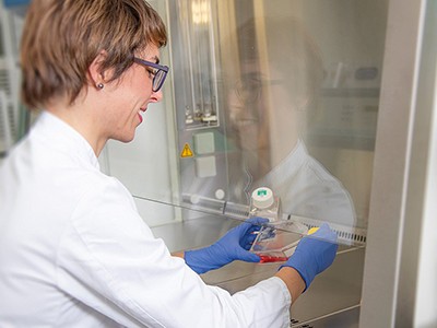
    Amir Minerbi prepares gut microbiome samples for analysis.Credit: Rambam Health Campus.

    Minerbi and his team have tried transplanting faecal matter from healthy donors into 14 women with fibromyalgia to address such gut-microbiome imbalances. After five fortnightly treatments, 12 of the volunteers in this pilot study, which has not been peer-reviewed, reported less severe pain than before9. A randomized, placebo-controlled trial is next.

    Minerbi’s group is also developing a machine-learning algorithm to relate chronic pain to gut microbiome profiles and other blood markers. So far, the tool differentiates only between people with fibromyalgia and those who do not have chronic pain14,15. The next step, Minerbi says, is to see “whether we can take someone with chronic pain and say what type of chronic pain they have, which is really the clinical question”. To that end, researchers are investigating the gut microbiome in various chronic pain conditions, hoping to find commonalities and distinct changes between them, as one 2024 study has done16.

    This work has just begun. So far, most human studies have involved broad-stroke characterizations of the gut microbiomes of people with chronic pain conditions and those without. These differences — some subtle, others striking — implicate the gut microbiome in a gamut of chronic conditions, from inflammatory arthritic pain and migraine headaches to nerve-injury pain. Whether these observed differences are an underlying cause of those conditions or a knock-on effect, however, remains unclear.

    Hurdles ahead

    Most studies capture only a snapshot of the gut microbiome. Therefore, Cryan says, large, longitudinal studies are needed to track microbiome changes in response to symptoms and treatment. His research in animals shows that early life stress affects the gut microbiome in ways that lead to persistent visceral pain in adulthood, even though the microbiome itself recovers17. “When you’re looking at pain, the microbiome may not be a reflection of that pathology; it might be something that happened way earlier that affected pain processes,” he says.

    Still, Cryan thinks that modifying the gut microbiota could help to relieve chronic pain. He cites animal studies showing that specific probiotic strains can reverse well-established pain, even when given in adulthood18. That apparent plasticity offers some hope, but he says it’s essential for researchers to investigate which strains of bacteria relieve chronic pain in humans — and how long that effect lasts.

    Despite these challenges, researchers are keeping an open mind that the gut microbiome could help to ease chronic pain. So immense is the burden, Maddern says, that “everything is worth trying at this point”.

    [ad_2]

    Source link

  • Commensal consortia decolonize Enterobacteriaceae via ecological control

    [ad_1]

    Mice

    C57BL/6 mice, maintained under GF conditions, were purchased from Sankyo Laboratories Japan, SLC Japan or CLEA Japan. GF and gnotobiotic mice were bred and maintained within the gnotobiotic facility of Keio University School of Medicine or the JSR-Keio University Medical and Chemical Innovation Center. Il10−/− and Ifngr1−/− mice were purchased from Jackson Laboratories. Myd88−/−Ticam1−/− and Rag2−/−Il2rg−/− mice were purchased from Oriental Bio Service. All mice were maintained under a 12-h light–dark cycle. A temperature of 20–24 °C and a humidity of 40–60% were used for the housing conditions. All animal experiments were approved by the Keio University Institutional Animal Care and Use Committee.

    Human faecal samples and isolation of bacterial strains

    Human faecal samples were obtained from healthy human donors, patients with ulcerative colitis and patients with Crohn’s disease following the protocol approved by the Institutional Review Board of Keio University School of Medicine (approval numbers 20150075, 20140211, and 20150075). Informed consent was obtained from each individual. Faecal samples were mixed with PBS (containing 20% glycerol) and stored at –80 °C. An aliquot of each sample was diluted with PBS in an anaerobic chamber (80% N2, 10% H2 and 10% CO2; Coy Laboratory Products) and plated onto different agar plates (EG, mGAM, BHK, CM0151, MRS or BL media). After incubating for 2–7 days, colonies with different appearances were transferred to liquid media (EG, mGAM, HK or CM0149), incubated for 24–48 h, mixed with glycerol (final concentration 20% (v/v)), and stored at –80 °C. Bacterial genomic DNA was extracted from the isolated strains using the same protocol as DNA isolation from faecal samples. The 16S rRNA gene locus was amplified by PCR using the KOD plus Neo kit (TOYOBO) according to the manufacturer’s protocol. DNA sequencing was performed by Eurofins. Sequences were aligned using the BLAST program of NCBI and the Ribosomal Database Project (RDP) databases. Primers used for DNA sequencing were as follows: F27 primer: 5′-AGRGTTTGATYMTGGCTCAG-3′; R1492 primer: 5′-TACGGYTACCTTGTTACGACTT-3′. Individual isolates in the culture collection were grouped as ‘strains’ if their 16S rRNA gene sequences shared >98.0% homology.

    To prepare the bacterial mixture for inoculation, isolated strains were individually cultured in the appropriate broth at 37 °C for 1–2 days (mGAM broth was used for culturing the F18 strains). Bacterial density was adjusted based on absorbance at 600 nm, and equal volumes of the cultured strains were mixed and centrifuged at 3,000g for 10 min at 4 °C to concentrate fivefold. Thereafter, GF mice were administered 200 μl of the bacterial mixture per mouse (approximately 1–2 × 109 CFU of total bacteria) by oral gavage. The bacterial mixture was administered into GF mice (200 μl per mouse, approximately 1–2 × 109 CFU of total bacteria) by oral gavage. In Extended Data Fig. 3e, f37_E. coli strain was swapped out for the E. coli Nissle1917 strain (Mutaflor, DSM 6601).

    Effect of defined consortia on pathogenic and commensal bacterial strains

    To examine the effects of defined consortia on pathogenic bacteria, C57BL/6 GF mice (8–14 weeks of age, housed in separate GF isolators) were inoculated with K. pneumoniae 2H7 (Kp-2H7), carbapenem-resistant K. pneumoniae (CPM+ Kp, ATCC BAA1705), K. aerogenes (strain Ka-11E1217), extended-spectrum-β-lactamase producing E. coli (ESBL+ E. coli, ATCC BAA2777), adherent-invasive E. coli (AIEC, strain LF82, provided by N. Barnich23), P. aeruginosa (ATCC 10145), vancomycin-resistant E. faecium (VRE Ef, ATCC 700221), C. upsaliensis (ATCC BAA1059), or C. difficile (strain 630, ATCC BAA1382) by oral gavage (2 × 108 CFU per mouse). Seven days after colonization with pathogenic microbes, the mice were administered 200 μl of isolated bacterial strain mix (total 109 CFU) or 200 μl of human faecal suspension by oral gavage. Faecal samples were collected from mice every three or four days, suspended in PBS (containing 20% glycerol), and cultured on selective media (DHL agar with 30 mg l−1 ampicillin and 30 mg l−1 spectinomycin for Kp-2H7, CPM+ Kp, Ka-11E12 and P. aeruginosa, MacConkey agar with 1 mg l−1 cefotaxime, and VRE-selective agar plates (BD 251832) for VRE). After 24–48 h of incubation, the CFUs were counted. In cases where evaluation was not possible by counting CFUs, bacterial DNA extracted from faeces was evaluated by quantitative real-time PCR (qPCR). To evaluate colonization resistance activity against Kp-2H7, C57BL/6 GF mice were first colonized with either F18-mix or F13-mix and then inoculated with Kp-2H7 on day 7. Faecal samples were collected every three or four days to count Kp-2H7 CFUs. To examine the cumulative effect of the 18 strains on Kp-2H7, C57BL/6 GF mice were inoculated with Kp-2H7 (2 × 108 CFU per mouse) by oral gavage, followed by oral administration of each strain of the F18-mix one by one every five days for 95 days. Faecal samples were collected every five days to count the CFUs of Kp-2H7 as well as to quantify the levels of gluconate.

    To investigate the influence of F18-mix on commensal strains, 7 strains (Dorea longicatena, Eubacterium rectale, C. scindens, Bacteroides thetaiotaomicron, Bacteroides uniformis, Bifidobacterium adlescentis and Collinsella aerofaciens) were selected from our culture collection. C57BL/6 GF mice were colonized with the 7 commensal strains together with Kp-2H7 (each 2 × 108 CFUs). Fourteen days later, mice were administered with F18-mix (total 109 CFUs in 200 µl) by oral gavage. Faecal samples were collected twice per week and subjected to quantification by CFU calculation and qPCR. To investigate the effect of F18-mix in the context of a more complex microbiota, C57BL/6 GF mice were colonized with either 41 strains from donor I, 46 strains from donor K, or a combination of both groups with C. scindens VE202-26 (totaling 88 strains). Subsequently, without any prior antibiotic treatment, these mice were orally inoculated with F18-mix. Faeces were collected for full-length 16S rRNA gene sequencing analysis.

    To examine the effects of F18-mix in the context of dysbiotic microbiota, GF B6 or Il10−/− (B6 background) mice were administrated with 200 µl of faecal suspension from patients with ulcerative colitis or Crohn’s disease containing high levels of ESBL+ E. coli or K. pneumoniae. Ten days after IBD microbiota inoculation, mice were treated with 500 μl of 1 g l−1 vancomycin by oral gavage. The mice were treated with or without F18-mix four times (at 4, 8, 24 and 48 h following the vancomycin treatment). Faeces were collected and subjected to 16S rRNA gene sequencing or counting CFUs of E. coli or K. pneumoniae.

    Dietary supplementation with carbohydrates

    Unless otherwise indicated, mice were fed a nutrient-rich diet (CL-2; CLEA Japan), which is high in gluconate. To assess the impact of dietary carbohydrate supplementation, a formula diet (AIN93G; Oriental Yeast Co.) was used, which is high in glucose but lacks gluconate, sorbitol, mannose, xylose, glucarate, galacturonate and xylose. This diet was supplemented with varying percentages of gluconate (0%, 2.5% or 10% of total calories) or with 10% of other carbohydrates, including glucarate, galactitol, sorbitol, cellobiose, glucosamine, xylose, mannose or galacturonate (Supplementary Table 8). The diets were sterilized by γ-irradiation (50 Gy).

    Bacterial DNA extraction and quantitative real-time PCR

    The frozen faecal samples were thawed and 50 μl of each sample was mixed with 350 μl TE10 (10 mM Tris-HCl, 10 mM EDTA) buffer containing RNase A (final concentration 100 μgml−1, Invitrogen) and lysozyme (final concentration 3.0 mg ml−1, Sigma). The suspension was incubated for 1 h at 37 °C with gentle mixing. Purified achromopeptidase (Wako) was added to a final concentration of 2,000 units ml−1, and the sample was further incubated for 30 min at 37 °C. Then, sodium dodecyl sulfate (final concentration 1%) and proteinase K (final concentration 1 mg ml−1, Nacalai) were added to the suspension and the mixture was incubated for 1 h at 55 °C. Thereafter, purified DNA was obtained from the samples using the Maxwell RSC cultured cell DNA kit, according to the manufacturer’s protocol. For quantifying the amount of bacterial DNA, real-time qPCR was performed using the Thunderbird SYBR qPCR Mix (TOYOBO) and LightCycler 480 (Roche). The primer pairs used in this study are listed in Supplementary Table 9.

    Full-length 16S rRNA gene amplicon sequencing

    Full-length 16S rRNA amplicon sequencing was performed according to the protocol prepared by PacBio with slight modifications. In brief, the full-length 16S rRNA gene (including hypervariable regions V1 to V9) was amplified using barcoded 27Fmod (5′-Phos-GCATCNNNNNNNNNNAGRGTTYGATYMTGGCTCAG-3′) and barcoded 1492R (5′-Phos-GCATCNNNNNNNNNNRGYTACCTTGTTACGACTT-3′) primers; ‘Phos’ indicates a 5′-phosphate modification, and ‘N’ represents a unique PacBio barcode sequence for each sample. The PCR conditions were as follows: an initial denaturation at 95 °C for 3 min, followed by 20 cycles of denaturation at 95 °C for 30 s, annealing at 57 °C for 30 s, and extension at 72 °C for 60 s. The PCR products were purified using AMPure magnetic beads and pooled at equimolar concentrations. The pooled amplicons were further purified with AMPure beads, and 1 μg was used for library preparation. The library was prepared according to the PacBio SMRTbell Prep Kit 3.0 protocol and sequenced on the PacBio Revio system. The HiFi reads were automatically generated using SMRT Link software (version 13.0) with default settings and demultiplexed using lima application in SMRT Tools with HIFI-ASYMMETRIC presets.

    Amplicon sequence variants analysis

    Full-length 16S rRNA gene amplicon sequence variants (FL16s-ASVs) were inferred from demultiplexed HiFi reads using the DADA2 package (version 1.30.0) in R (version 4.3.3) according to the previously described DADA2 for PacBio workflow53 with slight modifications. The reads were subjected to quality filtering and trimming using the filterAndTrim function with the following parameters: minQ=3, minLen=1300, maxLen=1600, maxN=0, rm.phix=FALSE, maxEE=2. To learn error rates using the learnErrors function without the ‘dada2:::PacBioErrfun’ option, due to the binned quality value adopted by the Revio system, with a maximum of QV40. FL16s-ASVs were then subjected to a homology search against 16S rRNA gene sequences extracted from publicly available genomes (downloaded from GenBank on 12 September 2023) using BLASTN with a maximum e-value cut-off of 1 × 10−10. Top hits were determined by the highest bitscore.

    Bacterial whole-genome sequencing

    The Illumina MiSeq and PacBio Sequel platforms were used for bacterial whole-genome sequencing. For Illumina sequencing, the library was prepared using the TruSeq DNA PCR-free library prep kit (Illumina), with a target insert size of 550 bp. All the Illumina reads were trimmed and filtered using the FASTX-toolkit (version 0.0.13). For the PacBio sequencing, the library was prepared using the SMRTbell template prep kit 1.0. Sequence data for both types of sequencing were assembled using the hybrid assembler Unicycler. Taxonomic assignment of the genomes was determined by classify_wf of GTDB-tk54 version 2.3.0 with GTDB55 database R214. NCBI taxonomy of fastANI reference genome related to the genome of each strain was retrieved using NCBI-genome-download version 0.3.3 (ncbi-genome-download; https://doi.org/10.5281/zenodo.8192432) and rankedlineage.dmp from NCBI taxonomy database56 (downloaded on 14 September 2023). The genes were predicted using Prokka version 1.14.0 with “–kingdom Bacteria –rnammer” options, and rnammer version 1.2. The homology search for the predicted genes was performed using diamond57 version 2.0.15 with “blastp –evalue 0.00001 –id 30 –query-cover 60 –ultra-sensitive” options, with KEGG (downloaded on 19 April 2022)58, COG (downloaded on 19 May 2021)59, VFDB (downloaded on 10 September 2022)60, and UniRef90 (downloaded on 24 May 2022; https://www.uniprot.org/help/uniref) databases. For homology search against KEGG DB, a database was manually constructed from protein sequences with KEGG Ontology (K number) which were extracted from KEGG non-redundant datasets at the species level. We also added homology search for gluconate metabolism genes in our isolated strains with “blastp –evalue 0.00001 –id 20 –query-cover 60 –ultra-sensitive” options. The sequences of gluconate kinase (gntK, MKMCEHOJ_02531) and gluconate transporters (MKMCEHOJ_02530 and MKMCEHOJ_02505) from f37_E. coli strain, and gluconate dehydratase (gad, EAOGLLOI_00767), gluconate transporters (EAOGLLOI_00766 and EAOGLLOI_00912), 2-dehydro-3-deoxygluconokinase (kdgK, EAOGLLOI_00768), and 2-dehydro-3-deoxyphosphogluconate aldolase (eda, EAOGLLOI_00769) from f17_Blautia caecimuris strain were used as reference sequences.

    Ex vivo caecal suspension culture

    Caecal contents from GF or F31-mix, F18-mix and F13-mix colonized mice were anaerobically resuspended in water at a concentration of 100 mg ml−1. Caecal contents were either filtered through a 0.22-μm filter (Millex Millipore) after centrifuging at 10,000g for 5 min, heat-killed at 105 °C for 30 min, or left untreated. Thereafter, a diluted overnight culture of Kp-2H7 (103 CFU in 10 μl) was added to 200 μl of each caecal suspension. After incubating at 37 °C for 48 h under aerobic or anaerobic conditions, samples were serially diluted and plated on a selection agar plate (DHL with 30 mg l−1 ampicillin and 30 mg l−1 spectinomycin) for counting Kp-2H7 CFU.

    Bacterial growth monitoring

    The wild-type, ΔgntK or ΔgntR Kp-2H7 strain was cultured in M9 minimal medium for 24 h at 37 °C. Afterward, the culture was diluted 100-fold with sterile water. A 10-μl aliquot of the diluted culture was inoculated into 200 μl M9 medium supplemented with individual carbohydrates (final concentration of 2 mM) as the sole carbon source, or with a mock control. To examine the effect of metabolites on Kp-2H7 growth, 10 μl of wild-type Kp-2H7 culture dilutions were inoculated into 200 μl M9 medium containing varying concentrations of 4-HBA (100, 10, 1 or 0.1 mM), cholic acid (500, 100, 20 or 4 μM), and acetate or butyrate (100, 25, 6.25, 1.56 or 0.39 mM). The pH of acetate and butyrate was adjusted to either 5.0 or 7.0. Bacterial growth was monitored by measuring absorbance at 600 nm every 30 min using a microplate reader (Sunrise Thermo (Tecan) for anaerobic conditions and Infinite 200 PRO (Tecan) for aerobic conditions) at 37 °C with 100 s shaking before each time point.

    Transcriptome analysis of epithelial cells

    Total RNA was isolated from colonic epithelial cells using NucleoSpin RNA (Macherey-Nagel), according to the manufacturer’s instructions. Libraries for RNA sequencing were prepared using TruSeq Stranded mRNA Library Prep (Illumina), according to the manufacturer’s instructions. The libraries were sequenced using NovaSeq 6000 (Illumina) with the mode of 150-bp paired-end. The sequenced paired-end reads were quality-controlled using Trimmomatic61 version 0.39 with “2:30:10 LEADING:3 TRAILING:20 SLIDINGWINDOW:4:15 MINLEN:5” options and FASTX-Toolkit version 0.0.13 (https://github.com/agordon/fastx_toolkit) with “-q 20 -p 80” options. Unpaired reads and reads mapped to the PhiX reference genome using minimap262 version 2.17-r941 were excluded from further analyses. The remaining quality-controlled reads were mapped to the mouse reference genome (mm10) using STAR63 version 2.7.2b. The mapped reads were counted for each gene using featureCounts64 version 1.5.2 with “-t exon -p -B -Q 1” options. the transcripts per million (TPM) values of each gene in each sample were calculated. The differential expression analysis was performed using DESeq265 version 1.28.1, and the P values were corrected by the Benjamini–Hochberg method to maintain the false discovery rate (FDR) below 5%.

    Transcriptome analysis of Kp-2H7

    To examine the transcriptome landscape of Kp-2H7 in vivo, GF mice were monocolonized with Kp-2H7, followed by oral administration of F18-mix or vehicle control. Two days after F18-mix administration, faecal samples were collected, and total RNA was extracted. For the in vitro examination of the transcriptomes of Kp-2H7 strains, wild-type, ΔgntK, and ΔgntR Kp-2H7 were cultured at 37 °C in M9 minimal medium supplemented with either glucose or gluconate. Bacteria were collected during the early log phase (absorbance at 600 nm = 0.35), and total RNA was extracted. Isolation of total RNA from in vivo faecal samples or in vitro culture samples was conducted using the NucleoSpin RNA kit (Macherey-Nagel), according to the manufacturer’s instructions. Libraries for RNA sequencing were prepared using TruSeq Stranded mRNA Library Prep (Illumina) and sequenced using HiSeq X (Illumina) with the mode of 150-bp paired-end. To analyse the in vivo transcriptome profiles of Kp-2H7 in the presence or absence of F18-mix, a reference genome was created by concatenating the genome sequence of Kp-2H7 with the genome sequences of the F18-mix. The sequenced paired-end reads were quality-controlled using Trimmomatic61 version 0.39 with “2:30:10 LEADING:3 TRAILING:20 SLIDINGWINDOW:4:15 MINLEN:5” options and FASTX-Toolkit version 0.0.13. Unpaired reads and reads mapped to the mouse (mm10) or PhiX reference genome using minimap262 version 2.17-r941 (in vivo) or 2.24-r1122 (in vitro) were excluded from further analyses. The quality-controlled reads were mapped to the concatenated or Kp-2H7 reference genome using bowtie266 version 2.3.4.1. (in vivo) or 2.4.4 (in vitro). For in vivo mice faecal samples, the read counts for each Kp-2H7 gene were obtained by counting uniquely mapped reads and then distributing and summing multi-hit read counts based on the number of uniquely mapped reads. For in vitro culture samples, the read counts for each Kp-2H7 gene were obtained using featureCounts64 version 2.0.1 with “-t CDS -p -B -Q 10” options. The differential expression analysis was performed using DESeq265 version 1.28.1 (in vivo) or 1.30.1 (in vitro) with “fitType = local” option and Benjamini–Hochberg correction method to maintain the FDR below 5%. The heat map was obtained from the variance-stabilizing transformations values obtained from the DESeq2 output.

    For real-time qPCR analysis, cDNA was synthesized using ReverTra Ace qPCR RT Master Mix (TOYOBO), and qPCR was performed using Thunderbird SYBR qPCR Mix (TOYOBO) on a LightCycler 480 (Roche).

    Construction of transposon mutant library

    A transposon insertion library of Kp-2H7 was constructed using the EZ-Tn5TM <KAN-2> Tnp Transposome kit (Lucigen). In brief, 80 μl (109 CFU) of Kp-2H7 suspension was mixed with 0.5 μl of EZ-Tn5TM <KAN-2>, transferred to a 1-mm gap width electroporation cuvette, and subjected to electroporation using ELEPO21 (Nepa Gene Co.) with the following parameters: poring pulse; voltage: 1,800 V, pulse length: 5.0 ms, pulse interval: 50 ms, number of pulses: 1, and polarity: +, and transfer pulse; voltage: 150 V, pulse length: 50 ms, pulse interval: 50 ms, number of pulses: 5, and polarity: ±. Transformed Kp-2H7 cells were incubated in 1 ml LB broth for 3 h at 37 °C, and then selected on LB agar plates containing kanamycin (90 mg l−1) at 37 °C. Thereafter, approximately 8 × 105 transposon mutant colonies were collected and stored at –80 °C in LB containing 20% glycerol.

    Transposon sequencing

    GF mice were colonized with the pool of 8 × 105 Kp-2H7 transposon mutants. Faecal samples were collected on days 0, 4, 10 and 28 following colonization, suspended in PBS (50 mg ml−1) containing 20% glycerol, and cultured overnight at 37 °C on LB agar plates containing kanamycin (90 mg l−1). Kp-2H7 mutant colonies were scraped together and DNA was extracted by the method described above. Transposon sequencing was carried out according to the method described by Kazi et al.67. In brief, genomic DNA was fragmented via sonication. Then, a poly-C tail was added to the 3′ end of the DNA fragment by terminal deoxynucleotidyl transferase. The transposon junctions were amplified using a biotinylated primer, which was then enriched using streptavidin beads. By performing a second nested PCR, a single barcode was added to each sample. The libraries were sequenced using HiSeq 2500 (Illumina) with the mode of 50-bp single-end. The first 24 bases of each sequenced read were trimmed to exclude primer and mosaic end sequences. The trimmed reads were quality-controlled using Trimmomatic61 version 0.39 and FASTX-Toolkit version 0.0.13. The remaining reads were mapped to the PhiX reference genome (mm10) using minimap2 version 2.17-r941 to exclude those that align with the PhiX genome. Then, the analysis-ready reads were mapped to the Kp-2H7 genome using bowtie2 version 2.4.266. The mapped reads were counted for each gene using featureCounts64 version 1.5.2 with “-t CDS -p -B -Q 1” options, and the TPM of each gene was calculated as the relative abundance of a gene mutant in a sample by assuming that each transposon mutant has a single insertion. The differential abundance mutants were detected by Welch’s t-test for log-scaled TPM with Benjamini–Hochberg correction method to maintain the FDR below 5%.

    Generation of Kp-2H7 mutants

    The Kp-2H7 deletion mutants were generated as shown in Supplementary Fig. 3 using the Quick and Easy E. coli Gene Deletion Kit (Gene Bridges) according to the manufacturer’s protocol. In brief, Kp-2H7 cells were transformed with the pRED/ET plasmid harbouring the tetracycline-resistant gene by electroporation. Bacteria with pRED/ET were selected on LB plates containing tetracycline (30 mg l−1) at 30 °C. Thereafter, these cells were incubated in LB broth with appropriate antibiotics at 30 °C until absorbance at 600 nm reached 0.2, followed by an additional 1 h of incubation with 0.3% l-arabinose at 37 °C to induce the expression of the recombinant proteins. These cells were used to prepare electrocompetent cells and were transformed with the linear DNA fragment (the FRT-PGK-gb2-neo-FRT cassette)-flanked homology arms. The functional cassettes were generated by PCR, according to the manufacturer’s protocol. The primers with homology arms are listed in Supplementary Table 9. The electroporated cells were incubated in 1 ml LB broth for 3 h at 37 °C. Gene deletion strains were selected on LB agar plates with kanamycin (90 mg l−1) after overnight growth at 37 °C. The double-knockout strains were generated by removing the kanamycin selection marker through electroporation of the FLP expression plasmid (707-FLPe) and repeating the above-mentioned protocol. The deletions were confirmed by DNA sequencing.

    Isolation of lymphocytes and flow cytometry

    Lymphocytes were collected from the large intestines and analysed according to previously described protocols17,68. In brief, the intestines were dissected longitudinally and washed with PBS to remove all luminal contents. All samples were incubated in 15 ml Hanks’ balanced salt solution (HBSS) containing 5 mM EDTA for 20 min at 37 °C in a shaking water bath to remove epithelial cells. Thereafter, after removal of any remaining epithelial cells, muscular layers and fat tissues using forceps, the samples were cut into small pieces and incubated in 10 ml RPMI1640 containing 4% foetal bovine serum (FBS), 0.5 mg ml−1 collagenase D (Roche Diagnostics), 0.5 mg ml−1 dispase II (Roche Diagnostics), and 40 μg ml−1 DNase I (Roche Diagnostics) for 50 min at 37 °C in a shaking water bath. Thereafter, the resultant digested tissues were washed with 10 ml HBSS containing 5 mM EDTA, resuspended in 5 ml of 40% Percoll (GE Healthcare), and underlaid with 2.5 ml of 80% Percoll in a 15-ml Falcon tube. Percoll gradient separation was performed by centrifugation at 850g for 25 min at 25 °C. Lymphocytes were collected from the interface of the Percoll gradient and washed with RPMI1640 containing 10% FBS, and then stimulated with 50 ng ml−1 PMA and 750 ng ml−1 ionomycin (both from Sigma) in the presence of Golgistop (BD Biosciences) at 37 °C for 4 h. After labelling of the dead cells with Ghost Dye Red 780 Viability Dye (Cell Signaling Technology), the cells were permeabilized and stained with anti-CD3e (BUV395; BD Biosciences), anti-CD4 (BUV737; BD Biosciences), anti-TCRβ (BV421; Biolegend) and anti-IFNγ (FITC; Biolegend) at 1:1,000 dilution using the Foxp3/Transcription Factor Staining Buffer Kit (Tonbo Biosciences), according to the manufacturer’s instructions. All data were collected on a BD LSRFortessa (BD Biosciences) and analysed using Flowjo software (TreeStar). CD4+ T cells were defined as a CD4+TCRβ+CD3e+ subset within the live lymphocyte gate.

    Measurement of lipocalin-2 and calprotectin

    The faecal pellets from Il10−/− mice were vortexed, suspended in PBS (5% w/v) with Complete Protease Inhibitor Cocktail (1 tablet dissolved in 50 ml PBS; Roche) and centrifuged, and supernatants were collected. The concentration of lipocalin-2 and calprotectin in faecal supernatants was measured by ELISA (Mouse Lipocalin-2 Matched Antibody Pair Kit; Abcam, Mouse S100A8/S100A9 Heterodimer DuoSet; R&D), according to the manufacturer’s protocol.

    Histological analysis

    Colon tissue samples were dissected longitudinally and swiss-rolled, fixed with 4% paraformaldehyde, embedded in paraffin, sliced to 5-μm sections and stained with hematoxylin and eosin. The degrees of colitis were graded by the mouse colitis histology index69. The histological slides were evaluated blind by two investigators.

    Non-targeted metabolomics analysis

    C57BL/6 GF mice were monocolonized with Kp-2H7, followed by oral administration of bacterial mix. Caecal contents were collected on day 28 after administration of isolated bacterial mix and stored at –80 °C until use. Frozen caecal contents were homogenized by shaking with metal corn using a multi beads shocker as previously described70. Then, the samples were suspended in 400 μl of methanol per 100 mg caecal contents, and a 40 μl aliquot was subjected to the single layer extraction and untargeted LC–QTOF/MS analysis70. SCFAs were simultaneously extracted and derivatized from 20 μl of the suspension by using pentafluorobenzyl bromide alkylation reagent (Thermo Fischer Scientific), and analysed by gas chromatography–mass spectrometry (GC–MS) as previously described71. Water-soluble metabolites were extracted by first mixing 4 μl of the suspension, 196 μl of methanol, 200 μl of chloroform, 70 μl of water, and 10 μl of internal standards mix (100 μM cycloleucine, 500 μM citric acid-d4, and 1.0 mM ornithine-d7 (Cambridge Isotope Laboratories)). After vortexing for 1 min and centrifugation at 15,000g for 5 min at 4 °C, 100 μl of supernatant was evaporated to dryness. The dried samples were derivatized via methoxyamination, trimethylsilylation, or tert-butyldimethylsilylation, and then analysed by GC–MS/MS using Smart Metabolite Database (Shimadzu) or GC–MS operated in selected ion monitoring mode, as described previously72. Bile acids were extracted from 4 μl of the suspension mixed with deuterium-labelled internal standard mix (1.0 μM cholic acid-d4, 1.0 μM lithocholic acid-d4, 1.0 μM deoxycholic acid-d4, 1.0 μM taurocholic acid-d4, and 1.0 μM glycocholic acid-d4 (Cayman Chemical)) using the Monospin C18 column (GL science). The column was washed with 300 μl water (×2) and 300 μl of hexane (×1). Bile acids were eluted with 100 μl methanol, then subjected to LC–MS/MS analysis using an UPLC I class (Waters) with a linear ion-trap quadrupole mass spectrometer (QTRAP 6500; AB SCIEX) equipped with an Acquity UPLC BEH C18 column (50 mm, 2.1 mm, and 1.7 μm; Waters). Samples were analysed with a mobile phase consisting of water:methanol:acetonitrile (14:3:3 (vol:vol:vol)) and acetonitrile, both containing 5 mM ammonium acetate, for 4 min, which was changed to 40:60 after 12 min, to 5:95 after 2 min, and then held for 2 min; with flow rates of 300 μl min−1. Bile acids were detected by multiple-reaction monitoring in negative mode. Ions of [M-H], taurine (m/z = 124), and glycine (m/z = 74), generated from the precursor ion, were monitored as product ions for non-conjugated, taurine-conjugated, and glycine-conjugated bile acids, respectively. MS/MS settings were as follows: ion source, turbo spray; curtain gas, 30 psi; collision gas, 9 psi; ion spray voltage, –4,500 V; source temperature, 600 °C; ion source gas 1, 50 psi; and ion source gas 2, 60 psi.

    Measurement of carbohydrate levels

    To evaluate bacterial gluconate utilization in vitro, isolated strains were cultured in mGAM broth or RCM containing 300 μM gluconate for 48 h at 37 °C under anaerobic conditions. Supernatant of each culture broth was collected, and the concentration of gluconate was measured by the ExionLC AD and SCIEX Triple Quad 6500+ LC–MS/MS system. To evaluate carbohydrate levels in faeces or intestinal contents, each sample was suspended in water (50 mg ml−1), and the carbohydrate levels in the supernatant were measured by LC–MS/MS. The measurement conditions for gluconate, glucuronate, and galacturonate were as follows: chromatographic separation was performed using the Intrada Organic Acid column, 150 × 2 mm (Imtakt); column temperature was 40 °C; and the volume of each injection was 5 μl. The mobile phase comprising A (acetonitrile/water/formic acid, 10/90/0.1) and B (acetonitrile/100mM ammonium formate, 10/90) was used under gradient conditions: 0–1.5 min, A 100%, B 0%; 1.6–7 min, A 70%, B 30%; 10–13 min, A 0%, B 100%; and 13.1–18 min, A 100%, B 0%); and the flow rate was 0.2 ml min−1. Detailed MS conditions were as follows: curtain gas, 30 psi; collision gas, 6; ion spray voltage, –4,500 V; temperature, 550 °C; ion source gas 1, 50 psi; and ion source gas 2, 60 psi. The retention time and multiple-reaction monitoring transitions are listed in Supplementary Table 10. The measurement conditions for other carbohydrates were as follows: chromatographic separation was performed using the UK-Amino column (UKA26), 250 × 2 mm, (Imtakt); column temperature was 65 °C and the volume of each injection was 2 μl. The mobile phase comprising A (5 mM ammonium acetate, 0.05% formic acid) and B (acetonitrile) was used under gradient conditions: 0–10 min, A 5%, B 95%; 35 min, A 15%, B 85%; 50 min, A 40%, B 60%; 50.1–55 min, A 80%, B 20%; 55.1–60 min, and A 5%, B 95%); and the flow rate was 0.25 ml min−1. Detailed MS conditions were as follows: curtain gas, 25 psi; collision gas, 9; ion spray voltage, –4,500 V in negative mode and 5,500 V in positive mode; temperature, 250 °C, ion source gas 1, 50 psi; and ion source gas 2, 70 psi. Multiple-reaction monitoring parameters are listed in Supplementary Table 10. Data were obtained using Analyst software version 1.7.1 and analysed using SCIEX OS-MQ software version 2.1.0.55343.

    Metagenomic analysis of IBD cohorts

    To explore established and novel microbial taxa possessing gluconate operon genes, gene catalogues were acquired from two cohorts with IBD aetiology: the paediatric PROTECT and adult HMP2 cohorts, comprising 240 and 1,638 longitudinal metagenomic samples from 94 and 91 individuals, respectively. MSPs were constructed via the co-abundant gene binning (MSPminer73), followed by quality assessment (CheckM74), as described by Schirmer et al.3 (PROTECT) and Kenny et al.75 (HMP2). A targeted screening of these bins with DIAMOND BLASTP version 0.9.1476 was conducted to identify putative gluconate transport and metabolism genes, retaining hits with an e-value <0.01 and sequence identity ≥60%. MSPs were categorized based on the combinations of gluconate-related genes detected. A differential abundance analysis was performed on TPM-normalized and centred log-ratio-transformed MSP counts to control for sequencing depth, gene length, and compositional biases. Statistical significance was ascertained through a non-parametric, two-sided Mann–Whitney U test with Benjamini–Hochberg correction. Effect sizes (r), calculated as the test statistic divided by the square root of the sample size, along with bootstrapped confidence intervals, were computed to account for unbalanced group sizes, offering insights into the robustness and directionality of the observed effects.

    For PROTECT, comparative analyses were iteratively repeated with varying seed values for random sample selection from longitudinal data pools of mild (n = 64), moderate/severe (n = 57), and non-IBD samples (n = 119). Within HMP2, inclusion was limited to cross-sectional samples accompanied by calprotectin data. In response to the attenuated disease signal observed in the study cohort77, a targeted inflammation-specific selection approach was utilized. For IBD cases the sample with maximal calprotectin value per patient was included (Crohn’s disease, n = 41; ulcerative colitis, n = 26). Conversely, for the non-IBD control group, the sample with the minimal calprotectin value per patient was chosen (n = 24). Statistical analyses were conducted using R software version 4.2.1 (Ubuntu 20.04.5 LTS).

    Untargeted stool metabolomics and gluconate intensity estimation

    Untargeted stool metabolomics of faecal samples from the PROTECT cohort was performed using LC–MS in negative mode, and calprotectin was measured by ELISA. In brief, hydrophilic interaction liquid chromatography (HILIC) analyses of water-soluble metabolites in the negative ionization mode were conducted using Shimadzu Nexera X2 U-HPLC (Shimadzu) coupled to a Q Exactive Plus mass spectrometer (Thermo Fisher Scientific). Metabolites were extracted from plasma or stool (30 μl) using 120 μl of 80% methanol containing inosine-15N4, thymine-d4, and glycocholate-d4 internal standards (Cambridge Isotope Laboratories). The samples were centrifuged (10 min, 9,000g, 4 °C), and the supernatants were injected directly onto a 150 × 2.0 mm Luna NH2 column (Phenomenex). All masses detected in HILIC negative mode were matched via adduct subtraction and molecular formula match to compounds downloaded from the Human Metabolome Database (HMDB) on 10 October 2022. The measured m/z values were adjusted for [M-H]- adducts, and molecular formulae matching to within 5 ppm were selected as candidate identifiers. In cases where multiple molecular formulae matched the adduct-adjusted mass (as a result of multiple potential adducts), the one with a minimal ppm difference was selected. Out of 4,461 detected features (m/z, retention time pairs), a single feature 195.0512 m/z at 4.34 min resolved to the formula C6H12O7 (delta ppm = 0.89), related to a group of 5 compounds with canonical structure O=C(O)C(O)C(O)C(O)C(O)CO, which includes l-gluconic acid (HMDB0000625). The metabolic feature was subsequently validated with a reference standard (Sigma Aldrich, S2054) via retention time and MS/MS match against iHMP pooled stool samples, and aligned via global m/z and retention time matching with the PROTECT stool samples using Eclipse78. This led to annotating HNs_QI1923 (HILIC-neg 195.0512 m/z at 4.34 min) from PROTECT and QI11027 (HILIC-neg 195.0512 m/z at 4.48 min) from HMP2.

    Statistical analyses

    Statistical analyses were performed using GraphPad Prism software. Kruskal–Wallis test and the FDR method of Benjamini and Hochberg were used for multiple comparisons during CFU comparisons. Mann–Whitney U test with Welch’s correction was used for comparisons between the two groups. Spearman’s rank correlation was used to investigate the correlation between the relative abundance of Kp-2H7 and isolated strains.

    Reporting summary

    Further information on research design is available in the Nature Portfolio Reporting Summary linked to this article.

    [ad_2]

    Source link

  • Are you what you eat? Biggest-ever catalogue of food microbes finds out

    Are you what you eat? Biggest-ever catalogue of food microbes finds out

    [ad_1]

    Many women pack around a table to make kimchi at a public event.

    Fermented foods such as kimchi contain microbes that are also found in the human microbiome.Credit: Chung Sung-Jun/Getty

    You are what you eat — at least when it comes to the microbiome. A catalogue of microorganisms from more than 2,500 cheeses, meats and other foods suggests that a small portion of each person’s microbiome comes from the food they eat. The study1 is the largest-ever compendium of bacteria, fungi and other microbes found in foods.

    Some microbes are an essential ingredient of fermented foods — from salami to sauerkraut and kimchi to kefir. Other microorganisms in fermented and unfermented foods could be important to their taste and other properties such as how quickly they spoil, says microbiologist Nicola Segata at the University of Trento, Italy, who co-led the work, published in Cell on 29 August.

    Fermented foods

    Segata and his colleagues sequenced microbial DNA from nearly 2,000 foods and collated these data with almost 600 existing food microbiomes. Most of the foods were fermented — Segata was sure to include samples of Gorgonzola cheese, one of his favourites — but the study also included fresh meat, fish, fruit and vegetables.

    Similar foods tended to harbour similar microbes, although a closer look revealed some intriguing patterns. Lactic acid-making bacteria including Lactobacillus were especially prevalent in dairy products, but the composition varied. Dutch blue cheese harboured different Lactobacillus species from Italian fontina and mozzarella, for instance. Microbes from coffee, kombucha and pu’er — a fermented tea from Yunnan, China — resembled those in alcoholic beverages.

    Nearly every microbiome study uncovers organisms that have never been seen before, and this one was no different. About half the microbes the researchers identified were new. Pulque — a sour agave wine drunk in Mexico — was especially rich in this microbial dark matter, as were African palm wine and cheese brine.

    Microbe overlap

    When the researchers compared the food microbiomes with thousands of microbiomes from human guts and mouths, they found a degree of overlap. About 3% of the microbe species in adults’ guts, 8% of children’s and more than 50% of newborns’ were also found in food. This doesn’t necessarily mean that these microbes all came from foods people ate, Segata says: the overlap could also reflect instances in the past when food microbes became established in peoples’ guts and began circulating between humans. The food microbes in newborns’ microbiomes tended to be associated with dairy but are also found in breast milk.

    None of these findings is especially surprising, says Benjamin Wolfe, a microbiologist at Tufts University in Medford, Massachusetts. But the study lays the groundwork for detailed research into why various microbes — and communities of microbes — are in particular foods. He’s also intrigued by all the unknown microbes in what we eat. Mining these, Wolfe says, could lead to new kinds of food with novel properties.

    [ad_2]

    Source link

  • Metabolism of organic carcinogens by gut bacteria induces bladder tumours

    Metabolism of organic carcinogens by gut bacteria induces bladder tumours

    [ad_1]

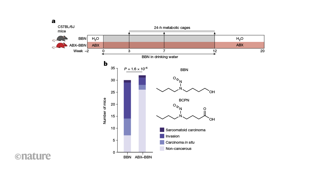
    Nature, Published online: 28 August 2024; doi:10.1038/d41586-024-02740-8

    Bacteria in the gut can affect the chemically induced growth of tumours in other parts of the body. Certain gut bacteria convert nitrosamine compounds, which are found in cigarette smoke and processed food, into metabolites that trigger the growth of tumours in the bladders of mice.

    [ad_2]

    Source link

  • The mysteries of inflammatory bowel disease are being cracked — offering hope for new therapies

    The mysteries of inflammatory bowel disease are being cracked — offering hope for new therapies

    [ad_1]

    An illustration of intestines affected by Crohn's disease highlighted in pink in a blue body

    Crohn’s disease, a form of inflammatory bowel disease, often affects the intestines (artist’s illustration).Credit: Sebastian Kaulitzki/Science Photo Library

    When geneticist James Lee and his colleagues published a paper in June linking a gene to inflammatory bowel disease (IBD), he didn’t expect the public to take much notice. Things did not go as planned.

    “I got inundated,” he says.

    By the end, Lee did more than 25 interviews for radio and print outlets around the world and received hundreds of e-mails from people with IBD. “It’s a testament to how common inflammatory bowel disease is,” says Lee, who works at the Francis Crick Institute in London. “And also a testament to how desperate people are for better treatments.”

    Lee’s paper, published in Nature1, is one of several recent reports offering hope that people with IBD could one day have better treatment options tailored to their disease. Lee and his colleagues found that changes in the activity of a gene that is important in the immune system could contribute to some cases of the disease. Another study found that some people with IBD make antibodies that disable a pivotal anti-inflammatory protein2 and a third study tracked how populations of gut bacteria adapt to an inflamed environment3.

    The papers look at IBD from different angles, but together offer a glimpse into the ways that physicians might one day be able to better match people with IBD to appropriate treatments, says David Artis, an immunologist at Weill Cornell Medicine in New York City. “Not every inflammatory bowel disease patient who walks in the door is the same,” he says. “If we can map that difference to some extent, I think we’re going to be able to better treat those people.”

    Life-altering diseases

    IBD is a painful condition that causes chronic inflammation of the digestive tract. Two of the most common forms of IBD are ulcerative colitis and Crohn’s disease. Both can cause diarrhoea, anaemia and abdominal cramping.

    Like many autoimmune disorders, IBD has an aetiology that is murky and complex, with contributions from both genetics and the environment. What is clear is that incidence of the disease is rising in many regions of the world4.

    Over the past decade, researchers have amassed a lengthy list of genetic variations that are linked to IBD. But Lee and his colleagues decided to examine a region of the genome where few geneticists had bothered to look: a “gene desert”, says Lee, so named because it is devoid of any recognizable genes. “We didn’t know what we were going to find,” he says. “And we ended up finding a master regulator of inflammatory responses.”

    Coloured scanning electron micrograph of round goblet cells shown in pink

    Mucus-producing cells (pink; artificially coloured) stud the intestines of a person with ulcerative colitis, a common form of inflammatory bowel disease.Credit: Steve Gschmeissner/Science Photo Library

    This master regulator is a stretch of DNA that controls the activity of a gene called ETS2, which is located far away from the gene desert. High levels of ETS2 activity, the team found, boost the ability of immune cells called macrophages to promote inflammation.

    The finding also indicated that a class of cancer drugs called MEK inhibitors might prevent the activation of ETS2. The team found that these drugs could block the effects of the ETS2 protein, including the release of inflammation-promoting molecules, in cells grown in the laboratory. But MEK inhibitors can become toxic to other cells if given over the long term, says Lee, and so the team is developing ways to deliver the inhibitors only to macrophages before testing the approach in people with IBD.

    Rogue antibodies

    Another study has found a select group of people with IBD who might have a new therapeutic option in the near future. Paediatric immunologist Sophie Hambleton at Newcastle University in Newcastle upon Tyne, UK, and her colleagues analysed samples from two children with IBD. The scientists discovered that the children were producing antibodies that block the activity of a protein called IL-102. This protein has anti-inflammatory effects in the gut.

    But the children’s antibodies meant that IL-10 was unable to dampen inflammation in their intestines, leading to IBD, the researchers reported in July in the New England Journal of Medicine. Once the link between IL-10 and their disease was identified, one of the children was treated with therapies to deplete the antibodies, easing their symptoms.

    It’s unclear how many people with IBD make antibodies against their own IL-10, says Hambleton. But when the team looked at a sample of adults with IBD, they found “a clear minority” who also produced the antibodies. “We are very confident that this is going to be a contributory mechanism in more patients,” she says.

    Microbiome reaction

    In addition to genetics and immune cells, microorganisms are thought to play a part in IBD. In the third study, Christopher Smillie, who studies the human microbiome at Harvard Medical School in Boston, Massachusetts, and his colleagues, looked at how chronic inflammation shapes evolution of the microorganisms living in the digestive track3.

    They identified 140,000 bacterial strains in stool samples from people with and without IBD. Hundreds of these strains were associated with IBD, and many appear to have adapted to living in inflamed tissue. Among those, several could be used to predict disease severity: for example, the abundance of some strains of Eggerthella lenta declined as the levels of a protein associated with inflammation rose. The results were published in Cell Host & Microbe in July.

    Ultimately, Smillie hopes that characterization of these microorganisms will lead to ways to monitor disease progression, and to sort people with IBD into groups on the basis of how likely they are to respond to potential therapies.

    Each of these studies could contribute to that goal, but the work is still preliminary, says Gabriel Nuñez, an immunologist at the University of Michigan Medical School in Ann Arbor. For example, the microbial study does not show that any of these organisms contribute to disease, he notes. And it is unclear what proportion of people with IBD have altered ETS2 activity or make autoantibodies against IL-10. “Perhaps these are rare patients, and only a handful in the world will benefit,” he says.

    Nevertheless, if only a handful of people find relief because of these results, that will be progress, he adds. “Even if you cure only one patient, it’s important for that person and their family.”

    [ad_2]

    Source link

  • How the stressed-out brain can weaken the immune system

    How the stressed-out brain can weaken the immune system

    [ad_1]

    Artist's illustration of the amygdaloid body (shown in red) in the brain on a black background

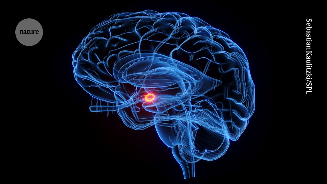
    The vagus nerve connects the brain region called the amygdala (red; artist’s illustration) to nerves for the Brunner’s glands in the gut.Credit: Sebastian Kaulitzki/Science Photo Library

    Stress can make people feel sick, and bacteria in the gut might be to blame, according to a study1 in mice. The research suggests that a stressed brain directly shuts down specific glands in the gut, affecting gut bacteria and the body’s broader immune system.

    The study “is a technical tour de force”, says neuroscientist John Cryan at University College Cork in Ireland, who reviewed the study. Most work on the gut–brain connection has focused on how bacteria affect the brain, so Cryan welcomes research into how psychological states can exert ‘top-down’ control of bacteria. “It’s a really cool part of the puzzle”, he says. The research was published on 8 August in Cell.

    Gut–brain chitchat

    Researchers have long known that the gut and brain ‘talk’ to each other. Under stress, the brain spurs the release of hormones that can trigger gut conditions such as inflammatory bowel disease. And certain bacteria in the gut can release chemical signals that affect the brain and behaviour.

    But the neural communication pathways are less understood. To find out more, neuroscientist Ivan de Araujo at the Max Planck Institute for Biological Cybernetics in Tübingen, Germany, and his colleagues focused on small organs called Brunner’s glands that are found in the walls of the small intestine. Little is known about these glands, other than that they produce mucus and contain numerous neurons.

    De Araujo’s team found that removing the Brunner’s glands of mice made the animals more susceptible to infection. It also raised markers of inflammation, a flood of immune chemicals and cells that can damage tissues. The team saw a similar effect in humans: people who’d had tumours removed from the part of the gut containing Brunner’s glands had higher levels of white blood cells — a marker of inflammation — than people who’d had tumours removed from other areas.

    Housekeeping bacteria

    Closer analysis showed that removing the Brunner’s glands from mice eliminates bacteria in the Lactobacillus genus, which live in the small intestine. In a healthy gastrointestinal tract, Lactobacilli stimulate production of proteins that act as grout between the cells lining the gut, keeping most of the gut’s contents inside while allowing certain nutrients to enter the bloodstream. But when Lactobacilli are gone, the gut becomes ‘leaky’ and “things that shouldn’t cross into the blood do so”, de Araujo says. The immune system attacks these foreign molecules, causing the inflammation and illness seen in mice without Brunner’s glands.

    The researchers then examined the glands’ neurons. They found that the neurons connect to fibres in the vagus nerve, a communications pathway between the gut and the brain. These fibres run directly to the brain’s amygdala, which is involved in emotion and stress response.

    Placing mice with intact Brunner’s glands under chronic stress had the same effect as removing the glands: Lactobacillus levels fell, and inflammation increased. This suggested that stress had shut down the Brunner’s glands.

    Lines of communication

    Asya Rolls, a neuroimmunologist at the Technion — Israel Institute of Technology in Haifa, is impressed by the direct line between the brain, Brunner’s glands, bacteria and immune system. “The specificity of the connection is amazing,” she says. But she cautions that the pathways in mice might not be identical to those in humans.

    “This paper is pretty inspiring,” says Christoph Thaiss, a microbiologist and neuroscientist at the University of Pennsylvania in Philadelphia. Understanding the specific pathways that connect the brain and gut, he says, could help researchers to study questions such as why some people are more resilient to stress than others.

    De Araujo says the study could have implications for treating stress-related disorders such as inflammatory bowel disease. His group is now studying whether chronic stress affects this pathway in infants, who receive their Lactobacillus through breast milk. “We are excited about the idea that these glands are important for normal development and immune function early in life,” de Araujo says.

    [ad_2]

    Source link