[ad_1]
- NEWS AND VIEWS
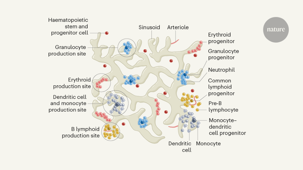
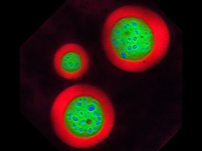
A method for imaging the production of blood cells in the bones of mice has revealed the organization of cell lineages, both in a steady state and in response to stressors, such as bleeding and infection.
[ad_2]
Source link
[ad_1]
A method for imaging the production of blood cells in the bones of mice has revealed the organization of cell lineages, both in a steady state and in response to stressors, such as bleeding and infection.
[ad_2]
Source link

[ad_1]
A study introduces a novel method for calibrating the spring constant of FluidFM micropipette cantilevers, crucial for the accurate measurement of forces in microfluidic environments. This method addresses the limitations of current calibration techniques, offering a significant advancement in the field of force microscopy.
Fluidic force microscopy (FluidFM) combines the sensitivity of atomic force microscopy with microfluidics’ capabilities, necessitating precise calibration of its cantilevers for reliable data. Traditional methods, however, struggle with the unique internal structure of FluidFM cantilevers, leading to inaccuracies.
A recent study (https://doi.org/10.1038/s41378-023-00629-6) published on February 18, 2024, in the journal Microsystems & Nanoengineering, researchers unveiled an innovative calibration technique for FluidFM micropipette cantilevers, pivotal for exact force measurements in microfluidic environments.
The FluidFM is a tiny tool used in microscopic environments to measure forces with high precision. Unlike traditional methods that often fall short due to the complex inner structure of FluidFM cantilevers, this new approach leverages the cantilever’s resonance frequencies in both air and liquid environments. By focusing on these frequencies, the method circumvents the common pitfalls of the widely-used Sader method, which can introduce errors due to its reliance on geometric and fluidic assumptions that don’t hold up well for FluidFM’s unique cantilever designs. This innovative calibration technique was meticulously tested and validated on data obtained by the HUN-REN Nanobiosensorics Lab, Cytosurge, Nanosurf and Bruker, showing that it not only provides more accurate measurements but also simplifies the calibration process by reducing the effects of noise and eliminating the need for intricate experimental setups.
Our method simplifies the calibration process, significantly reducing the influence of noise and eliminating the need for complex measurements, marking a significant step forward in the practical application of FluidFM technologies.”
Dr. Attila Bonyár, study’s lead author
The new calibration method promises enhanced accuracy in force measurements, with profound implications for biological, biophysical, and materials science research. It enables the precise manipulation of cells and nanoparticles, opening new avenues for investigation in these fields.
Source:
Journal reference:
Bonyár, A., et al. (2024). Hydrodynamic function and spring constant calibration of FluidFM micropipette cantilevers. Microsystems & Nanoengineering. doi.org/10.1038/s41378-023-00629-6.
[ad_2]
Source link

[ad_1]

High-speed atomic force microscopy combined with a laser irradiation system for the in-situ real-time observation of azo-polymer deformation process. Credit: Osaka University
Expanding our scientific understanding often comes down to getting as close a look as possible at what is happening. Now researchers from Japan have observed the nanoscale behavior of azo-polymer films while triggering them with laser light.
In a study recently published in Nano Letters the researchers from Osaka University used tip-scan high-speed atomic force microscopy (HS-AFM) combined with an optical microscope to create movies as the polymer films changed.
Azo-polymers are photoactive materials, meaning they undergo changes when light is shined on them. Specifically, light changes their chemical structure, which alters the surface of the films. This makes them interesting for applications such as optical data storage and providing light-triggered motion.
Being able to initiate these changes with a focused laser light while capturing images is known as in situ measurement.
“It is usual to investigate changes in polymer films by subjecting them to a treatment, such as irradiating with light, and then making measurements or observations afterward. However, this provides limited information,” explains study lead author Keishi Yang. “Using an HS-AFM setup including an inverted optical microscope with a laser, allowed us to trigger changes in azo-polymer films while observing them in real-time with high spatiotemporal resolution.”
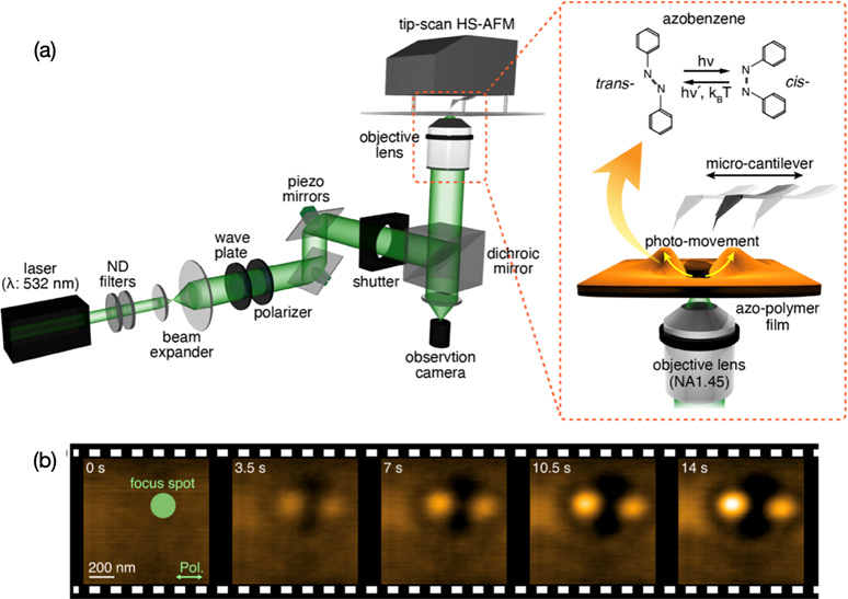
(a) Overview of the high-speed atomic force microscopy integrated with the laser irradiation system (b) High-speed atomic force microscope images of the azo-polymer deformation. Credit: The American Chemical Society
The HS-AFM measurements were able to track the dynamic changes in the surfaces of the polymer films in movies with two frames per second. It was also found that the direction of the polarized light used had an influence on the final surface pattern.
Further investigation using the in situ approach is expected to lead to a thorough understanding of the mechanism of light-driven azo-polymer deformation, allowing the potential of these materials to be maximized.
“We have demonstrated our technique for observing polymer film deformation,” says study senior author Takayuki Umakoshi. “However, in doing so, we have shown the potential of combining tip-scan HS-AFM and a laser source for use across materials science and physical chemistry.”
Materials and processes that respond to light are important in a wide range of fields in chemistry and biology, including sensing, imaging, and nanomedicine. The in situ technique provides an opportunity to deepen understanding and maximize potential and are hence expected to be applied to various optical devices.
Reference: “In Situ Real-Time Observation of Photoinduced Nanoscale Azo-Polymer Motions Using High-Speed Atomic Force Microscopy Combined with an Inverted Optical Microscope” by Keishi Yang, Feng-Yueh Chan, Hiroki Watanabe, Shingo Yoshioka, Yasushi Inouye, Takayuki Uchihashi, Hidekazu Ishitobi, Prabhat Verma and Takayuki Umakoshi, 26 February 2024, Nano Letters.
DOI: 10.1021/acs.nanolett.3c04877
The study was funded by the Japan Society for the Promotion of Science and the Ministry of Education, Culture, Sports, Science and Technology.
[ad_2]
Source link
[ad_1]
Researchers at the National Institutes of Health have identified antibodies targeting a hard-to-spot region of the influenza virus, shedding light on the relatively unexplored “dark side” of the neuraminidase (NA) protein head. The antibodies target a region of the NA protein that is common among many influenza viruses, including H3N2 subtype viruses, and could be a new target for countermeasures. The research, led by scientists at the National Institute of Allergy and Infectious Diseases’ Vaccine Research Center, part of NIH, was published today in Immunity.
Influenza, or flu, sickens millions of people across the globe each year and can lead to severe illness and death. While vaccination against influenza reduces the burden of the disease, updated vaccines are needed each season to provide protection against the many strains and subtypes of the rapidly evolving virus. Vaccines that provide protection against a broad range of influenza viruses could prevent outbreaks of new and reemerging flu viruses without the need for yearly vaccine reformulation or vaccinations.
One way to improve influenza vaccines and other countermeasures is to identify new targets on the virus’s surface proteins in “conserved” regions-;portions that tend to be relatively unchanged between different strains of the virus. Influenza NA is a surface protein containing a globular head portion and a narrow stalk portion. The underside of the NA head contains a highly conserved region with targets for antibodies-;known as epitopes-;that make it vulnerable to antibody binding and inhibition of the virus, as well as not being impacted by mutations common in drug-resistant strains. This region is termed the “dark side” due to its partially hidden location and relatively unexplored characteristics.
The researchers isolated human antibodies that target the NA dark side from the blood of two people who had recovered from influenza type A subtype H3N2, a major subtype of seasonal flu viruses. In lab tests, the antibodies inhibited propagation of viruses from subtype H2N2, the subtype that caused pandemic influenza in 1957-58, and H3N2 viruses from humans, swine, and birds. The antibodies also protected mice from lethal infection by a subtype H3N2 virus when given to the animals either one day before or two days after infection, showing that the antibody may treat and prevent influenza in this model.
The scientists analyzed the structure of two of the antibodies while bound to NA using advanced microscopy techniques known as cryogenic electron microscopy. Each antibody targeted different, nonoverlapping regions of the dark side, demonstrating that this region has multiple areas that may be useful to explore for countermeasure development.
These findings show that the NA dark side has unique, previously untapped epitopes that could be applied to the development of new vaccine and therapeutic strategies. They suggest that antibodies targeting the NA dark side could be useful in combination with antivirals or other types of antibodies for interventions against influenza, as they are effective against influenza viruses with drug-resistant mutations. The researchers also note that NA dark side targets could be included in the next generation of broadly protective vaccines against influenza.
Source:
Journal reference:
Lederhofer, J., et al. (2024) Protective human monoclonal antibodies target conserved sites of vulnerability on the underside of influenza virus neuraminidase. Immunity. doi.org/10.1016/j.immuni.2024.02.003.
[ad_2]
Source link

[ad_1]
For centuries, coronaviruses have triggered health crises and economic challenges, with SARS-CoV-2, the coronavirus that spreads COVID-19, being a recent example. One small protein in SARS-CoV-2, the Membrane protein, or M protein, is the most abundant and plays a crucial role in how the virus acquires its spherical structure. Nonetheless, this protein’s properties are not well understood.
A research team led by a physicist at the University of California, Riverside, has devised a new method to make large quantities of M protein, and has characterized the protein’s physical interactions with the membrane -; the envelope, or “skin,” -; of the virus. The team’s theoretical modeling and simulations show how these interactions are likely contributing to the virus assembling itself.
The researchers report in their paper published today in Science Advances that when the M protein, which is adjacent to the spike protein on SARS-CoV-2, gets lodged in the membrane, it coaxes the membrane to curve by locally reducing the membrane thickness. This induction of curvature leads to SARS-CoV-2’s spherical shape.
“If we can better understand how the virus assembles itself, then, in principle, we can come up with ways to stop that process and control the virus’ spread,” said Thomas E. Kuhlman, an assistant professor of physics and astronomy, who led the research project. “M protein has previously resisted any kind of characterization because it is so hard to make.”
Kuhlman and his colleagues overcame this difficulty by using Escherichia coli bacteria as a “factory” to make the M protein in large numbers. Kuhlman explained that although E. coli can make copious amounts of M proteins, the proteins tend to clump together in the E. coli cells, eventually killing them. To circumvent this challenge, the researchers induced the E. coli cells to produce the protein Small Ubiquitin-related Modifier, or SUMO, along with the M protein.
In our experiments, when E. coli makes M protein, it makes SUMO at the same time. The M protein fuses with the SUMO protein, which prevents the M proteins from sticking to one another. The SUMO protein is relatively easy to remove via another protein that simply cuts it off. The M protein is thus purified and separated from SUMO.”
Thomas E. Kuhlman, assistant professor of physics and astronomy, UCR
The work provides fundamental insights into the mechanisms driving SARS-CoV-2 viral assembly.
“As M proteins are an integral component of other coronaviruses as well, our findings provide useful insights that can enhance our understanding and potentially enable interventions in viral formation not only in SARS-CoV-2 but also in other pathogenic coronaviruses,” Kuhlman said.
Next, the researchers plan to study the interactions of the M protein with other SARS-CoV-2 proteins to potentially disrupt these interactions with drugs.
Kuhlman was joined in the research by fellow-UCR physicists Roya Zandi and Umar Mohideen. Kuhlman was charged with making the M proteins. Mohideen, a distinguished professor of physics and astronomy, used atomic force microscopy and cryogenic electron microscopy to measure how the M protein interacts with the membrane. Zandi, an expert on virus assembly and a professor of physics and astronomy, developed simulations of how the M proteins interact with each other and with the membrane.
Other coauthors on the paper are Yuanzhong Zhang, Siyu Li, Michael Worcester, Sara Anbir, Joseph McTiernan, Pratyasha Mishra, and Ajay Gopinathan of UCR; and Michael E. Colvin of UC Merced. Co-first authors Zhang and Anbir contributed equally to the work.
The research was supported by a grant from the University of California Office of the President to investigate how the COVID-19 virus assembles itself.
Source:
Journal reference:
Zhang, Y., et al. (2024) Synthesis, insertion, and characterization of SARS-CoV-2 membrane protein within lipid bilayers. Science Advances. doi.org/10.1126/sciadv.adm7030.
[ad_2]
Source link

[ad_1]
In a recent study published in the journal Proceedings of the National Academy of Sciences, researchers demonstrated that human transferrin receptor (TfR) mediates severe acute respiratory syndrome coronavirus 2 (SARS-CoV-2) infection.
Coronavirus disease 2019 (COVID-19), caused by SARS-CoV-2, presents influenza-like manifestations, including mild-to-severe pneumonia, acute respiratory distress syndrome, multiorgan failure, and fatal lung injury. Further, the etiology and pathogenesis of COVID-19 are not entirely understood and targeted therapies remain inadequate.
The viral spike protein binds to the host receptor, angiotensin-converting enzyme 2 (ACE2), for cellular entry. Although SARS-CoV-2 preferentially infects cells in the respiratory tract, the virus has been detected in virtually all organs. Studies have revealed the presence of SARS-CoV-2 RNA in diverse cells lacking ACE2, suggesting that other receptors or co-receptors may mediate viral entry.
 Study: Human transferrin receptor can mediate SARS-CoV-2 infection. Image Credit: Kateryna Kon / Shutterstock
In the present study, researchers identified TfR as an alternative receptor mediating the cellular entry of SARS-CoV-2. First, they used co-immunoprecipitation (Co-IP) to identify host proteins interacting with the viral spike in Calu-3 cells. This revealed 293 proteins, including 42 transmembrane proteins; two proteins were associated with entry (ACE2 and TfR). Next, the team evaluated TfR expression in the respiratory tract and liver in mice.
TfR expression, both transcript and protein levels, was substantially higher in the lungs and trachea than in other tissues. Using immunohistochemical analysis, the researchers investigated the effects of SARS-CoV-2 on TfR expression in the lungs of humanized ACE2 (hACE2) mice and monkeys. This revealed a 1.5- and 1.8-fold increase in TfR expression in mice and monkeys, respectively.
In addition, surface plasmon resonance revealed direct interactions between the viral spike and human TfR. Notably, the spike protein lacked interactions with Syrian hamster or mouse TfR. Docking analysis predicted two peptide sequences (QK8: QDSNWASK and SL8 SKVEKLTL) in TfR to be involved at the interface of TfR-spike interactions.
Mutagenesis and Co-IP revealed that the A529 residue in TfR was essential for interactions with the spike. Further analysis indicated that physiological interactions between spike and TfR occurred at the cellular surface and during endocytosis. This was confirmed by electron microscopy using SARS-CoV-2 pseudoviral spike and HEK293/hACE2 and BHK-21/TfR cells.
Next, the team evaluated the effects of soluble TfR, anti-TfR antibody, and SL8 and QK8 peptides on SARS-CoV-2 infection using reverse-transcription polymerase chain reaction (RT-PCR) and plaque assays. Results showed their inhibitory effects on SARS-CoV-2 in Vero E6 and Calu-3 cells. Cytotoxicity was not observed even at 1,000 nM.
Confocal microscopy revealed that TfR was widespread on the surface of Calu-3 and Vero E6 cells, with the colocalization of TfR and SARS-CoV-2 at the surface and during endocytosis. Notably, treatment with the anti-TfR antibody inhibited the colocalization. Further, electron microscopy showed that viral particles were present in the cytosol and clathrin-coated pits in Vero E6 cells; likewise, treatment with anti-TfR antibody inhibited viral internalization.
Next, ACE2 was knocked out (KO) from Calu-3 and Vero E6 cells and the cells were infected with SARS-CoV-2. This inhibited infection by 40% to 50%, suggesting that ACE2 might not be the only receptor mediating infection. In addition, TfR knockdown (KD) inhibited infection by 30%, whereas its overexpression (OE) promoted infection. TfR KO was not performed as it is lethal. TfR OE or KD did not impact ACE2 expression.
Further, the team transfected C57 mice with adenovirus vector (Ad5) expressing hACE2 or humanized TfR (hTfR) and infected them with SARS-CoV-2. Viral load in the lungs in Ad5-hTfR and Ad5-hACE2 mice was significantly higher than in Ad5-empty mice. Finally, the researchers evaluated the effects of the anti-TfR antibody on infection in rhesus macaques. Anti-TfR antibody inhibited viral replication and reduced pneumonia.
Viral load in the respiratory epithelium was also significantly lower between 3- and 7 days post-infection (dpi) compared to controls. Radiographs taken at 0 and 5 dpi revealed significantly less severe pulmonary infiltration in antibody-treated macaques relative to controls. Antibody-treated animals had no significant pulmonary lesions, while controls showed lung lesions of varying degrees.
Taken together, the study described the human TfR as a receptor for SARS-CoV-2. TfR can directly bind to the viral spike at an affinity comparable to that of ACE2. Notably, mouse TfR and the viral spike lacked interactions. Soluble TfR, SL8, and QK8 peptides and anti-TfR antibodies can inhibit infection. The team also illustrated the antiviral effects of the anti-TfR antibody in rhesus macaques. Overall, TfR could serve as an alternative infection pathway, facilitating viral entry through endocytosis.
[ad_2]
Source link

[ad_1]
The cells glow green under the high-powered microscope, each bedazzled with a constellation of luminous proteins and RNA that, like oil droplets in water, have huddled together through a process known as phase separation.
A foundational concept in the fields of engineering, chemistry and physics, phase separation — the mechanism by which complex mixtures segregate into distinct components — is beginning to revolutionize biology as well. The process is being hailed as a key organizing principle of the cell.
In a fourth-floor laboratory at the Whitehead Institute in Cambridge, Massachusetts, chemical biologist Henry Kilgore has tagged a protein to emit a verdant fluorescent signal wherever throngs of RNA-splicing factors, unbound by membranes, congregate in the cell nucleus to influence gene regulation. “Oh, these look really nice,” he murmurs.
Far from being a disorganized warehouse, the cell more closely resembles an intricate and efficient logistics hub. Clusters of proteins and nucleic acids are compartmentalized into specialized units — called condensates — each with an invisible boundary that is shaped by biochemical affinity and the unique features of phase separation.
The molecules inside are actively sorted, brought together and dispersed, mirroring the bustling efficiency of a dynamic distribution centre. This orderly orchestration underpins the cell’s capacity to regulate its internal environment with precision, allowing for everything from gene control and stress responses to DNA repair and cell division. When this sorting mechanism goes awry, diseases such as cancer, diabetes and neurodegenerative conditions can arise.
Kilgore blasts one condensate with a laser, wiping out its fluorescent signal. Then, he records how quickly the green glow comes back. Called fluorescence recovery after photobleaching, or FRAP, the technique is a popular choice for studying the molecular and fluid dynamics of phase-separated droplets. “This is one of the more convincing demonstrations that a molecule’s mobility can be changed in condensates while doing condensate things,” Kilgore says.
But it’s not the only way to do so. Condensate researchers now have a range of molecular, biophysical and computational tools at their disposal — “technologies that allow us to precisely measure and control phase behaviour in living cells”, says biophysicist Cliff Brangwynne at Princeton University in New Jersey.
A growing number of biotechnology start-up companies — including one that Brangwynne founded, called Nereid Therapeutics in Boston, Massachusetts — now aims to harness these techniques for drug discovery. Academic researchers are playing their part, too. By leveraging these technologies to explore new frontiers in condensate biology, they are transforming the understanding of cellular operations and opening pathways for medical intervention.
“The tools are going to validate themselves,” says Tuomas Knowles, a biophysicist at the University of Cambridge, UK, who doubles as chief executive of Transition Bio in Cambridge, Massachusetts. “These are just incredibly powerful techniques, and they are going to really drive this field forward.”
The pioneering US cell biologist Edmund Beecher Wilson had none of these tools when, in 1899, he conjectured1, on the basis of crude imaging of sea-star eggs, that cells might harbour “a mixture of liquids” interspersed with “suspended drops … of different chemical nature”.
Yet, it would take more than a century for scientists to prove Wilson right.
The shape-shifting blobs that shook up cell biology
In 2008, at a summer workshop in Woods Hole, Massachusetts, a team led by Brangwynne and his postdoctoral adviser, cell biologist Tony Hyman at the Max Planck Institute of Molecular Cell Biology and Genetics in Dresden, Germany, noticed that clusters of RNA and protein found in worm embryos behave like liquids, even though they were thought to be solid2.
Like oil droplets in a well-mixed vinaigrette, the structures seemed to coalesce and dissolve, choreographed by some kind of intrinsic molecular or biophysical pull.
“It was surprising and cool,” says Brangwynne, who, together with Hyman, was awarded the 2023 Breakthrough Prize in Life Sciences, in part for this work.
Researchers around the world jumped on the discovery, delineating condensates in their own model systems and seeking the rules that govern their assembly. Many condensates seemed to be built around disordered proteins, which, although lacking a rigid structure, have the flexibility to interact with other molecules in versatile and dynamic ways.
Scientists can visualize these structures by tagging proteins with light-emitting markers — as Kilgore did with his splicing-factor protein — and tracking where the glowing signal aggregates in cells. But that method only works for naturally occurring condensates. And as Knowles points out: “You can’t really understand something unless you can take it apart and control it.”
A major advance, therefore, has been the ability to toggle condensate formation at will, a capability that became possible after the introduction of light-tunable technologies with playful names such as optoDroplet3 and OptoGranules4.
These ‘optogenetic’ platforms take advantage of special light-responsive domains that are fused onto condensate-prone proteins of interest. When exposed to a specific wavelength of light, these engineered proteins self-aggregate. This triggers condensate assembly and provides a powerful tool for researchers to observe and analyse the process in real time.
Academic scientists have embraced these tools, and used them to dissect the material properties of condensates and the molecular interactions that propel phase separation. “And as time goes by, we’ll be able to design these tools more carefully such that we can interrogate the real driving forces underlying endogenous condensates,” says Dan Bracha, a bioengineer at the Technion—Israel Institute of Technology in Haifa.
At Nereid, scientists are leveraging Corelet, an optogenetic technology co-developed5 by Bracha during his postdoctoral tenure in Brangwynne’s lab, to initiate condensate formation in cellular models. The technology is key to the firm’s strategy of identifying therapeutic compounds that can alter the dynamics of phase separation, says Nereid chief scientific officer John Reilly.
For neurodegenerative disease, in which abnormal condensate formation is implicated, the company’s objective is to find molecules that can prevent these condensates from assembling. Conversely, for scenarios in which condensates are beneficial, such as in enhancing the expression of genes that fight tumours, Nereid focuses on compounds that promote phase separation.
“It’s a great screening tool,” Reilly says. “It has given us actionable small molecules that we can now turn into drugs.”

Optogenetic technology Corelet initiates condensate formation in the nucleus. Phase separation occurs after activation with light.Credit: Dan Bracha and Cliff Brangwynne
But optogenetic systems like Corelet only work with proteins that are already known to form condensates. That’s not a problem for drug hunters such as Reilly, who want to perturb condensate dynamics in natural settings for therapeutic gain. But many synthetic biologists are also seeking to harness condensate-mediated organization to imbue cells with new and interesting capabilities, such as enhanced drug production or the formation of super-crops and microorganisms.
“We want to build simple, engineerable, modular systems,” says Ashutosh Chilkoti, a biomedical engineer at Duke University in Durham, North Carolina.
In 2023, Chilkoti unveiled a series of artificial proteins that provide this level of control over condensate formation in bacterial cells. Guided by the molecular principles of phase separation, he and his colleagues — including Duke synthetic biologist Lingchong You and chemical biologist Yifan Dai, who is now at Washington University in St. Louis, Missouri — crafted shape-shifting proteins with repeating peptide sequences. These not only mimic the structure of disordered proteins found in natural condensates, but can also be fine-tuned to enable precise command over what they do.
Some of the bespoke condensates the researchers made boosted gene expression, whereas others effectively isolated target proteins from degradation components, extending their half-life6. A few even modified the distribution of charged particles in the cell, leading to electrochemical shifts that influenced cellular stress responses and overall gene-activity patterns7. “It’s programmable in a way that you can have precise regulation,” explains You.
What lava lamps and vinaigrette can teach us about cell biology
The team behind these configurable condensates is mostly looking to harness the structures for synthetic-biology applications. But, as Dai points out, the same tools are also revealing the biochemical features that govern phase separation inside cells and suggesting never-before-seen functions for condensates — for example, creating pH gradients and electrical potentials — that others can now test. “It’s generating new hypotheses,” he says.
Similarly, biochemist Dek Woolfson and his colleagues at the University of Bristol, UK, have crafted synthetic proteins that are capable of co-condensing with one another. Each protein in the researchers’ design gets strategically fused to different enzymes that act in tandem to carry out a common biochemical function. Phase separation brings these proteins together, enhancing the efficiency of the associated pathway.
As a proof of concept, the researchers engineered condensates that would co-localize a pair of enzymes involved in the two-step conversion of the amino acid tryptophan to indigo, a blue dye. Bacteria expressing these designer condensates produced up to six times more indigo compared with bacteria with free-floating enzymes dispersed separately throughout the cell8.
According to co-author Alex Hilditch, a former doctoral student in Woolfson’s lab who is now at the Swiss Federal Institute of Technology in Lausanne (EPFL), this synthetic co-condensation strategy has the potential to optimize a wide range of enzymatic processes in bacterial-cell manufacturing. “And the real benefit,” he says, “is that, because you are just co-localizing things” — rather than altering their expression levels — “hopefully you shouldn’t increase the metabolic burden on your chassis organism”.
Even with these newer technologies, the mainstays of condensate research remain older methods such as FRAP that have been adapted for phase separation but originated in other fields of enquiry. “There’s a lot of repurposing,” says Sua Myong, a molecular biophysicist at Boston Children’s Hospital. And a lot of poking, prodding, and mixing and matching of different techniques, often in both test tubes and living cells.
“Every procedure has its own caveats,” explains Alessandra Dall’Agnese, a cell biologist at the Whitehead. “This is why it is important to accumulate orthogonal lines of evidence that support, or not, your hypothesis.”

Phase separation inside the nucleolus.Credit: Marina Feric and Cliff Brangwynne
For molecular imaging, for example, researchers often tag proteins of interest with fluorescent markers. But these biochemical dongles can change the solubility or charge distribution of a protein, altering the kinetics of phase separation. “You have to be mindful of picking the right fluorescent protein to minimize these confounding effects,” says Jonathon Ditlev, a cellular biophysicist at the Hospital for Sick Children in Toronto, Canada.
Yet another consideration is that many of today’s laboratory techniques only work with big condensates — those that either can be seen under a microscope, or that contain artificially high concentrations of their constituent parts. Condensates come in a range of sizes, however. And it is quite possible, says Steph Weber, a cell biologist at McGill University in Montreal, Canada, that condensation inside small droplets happens by a completely different means, with different functional consequences, than it does for visible conglomerates.
“We’ve been looking at all these big things,” Weber says. But what happens inside smaller clusters could be equally, or more, important determinants of cellular order. “That might be where the action is.”
Translating any experimental insight into actionable treatments is a complex task — a challenge well known to the team at one-time biomolecular firm Faze Medicines.
Company scientists had been trying to develop small molecules that could rectify the aberrant phase changes associated with cancer and motor neuron disease (also known as amyotrophic lateral sclerosis). They designed high-throughput assays to identify drug candidates that are capable of disrupting harmful interactions in condensates in a controlled laboratory setting9. Promising compounds could then be advanced to testing in living cells.
But, as Rachel Meyers, the company’s former chief scientific officer, points out: “It’s complicated biology.” And when broader economic pressures led investors to consolidate their holdings, the company was forced to cease operations. “We just weren’t far enough along,” Meyers says.

NatureTech hub
One company that is further along is Dewpoint Therapeutics in Boston. Last year, the firm, which was co-founded by Hyman and Whitehead biologist Rick Young (Dall’Agnese and Kilgore are both members of Young’s lab and consult for Dewpoint), unveiled a series of condensate-modifying drugs capable of rescuing motor neurons from the ravages of motor neuron disease, at least in cell culture. When administered at therapeutically relevant concentrations, these drugs helped to restore gene activity to normal levels and reduced the harmful effects of stress on the neurons, specifically preventing the cellular extensions known as axons and dendrites from shrinking, a common issue in the disease.
The company found these molecules through a phenotypic screening strategy, using robotic systems to bombard cells with an array of 370,000 unique molecules from Dewpoint’s chemical library. The process uses automated imaging systems and advanced artificial intelligence (AI) software to capture, quantify and sort through high-resolution pictures of the cells, looking for changes in the size, position or composition of condensates that could signal potential therapeutic benefits.
“It’s a discovery and technology platform,” explains chief scientific officer Isaac Klein — one that allows for “precision alteration of condensate behaviour”.
AI also underpins the drug-discovery philosophy at Transition Bio, which has branded its research platform Condensomics. The company uses a microfluidic system developed in Knowles’s academic laboratory for studying condensate dynamics under tens of thousands of conditions10. Data are then fed into machine-learning algorithms designed to tease apart the molecular ‘grammar’ of phase separation, aiding target prediction and drug design11.
“There’s enormous potential to include these techniques now in almost all aspects of our work,” says Knowles, who last year described a series of antimicrobial peptides discovered in this way that phase separate together with nucleic acids inside bacteria to exert their inhibitory effects12. “If you want to understand the sequence grammar,” he adds, “AI is really the only way to do that.”
Knowles’s AI model, termed DeePhase, forecasts protein entry into condensates on the basis of their structural pliability, fostering interactions at disordered regions that lead to phase separation. Yet, as Kilgore points out, these regions are not the only drivers of phase separation; other structures play a part as well.
Together with members of Regina Barzilay’s computer science lab at the Massachusetts Institute of Technology in Cambridge, Kilgore has been developing an AI tool that takes a holistic view of proteins and can even suggest sequences that should undergo phase separation inside cells. By integrating machine learning with these kinds of AI-generated sequence, Kilgore foresees a deeper understanding of the fundamental mechanisms and chemical principles that govern these enigmatic cellular structures, ushering in a new era of discovery for academics and drug developers alike.
Back in his laboratory, Kilgore zaps another condensate. As he considers the technical toolbox slowly taking shape in his field, he muses aloud: “It’s really going to be a game changer.”
[ad_2]
Source link

[ad_1]
The fragmentation of type I collagen has been seen to impair the integrity of the dermal extracellular matrix (ECM). This results in lower type I procollagen synthesis and contracted or collapsed fibroblasts.
A recent Experimental Dermatology study assessed how these deleterious changes could be reversed by injecting cross-linked hyaluronic acid (CL-HA).
 Study: Implications for cumulative and prolonged clinical improvement induced by cross-linked hyaluronic acid: An in vivo biochemical/microscopic study in humans. Image Credit: Ground Picture/Shutterstock.com
Photoaging of the human skin is caused by chronic exposure to ultraviolet irradiation. This is characterized by fragility, wrinkles, and low elasticity, mainly caused by dermal deleterious molecular alterations.
ECM in the dermis comprises 80%–90% type I collagen, which supports and strengthens the human skin. Dermal fibroblasts produce type I collagen, which forms an interwoven mesh in the ECM. The interactions between the ECM and dermal fibroblasts determine cell functions.
As the skin photoages, fragmentation of type I collagen occurs due to a greater expression and enzymatic activity of metalloproteinases (MMPs). This weakens the collagenous scaffolding, making it difficult for fibroblasts to bind.
Prior research has shown that cross-linked hyaluronic acid (CL-HA) injection can reverse these changes.
To delve further into the biochemical mechanism of CL-HA action, CL-HA and vehicle (saline) were injected into the photoaged skin of human subjects aged 60 years and above.
To assess whether fibroblast activation led to the deposition/accumulation of dermal collagen, biochemical/microscopic analyses were performed.
From 1 week to 6–9 months post-injection, fibroblasts demonstrated activation. Multiphoton microscopy at 1-week post-injection showed stretching of fibroblasts. This indicated greater dermal mechanical support.
A second harmonic generation microscopy analysis at four weeks post-injection showed densely packed thick collagen bundles around pools of injected CL-HA.
At 12 months post-injection, it was noted that thick collagen bundles accumulated, and a significant amount of CL-HA was also present.
Therefore, it was concluded that CL-HA enhanced mechanical support rapidly and durably by occupying space in the dermal ECM.
CL-HA prompted fibroblast activation and elongation, resulting in densely packed and thick type I collagen bundles at four weeks post-injection and continuing till at least week 52.
Important components of the TGF-β pathway were also stimulated post-injection, and this pathway is crucial for the synthesis of type 1 procollagen by fibroblasts.
The elements of the TGF-β pathway are decreased in photoaged skin, which reduces the synthesis of type 1 procollagen. The findings of this study suggest that the activation of the TGF-β pathway could regulate the response of fibroblasts to higher dermal mechanical support.
It was noted that post-injection with CL-HA, procollagen N- and C- proteinase enzymes increased rapidly. These enzymes aid in the assembly of intact type 1 collagen.
Immediately after the injection, clinical improvement of the skin could be due to the filler, which occupies space in the dermal ECM and provides mechanical support.
Considering the fact that collagen bundles accumulated till at least week 52, it could be the case that following the CL-HA injection, the type 1 procollagen is converted to
durable and stable dermal type 1 collagen. Furthermore, the presence of the injected filler after 12 months suggests the are long-lasting clinical benefits of CL-HA.
Future research should investigate which properties of HA contribute to the clinical benefits of CL-HA.
The findings documented here have other clinical implications as well. Intact and mature type 1 dermal collagen has an extremely stable half-life of approximately 15 years.
This suggests that collagen bundles should continue accumulating for years after a single CL-HA injection, reducing the need for subsequent treatment following each re-injection.
The observations made here also support the theory that the integrity of the dermal ECM heavily determines functional decline and fibroblast collapse in photoaging.
Interestingly, these functional and cellular changes are reversible by stimulating fibroblasts in photoaged skin by augmenting dermal mechanical support.
In sum, the findings in this study suggest that interventions that provide more mechanical support to photoaged can activate fibroblasts durably and rapidly, thereby leading to collagen deposition.
More specifically, a CL-HA injection stimulated synthetic activation and fibroblast stretching durably (for 6-9 months) and rapidly (within one week).
The result was the accumulation of densely packed and thick type 1 collagen bundles. This accumulation started as early as four weeks and lasted at least a year post-injection.
A sustained clinical improvement is obtained by the accumulation of dermal collagen, which can last for years. The findings of this study pave the way for future research on the frequency and timing of repeat CL-HA injections.
Journal reference:
Wang, F., Do, T. T., Smith, N., Orringer, J. S., Kang, S., Voorhees, J. J., and Fisher, G. J. (2024) Implications for cumulative and prolonged clinical improvement induced by cross-linked hyaluronic acid: An in vivo biochemical/microscopic study in humans. Experimental Dermatology. 33(1), e14998. doi: https://doi.org/10.1111/exd.14998. https://onlinelibrary.wiley.com/doi/10.1111/exd.14998.
[ad_2]
Source link

[ad_1]
In a recent report published in the Morbidity and Mortality Weekly Report (MMWR), the United States (US) Centers for Disease Control and Prevention (CDC) shares its recommendations for the tests and protocols required to diagnose syphilis in the US. The recommendations build upon existing conventional serologic algorithms and involve the combined use of nontreponemal and treponemal tests to identify if the patient’s immune response indicates a current untreated infection or a past-treated one. Additionally, recommendations for the direct microscopic detection of Treponema pallidum, the causative pathogen of syphilis, are included. These recommendations will help clinical laboratory directors, clinicians, laboratory staff, disease control personnel, and patients combat this dreadful disease.
 CDC Laboratory Recommendations for Syphilis Testing, United States, 2024. Image Credit: Peddalanka Ramesh Babu / Shutterstock
Syphilis is a bacterial sexually transmitted infection (STI) that progresses in stages from a painless sore (stage 1) to a severe disease that may attack the brain, liver, nerves, eyes, or cardiovascular system (stage 3). Caused by the bacterium Treponema pallidum subspecies pallidum, the disease has been further associated with congenital complications such as stillbirths, spontaneous abortions, and miscarriages.
Alarmingly, the global burden of syphilis is rising rapidly, especially in wealthy and developed countries, with approximately 6 million new cases in 2021 alone. Reports from the United States of America (US) observe an increase of 2,140% in syphilis incidence rates between 2000 (n = 5,979) and 2020 (n = 133,945), suggesting a local epidemic within the country. Research has characterized this epidemic as displaying significant health disparities, with gender and sexual minorities being the worst affected.
The laboratory diagnosis of syphilis represents a crucial effort in the war against the disease. Timely diagnoses, especially before the onset of stage 3 syphilis, are critical for positive disease outcomes. Public health reporting by laboratories provides the Centers for Disease Control and Prevention (CDC) and other government agencies the information needed to enact policies to attenuate local outbreaks and monitor epidemic trends.
The current report details the CDC’s first-ever recommendations for syphilis testing and includes optimal methods for point-of-care (POC) tests, laboratory-based tests, sample processing, and reporting. These recommendations were formulated by CDC scientists in collaboration with the Association of Public Health Laboratories (APHL) after reviewing up-to-date peer-reviewed literature, especially publications published by the US Food and Drug Administration (FDA).
The first section of the report summarises progress in syphilis serological testing since the invention of the Wassermann test through to current nontreponemal (lipoidal antigen) and treponemal tests. In brief, when patients display symptoms or signs of syphilis or have had known sexual contact with a diagnosed syphilis patient, nontreponemal (lipoidal antigen) tests are recommended for laboratory screening. These tests are also recommended when assessing reinfections or reporting outcomes during clinical trials.
“Nontreponemal (lipoidal antigen) tests might be less sensitive than treponemal tests in early primary syphilis and tend to wane with time regardless of treatment. Before testing, test and specimen type should be carefully considered because serum and plasma cannot always be used interchangeably, and certain nontreponemal (lipoidal antigen) tests require heat treatment of specimens. One caveat of nontreponemal (lipoidal antigen) tests is that a reactive result could be a false positive because of recent conditions (e.g., infections, vaccinations or injection drug use, or underlying autoimmune or other chronic conditions).”
Nontreponemal tests are usually carried out manually, but some test versions (e.g., the rapid plasma reagin [RPR] test) may be automated to increase throughput. In the latter (automated) case especially, care must be taken to ensure that samples are maintained at optimal conditions and coinfections are accounted for to ensure diagnostic performance and prevent false positives. Treatment outcomes are best reported by comparing nontreponemal antibody titers at baseline and 12 months following treatment, at which time titers are expected to reduce fourfold.
Treponemal tests are used to validate nontreponemal test results and to diagnose early syphilis infections that cannot (yet) be detected by nontreponemal tests. Most of these tests are conducted manually, but some can be automated for high throughput. In most patients, treponemal antibodies are detectable for life and are helpful to indicate a past, successfully treated syphilis infection. All donations to blood banks must undergo treponemal screening to ensure that volunteered blood is free of syphilis antibodies.
The traditional algorithm for syphilis screening involved nontreponemal tests followed by treponemal tests to confirm any dubious or contentious nontreponemal results. This is because the former is comparatively inexpensive and rapid, while the latter is manually labor-intensive, expensive, and limited in number. However, recent advancements in automated treponemal immunoassays have bridged the time and monetary gap between these techniques, resulting in the more recent ‘reverse’ algorithm for syphilis screening, wherein nontreponemal tests are used to validate the results of treponemal tests further.
Building upon these methodologies, the report provides recommendations for the optimal screening, clinical care, and recording of patients suffering from neurosyphilis, ocular syphilis, otosyphilis, and congenital syphilis. Pregnancy status, in particular, is noted as a condition that should not influence the standard interpretation of nontreponemal and treponemal test results due to scientific evidence that their functioning is not altered during pregnancy.
Immunohistochemistry and microscopy techniques for detecting syphilis from biopsies and other preserved samples are outlined with a particular focus on darkfield microscopy (the most widely used method for direct syphilis detection) and silver staining. Darkfield microscopy has been found to outperform antibody screening approaches, especially in the early stages of infection, thereby presenting itself as an ideal tool for early disease detection.
While Nucleic Acid Amplification Tests (NAATs) have shown great potential in accurately diagnosing syphilis infection, no FDA-approved NAATs currently exist. These recommendations are expected to be updated once FDA-approved NAATs enter the market.
Journal reference:
[ad_2]
Source link

[ad_1]
A recent re-emergence and outbreak of Mpox brought poxviruses back as a public health threat, underlining an important knowledge gap at their core. Now, a team of researchers from the Institute of Science and Technology Austria (ISTA) lifted the mysteries of poxviral core architecture by combining various cryo-electron microscopy techniques with molecular modeling. The findings, published in Nature Structural & Molecular Biology, could facilitate future research on therapeutics targeting the poxvirus core.
Variola virus, the most notorious poxvirus and one of the deadliest viruses to have afflicted humans, wreaked havoc by causing smallpox until it was eradicated in 1980. The eradication succeeded thanks to an extensive vaccination campaign using another poxvirus, the aptly named Vaccinia virus. The 2022-2023 re-emergence and outbreak of Mpox virus reminded us once more that viruses find ways to return to the forefront as public health threats. Importantly, this has highlighted the fundamental questions about poxviruses that have remained unanswered to this day.
One such fundamental question lies, quite literally, at the core of the matter: “We know that for poxviruses to be infective, their viral core must be properly formed. But what is this poxviral core made of, and how do its individual components come together and function?” asks ISTA Assistant Professor Florian Schur, the corresponding author of the study.
Schur and his team now put their finger on the missing link: a protein called A10. Interestingly, A10 is common to all clinically relevant poxviruses. In addition, the researchers found that A10 acts as one of the main building blocks of the poxviral core. This knowledge could be instrumental for future research on therapeutics targeting the poxviral core.
The viral core is one of the factors common to all infectious poxvirus forms.
Previous experiments in virology, biochemistry, and genetics suggested several core protein candidates for poxviruses, but there were no experimentally-derived structures available.”
Julia Datler, ISTA PhD student, one of the co-first authors of the study
Thus, the team started by computationally predicting models of the main core protein candidates, using the now-famous AI-based molecular modeling tool AlphaFold. In parallel, Datler was setting the project’s biochemical and structural foundations by drawing on her background in virology and the Schur group’s main expertise: cryogenic electron microscopy, or cryo-EM for short. “We integrated many of the most advanced cryo-EM techniques available today with AlphaFold molecular modeling. This gave us, for the first time, a detailed overall view of the poxviral core–the ‘safe’ or ‘bioreactor’ inside the virus that encloses the viral genome and releases it in infected cells,” says Schur. “It was a bit of a gamble, but we eventually managed to find the right mix of techniques to examine this complex question,” says postdoc Jesse Hansen, the study’s co-first author whose expertise in various structural biology techniques and image processing methods was pivotal for the project.
The ISTA researchers examined “live” Vaccinia virus mature virions and purified poxviral cores under every possible angle–quite literally. “We combined the ‘classic’ single-particle cryo-EM, cryo-electron tomography, subtomogram averaging, and AlphaFold analysis to gain an overall view of the poxviral core,” says Datler. With cryo-electron tomography, researchers can reconstitute 3D volumes of a biological sample as large as an entire virus by acquiring images while gradually tilting the sample. “It’s like doing a CT scan of the virus,” says Hansen. “Cryo-electron tomography, our lab’s ‘specialty,’ allowed us to gain nanometer-level resolutions of the whole virus, its core, and interior,” says Schur. In addition, the researchers could fit the AlphaFold models into the observed shapes like a puzzle and identify molecules that make up the poxviral core. Among these, the core protein candidate A10 stood out as one of the major components. “We found that A10 defines key structural elements of the core of poxviruses,” says Datler. Schur adds, “These findings are a great resource to interpret bits of structural and virological data generated over the last decades.”
The path to these findings was all but straightforward. “We needed to find our own way from the start,” says Datler. Leveraging her expertise in biochemistry, virology, and structural biology, Datler isolated, propagated, and purified samples of Vaccinia virus and established the protocols to purify the complete viral core, all while optimizing these samples for structural studies. “Structurally, it was extremely hard to study these virus cores. But luckily, our perseverance and optimism paid off,” says Hansen.
The ISTA researchers are convinced that their findings could provide a knowledge platform for future therapeutics that seek to target poxviral cores. “For example, one could think of drugs that prevent the core from assembling – or even disassembling and releasing the viral DNA during infection. Ultimately, fundamental virus research, as done here, allows us to be better prepared against possible future viral outbreaks,” concludes Schur.
Source:
Journal reference:
Datler, J., et al. (2024). Multi-modal cryo-EM reveals trimers of protein A10 to form the palisade layer in poxvirus cores. Nature Structural & Molecular Biology. doi.org/10.1038/s41594-023-01201-6.
[ad_2]
Source link