Theory
In the section ‘TCMT: critical coupling for an isolated mode’, the local field enhancement at critical coupling for an isolated resonant mode is demonstrated. In the section ‘Open-resonator TCMT’, the non-Hermitian Hamiltonian formalism of TCMT for FW-BIC formation is used. It will be shown that, as the asymptotic condition of BIC cannot be ideally reached, the FW quasi-BIC originates from non-orthogonal modes. This understanding will then be used in the section ‘Supercritical coupling’ to evaluate the coupling between the dark FW quasi-BIC and the bright leaky partner, demonstrating the analogy to EIT and the equation for supercritical local field enhancement. In the section ‘RCWA validation’, we validate the TCMT results using RCWA.
TCMT: critical coupling for an isolated mode
The basic equation describing the evolution of the mode amplitude A1 (oscillator 1) in a resonating system with a characteristic angular frequency ω1 = 2πc/λ1, is
$$\frac{{\rm{d}}{A}_{1}}{{\rm{d}}t}=\,j{\omega }_{1}{A}_{1}-\left(\frac{1}{{\tau }_{{\rm{a}}}}+\frac{1}{{\tau }_{{\rm{r}}}}\right){A}_{1},$$
(2)
in which energy can be lost through absorption (or other additive non-radiative channels, such as scattering by dielectric fluctuations or in-plane leakage) with decay rate γa = 1/τa, as well as through direct far-field coupling with external radiation in the outer space with a decay rate γr = 1/τr. The amplitude is normalized such that |A1|2 represents the energy of the mode15. When adding the driving field of power |s+|2 and monochromatic time dependence exp(jωint), associated with the external excitation and coupled with the resonator with coefficient κi, the equation becomes
$$\frac{{\rm{d}}{A}_{1}}{{\rm{d}}t}=\,j{\omega }_{1}{A}_{1}-\left(\frac{1}{{\tau }_{{\rm{a}}}}+\frac{1}{{\tau }_{{\rm{r}}}}\right){A}_{1}+{\kappa }_{{\rm{i}}}\,{s}_{+}.$$
(3)
The solution is
$${A}_{1}({\omega }_{{\rm{in}}})=\frac{{\kappa }_{{\rm{i}}}\,{s}_{+}}{j({\omega }_{{\rm{in}}}-{\omega }_{1})+\left(\frac{1}{{\tau }_{{\rm{a}}}}+\frac{1}{{\tau }_{{\rm{r}}}}\right)}.$$
(4)
It is possible to demonstrate that the input power coupling must be related to the radiation decay as \({\kappa }_{{\rm{i}}}=\sqrt{2/{\tau }_{{\rm{r}}}}\) by invoking energy conservation and time-reversal symmetry of Maxwell’s equations. On resonance, that is, when the input frequency 2πc/λin = ωin is set at the peak ω1, it follows that
$${A}_{1}({\omega }_{1})=\frac{\sqrt{2/{\tau }_{{\rm{r}}}}{s}_{+}}{1/{\tau }_{{\rm{a}}}+1/{\tau }_{{\rm{r}}}}.$$
(5)
Now let us consider that the quality factor Q of a resonator is defined as the ratio between the stored (W) and the lost energy fractions. Indeed, for the absorption-related power loss Pabs (or, more generally, all non-radiative losses) and the radiation loss Prad, the following holds true
$$\frac{1}{Q}=\frac{1}{{Q}_{{\rm{a}}}}+\frac{1}{{Q}_{{\rm{r}}}}=\frac{{P}_{{\rm{abs}}}}{{\omega }_{1}W}+\frac{{P}_{{\rm{rad}}}}{{\omega }_{1}W}=\frac{2}{{\omega }_{1}}\left(\frac{1}{{\tau }_{{\rm{a}}}}+\frac{1}{{\tau }_{{\rm{r}}}}\right)=\frac{2}{{\omega }_{1}}\left({\gamma }_{{\rm{a}}}+{\gamma }_{{\rm{r}}}\right).$$
(6)
The driving field has amplitude Ei = s+/Ac, in which Ac is a normalized cross-section, which we define as Ac = 1, for simplicity. The local field of the resonant mode has amplitude given by \({E}_{{\rm{loc}}}={A}_{1}/\sqrt{{V}_{{\rm{eff}}}}\), with Veff the normalized effective mode volume. Thus, from equation (5), it follows that the local field enhancement G is given by
$$G=\frac{| {E}_{{\rm{loc}}}{| }^{2}}{| {E}_{{\rm{i}}}{| }^{2}}\simeq \frac{{Q}^{2}}{{Q}_{{\rm{r}}}{V}_{{\rm{eff}}}}=\frac{{Q}_{{\rm{a}}}^{2}{Q}_{{\rm{r}}}^{2}}{{Q}_{{\rm{r}}}{({Q}_{{\rm{r}}}+{Q}_{{\rm{a}}})}^{2}{V}_{{\rm{eff}}}},$$
(7)
and depends on the ratio between the total quality factor Q = 1/(1/Qa + 1/Qr) = QaQr/(Qa + Qr) and the radiation quality factor Qr. Clearly, if Qr ≫ Qa as for the ideal BIC with Qr → ∞, then asymptotically \(G\approx {Q}_{{\rm{a}}}^{2}/{Q}_{{\rm{r}}}\to 0\). The maximum enhancement Gcr is reached when Qr = Qa, at the critical coupling condition, for which
$${G}_{{\rm{cr}}}\approx \frac{{Q}_{{\rm{a}}}^{4}}{{Q}_{{\rm{a}}}^{3}{V}_{{\rm{eff}}}}=\frac{{Q}_{{\rm{a}}}}{{V}_{{\rm{eff}}}}.$$
(8)
The above result has been applied to BICs in several papers13 and its origin dates to the general theory of optical and electrical resonators discussed in textbooks15. Supposing a nearly ideal resonator with Qr = Qa, the maximum field enhancement would reach the physical capacity limit imposed by the unavoidable system losses represented by Qa. In dielectric resonators sustaining quasi-BICs, the critical coupling point can be approached by breaking the in-plane symmetry of the system to tune the radiation quality factor that scales quadratically with the asymmetry parameter5, which requires precise nanostructure engineering and knowledge of the system losses.
Open-resonator TCMT
The theory of FW-BIC formation owing to coupling of two leaky modes has been reviewed in ref. 47. The demonstration based on the non-Hermitian Hamiltonian of temporal coupled modes can be found in recent papers19. The formation of FW-BICs has gained attention particularly in the context of photonic-crystal slabs with vertical asymmetry, in which TM-like and TE-like modes couple and interfere48,49. However, it is worth noting that the existence of non-radiating modes arising from the interference of vector TE-like and TM-like eigenmodes was first discussed in ref. 17. It was found that, in 2D holey textured slabs, TE and TM modes can couple at virtually any point in the first Brillouin zone, leading to anticrossing of their dispersion and formation of a mode with zero imaginary part of its eigenfrequency, known as an FW-BIC2. In this study, a photonic-crystal slab placed over a dielectric waveguide substrate with air cladding was considered, breaking vertical symmetry and favouring the coupling of vector TE-like and TM-like modes. The same system was used in our previous work10, in which we experimentally observed and applied the FW quasi-BIC, and it is also used in the present study.
To develop what we term the ‘supercritical enhancement equation’, we start from the non-Hermitian Hamiltonian of coupled waves25,48. By generalizing equation (3), the dynamic equations for resonance amplitudes can be written in the following form
$$\frac{{\rm{d}}{\bf{A}}}{{\rm{d}}t}=(\,j\hat{\varOmega }-{\hat{\varGamma }}_{{\rm{r}}}-{\hat{\varGamma }}_{{\rm{a}}}){\bf{A}}+{\hat{K}}_{{\rm{i}}}^{{\rm{T}}}{{\bf{s}}}^{+},$$
(9)
$${{\bf{s}}}^{-}=\hat{C}{{\bf{s}}}^{+}+\hat{D}{\bf{A}},$$
(10)
in which both \(\hat{\varOmega }\) and \({\hat{\varGamma }}_{{\rm{r}}}\) matrices are Hermitian matrices representing the resonance frequencies and the radiation decay, respectively. On the other hand, \({\hat{\varGamma }}_{{\rm{a}}}\) represents non-radiative losses and is initially set to zero \({\hat{\varGamma }}_{{\rm{a}}}=0\) to isolate the radiative rate associated with an ideal BIC. The resonant mode is excited by the incoming far-field waves s+ coupled to the resonator with coefficients denoted by \({\hat{K}}_{{\rm{i}}}\). The outgoing waves s− depend on the direct scattering channel \(\hat{C}\) and the resonant modes A by means of the decay port coefficients in \(\hat{D}\). Energy conservation and time-reversal symmetry imply that \({\hat{K}}_{{\rm{i}}}=\hat{D}\) and that the coupling with the port is linked with radiation loss, implying that \({\hat{D}}^{\dagger }\hat{D}=2{\hat{\varGamma }}_{{\rm{r}}}\). These relationships determine the elements of \({\hat{K}}_{{\rm{i}}}^{{\rm{T}}}\) and imply that \({\rm{r}}{\rm{a}}{\rm{n}}{\rm{k}}({\hat{\varGamma }}_{{\rm{r}}})={\rm{r}}{\rm{a}}{\rm{n}}{\rm{k}}({\hat{D}}^{\dagger }\hat{D})={\rm{r}}{\rm{a}}{\rm{n}}{\rm{k}}(\hat{D})\). Also, \(\hat{D}=-\,\hat{C}\,{\hat{D}}^{\star }\). Let us consider a system denoted by A = (A1, A2)T, in which A1 and A2 represent the amplitudes of two modes with frequencies ω1 and ω2, respectively. These resonances have radiative lifetimes τr1 = 1/γr1 and τr2 = 1/γr2. Moreover, both resonances may experience absorption loss, characterized by 1/τa = γa. It is important to note that, for the specific case of the avoided crossing point, the absorption terms for both modes are the same, as we demonstrate below. Then, in general, γ1,2 = γr1,r2 + γa, but for now, let’s turn γa = 0.
Recall that the modes of the resonator are defined as the eigenmodes of the non-Hermitian Hamiltonian operator \(\hat{H}=j\hat{\varOmega }-{\hat{\varGamma }}_{{\rm{r}}}\) (neglecting non-radiative loss). Only Hermitian matrices allow for a diagonal representation with orthogonal eigenvectors, whereas non-Hermitian matrices may have linearly dependent or linearly independent but non-orthogonal eigenvectors, or they may have orthogonal eigenvectors depending on specific properties such as parity–time symmetry. The Hamiltonian and its eigenvalues are functions of the in-plane momentum k = ko(sinθcosϕ, sinθsinϕ). A previous study demonstrated that the eigenvectors of the non-Hermitian Hamiltonian are always non-orthogonal when the total number of independent decay ports is less than the number of optical modes and both modes are coupled to the decay ports25. The crucial concept here is that of independent decay ports, which are related to the sharing of the vertical symmetry of the modes. In the case of evolving TE-like and TM-like modes, the inversion of their character at the avoided crossing can occur at any point in energy–momentum space. We know that the eigenmodes of a matrix form an orthogonal basis if and only if \({\hat{H}}^{\dagger }\hat{H}=\hat{H}{\hat{H}}^{\dagger }\). Because both \(\hat{\varOmega }\) and \({\hat{\varGamma }}_{{\rm{r}}}\) are Hermitian, this is equivalent to the relation \(\hat{\varOmega }\,{\hat{\varGamma }}_{{\rm{r}}}={\hat{\varGamma }}_{{\rm{r}}}\,\hat{\varOmega }\), which implies that \(\hat{\varOmega }\) and \({\hat{\varGamma }}_{{\rm{r}}}\) can be simultaneously diagonalized. When considering two eigenmodes and a single independent radiation channel, in which \({\rm{r}}{\rm{a}}{\rm{n}}{\rm{k}}({\hat{\varGamma }}_{{\rm{r}}})=1\), one of the orthogonal eigenmodes of the matrix will have a pure imaginary eigenvalue. This indicates that one of the two modes has an infinite lifetime (BIC) and does not couple to the decay port. As a non-zero coupling with the single decay port exists, the two eigenvectors in the resonator system will always be non-orthogonal25. Therefore, the modes are generally non-orthogonal if a single radiation channel is involved. However, they can satisfy the orthogonality condition at a specific point in momentum space. This point is referred to as an ideal FW-BIC point kBIC when the Hamiltonian (\(\hat{H}=j\hat{\varOmega }-{\hat{\varGamma }}_{{\rm{r}}}\), defined below) has a purely imaginary eigenvalue (or, equivalently, \(\hat{\varOmega }+j{\hat{\varGamma }}_{{\rm{r}}}\) has a purely real eigenvalue). This allows for the simultaneous diagonalization of the Hermitian matrices \(\hat{\varOmega }\) and \({\hat{\varGamma }}_{{\rm{r}}}\).
FW condition
The Hamiltonian of a two-waves-two-ports system is represented as:
$$\hat{H}=j(\begin{array}{cc}{\omega }_{1} & \kappa \\ \kappa & {\omega }_{2}\end{array})-(\begin{array}{cc}{\gamma }_{{\rm{r}}1} & X\\ {X}^{\star } & {\gamma }_{{\rm{r}}2}\end{array})=j(\begin{array}{cc}{\omega }_{1}+j{\gamma }_{{\rm{r}}1} & \kappa +jX\\ \kappa +j{X}^{\star } & {\omega }_{2}+j{\gamma }_{{\rm{r}}2}\end{array})\equiv j(\begin{array}{cc}{\mathop{\omega }\limits^{ \sim }}_{1} & {\mathop{\omega }\limits^{ \sim }}_{12}\\ {\mathop{\omega }\limits^{ \sim }}_{21} & {\mathop{\omega }\limits^{ \sim }}_{2}\end{array}),$$
(11)
in which κ measures the near-field coupling and X represents the coupling mediated by the continuum between the two closed, uncoupled channel resonances of frequencies ω1 and ω2. Following the calculation in refs. 19,25, X can be expressed as
$$X=\sqrt{{\gamma }_{{\rm{r1}}}{\gamma }_{{\rm{r2}}}}{{\rm{e}}}^{j\psi },$$
(12)
in which the phase angle ψ describes the relative phase of the coupling with the open channel and in general with the two ports (up and down). The eigenvalues of the two diagonal frequency and decay matrices of the Hamiltonian at the BIC point, defined by
$$\hat{{H}^{{\rm{r}}}}({{\bf{k}}}_{{\rm{B}}{\rm{I}}{\rm{C}}})=\hat{\varOmega }+j{\hat{\varGamma }}_{{\rm{r}}}=(\begin{array}{cc}{\mathop{\omega }\limits^{ \sim }}_{+} & 0\\ 0 & {\mathop{\omega }\limits^{ \sim }}_{-}\end{array})+j(\begin{array}{cc}{\mathop{\gamma }\limits^{ \sim }}_{+} & 0\\ 0 & {\mathop{\gamma }\limits^{ \sim }}_{-}\end{array}),$$
(13)
and associated with the collective modes \({\widetilde{A}}_{+},{\widetilde{{\rm{A}}}}_{-}\), are related to the uncoupled mode frequency and decay rates by
$${\mathop{\omega }\limits^{ \sim }}_{\pm }+{j\mathop{\gamma }\limits^{ \sim }}_{\pm }=({\omega }_{1}+{\omega }_{2})/2+j({\gamma }_{{\rm{r}}1}+{\gamma }_{{\rm{r}}2})/2\,+$$
(14)
$$\pm \frac{1}{2}\sqrt{{\left[({\omega }_{1}-{\omega }_{2})+j({\gamma }_{{\rm{r}}1}-{\gamma }_{{\rm{r}}2})\right]}^{2}+4{\left(\kappa +j\sqrt{{\gamma }_{{\rm{r}}1}{\gamma }_{{\rm{r}}2}}{{\rm{e}}}^{j\psi }\right)}^{2}}.$$
(15)
This relation allows us to determine the asymptotic FW condition as a function of the uncoupled mode frequency, the decay rate and the coupling rate among closed channel modes κ
$$\kappa ({\gamma }_{{\rm{r}}1}-{\gamma }_{{\rm{r}}2})=\sqrt{{\gamma }_{{\rm{r}}1}{\gamma }_{{\rm{r}}2}}{{\rm{e}}}^{j\psi }({\omega }_{1}-{\omega }_{2}),$$
(16)
$$\psi =m{\rm{\pi }},\,m\in {\mathscr{Z}}$$
(17)
Substituting (γr1 − γr2) from equation (16) into equation (15), it is possible to find that the third term with the square root is exactly equal to the second term in equation (14) and cancels, or adds with it, depending on the sign ±. The dark mode acquires ideally zero radiation loss (say, \({\widetilde{\omega }}_{-}\) without loss of generality). At this condition, the eigenvalues are
$${\mathop{\omega }\limits^{ \sim }}_{+}{+j\mathop{\gamma }\limits^{ \sim }}_{+}=\frac{{\omega }_{1}+{\omega }_{2}}{2}+\frac{\kappa ({\gamma }_{{\rm{r}}1}+{\gamma }_{{\rm{r}}2})}{2\sqrt{{\gamma }_{{\rm{r}}1}{\gamma }_{{\rm{r}}2}}{{\rm{e}}}^{j\psi }}+\,j({\gamma }_{{\rm{r}}1}+{\gamma }_{{\rm{r}}2}),$$
(18)
$${\mathop{\omega }\limits^{ \sim }}_{-}+\,j{\mathop{\gamma }\limits^{ \sim }}_{-}=\frac{{\omega }_{1}+{\omega }_{2}}{2}-\frac{\kappa ({\gamma }_{{\rm{r}}1}+{\gamma }_{{\rm{r}}2})}{2\sqrt{{\gamma }_{{\rm{r}}1}{\gamma }_{{\rm{r}}2}}{{\rm{e}}}^{j\psi }},\,\,\,{\rm{w}}{\rm{i}}{\rm{t}}{\rm{h}}\,{\mathop{\gamma }\limits^{ \sim }}_{-}=0,$$
(19)
in which the wave of amplitude \({\widetilde{A}}_{-}\) has no radiative loss and becomes the ideal FW-BIC (ideally dark mode), whereas all radiative loss is transferred to the bright mode \({\widetilde{A}}_{+}\). At this point in momentum space (k = kBIC), \(\hat{\varOmega }\) and \({\hat{\varGamma }}_{{\rm{r}}}\) are both diagonal, and because \({\rm{r}}{\rm{a}}{\rm{n}}{\rm{k}}({\hat{\varGamma }}_{{\rm{r}}})=1\) (only a single independent decay port exists), the resonant states interfere to annihilate the coupling with the radiation channel of the BIC mode, which guarantees energy conservation, as any coupling among the final orthogonal modes asymptotically vanishes47.
However, arbitrarily close to the BIC point in the momentum, both modes experience non-zero radiative loss. The modes are coupled with a single independent radiation channel and, thus, are non-orthogonal because their coupling guarantees energy conservation. This behaviour holds true in any real system, particularly with momentum close to ideal FW-BICs, referred to as FW quasi-BICs. It is worth mentioning that, in the presence of non-negligible absorption loss, the modes are always non-orthogonal. If we perturb the ideal FW-BIC condition by moving in momentum space, in the representation in which \(\hat{\varOmega }\) is diagonal, in general, \({\hat{\Gamma }}_{r}\) must have non-zero off-diagonal terms to ensure energy conservation, or similarly, in the representation in which \({\hat{\varGamma }}_{{\rm{r}}}\) is diagonal, \(\hat{\varOmega }\) must have non-zero off-diagonal terms, κ12,21, which represent the near-field coupling. This is a key concept that implies that \(\forall {\bf{k}}:{\bf{k}}\simeq {{\bf{k}}}_{{\rm{BIC}}}\), the new perturbed Hamiltonian \(\hat{{H}^{{\rm{r}}}}({\bf{k}}\simeq {{\bf{k}}}_{{\rm{B}}{\rm{I}}{\rm{C}}})\) for the final coupled modes, the FW quasi-BIC \({A}_{-}({\bf{k}}\simeq {{\bf{k}}}_{{\rm{B}}{\rm{I}}{\rm{C}}})\) and bright \({A}_{+}({\bf{k}}\simeq {{\bf{k}}}_{{\rm{B}}{\rm{I}}{\rm{C}}})\) modes, can be represented with non-zero off-diagonal terms in \(\hat{\varOmega }({\bf{k}}\simeq {{\bf{k}}}_{{\rm{B}}{\rm{I}}{\rm{C}}})\), when \({\hat{\varGamma }}_{{\rm{r}}}\) is diagonal because of energy conservation, as described below (Extended Data Fig. 1a).
The same non-Hermitian Hamiltonian can also describe the effect of coupled-resonance-induced transparency resulting from the interference of non-orthogonal eigenvectors, that is, at a wavevector different from the ideal FW-BIC condition. Hsu et al. demonstrated that, when several resonances (two or more) are connected to a single independent decay port, a transparency window, known as coupled-resonance-induced transparency, always occurs regardless of the radiation loss values of the resonances because of the off-diagonal terms21. Therefore, this coupling, also necessary for any FW quasi-BIC point, can give rise to coupled-resonance-induced transparency in special cases. The condition for EIT can, in principle, also occur with momentum near the ideal FW-BIC point, for example, when kEIT = kBIC + δk (Extended Data Fig. 1a). At the EIT point, the slow light condition increases the photon–matter interaction time, enhancing emission properties.
Supercritical coupling
Coupled-resonance-induced transparency in far-field representation
We first describe the occurrence of the transparency condition in the far-field representation and its link with the near-field representation. We then consider the perturbation of the Hamiltonian close to the FW-BIC to explicitly demonstrate that the FW quasi-BIC, despite being a quasi-dark mode, can reach the maximum physical limit of the local field enhancement under the supercritical coupling condition, thanks to the near-field coupling with its bright partner. The calculations presented here follow refs. 21,25 for clarity of description, but with harmonic time dependence convention exp(jωint). Let us first restate the TCMT problem by writing the dynamical equations for the two modes that are non-orthogonal in the representation in which \(\hat{\varOmega }({\bf{k}})\) is diagonal, with a single radiation channel. Because the representation is changed with respect to equation (11), we consider different symbols for elements in the matrices and we adopt this representation only because the condition for EIT emergence is rather simple to show:
$$\frac{{\rm{d}}}{{\rm{d}}t}\left(\begin{array}{c}{A}_{1}\\ {A}_{2}\end{array}\right)=\left[j\left(\begin{array}{cc}{\bar{\omega }}_{1} & 0\\ 0 & {\bar{\omega }}_{2}\end{array}\right)-\left(\begin{array}{cc}{\bar{\gamma }}_{{\rm{r}}1} & {\gamma }_{12}\\ {\gamma }_{12} & {\bar{\gamma }}_{{\rm{r}}2}\end{array}\right)-\left(\begin{array}{cc}{\gamma }_{{\rm{a}}} & 0\\ 0 & {\gamma }_{{\rm{a}}}\end{array}\right)\right]\left(\begin{array}{c}{A}_{1}\\ {A}_{2}\end{array}\right)+\left(\begin{array}{c}{d}_{1}\\ {d}_{2}\end{array}\right){s}^{+},$$
(20)
$${s}^{-}={c}_{21}{s}^{+}+{d}_{1}{A}_{1}+{d}_{2}{A}_{2}.$$
(21)
In equation (20), the off-diagonal terms γ12 in the radiative decay matrix must be non-zero for energy conservation if both modes decay in the channel, meaning that the decay matrix and the frequency matrix cannot have diagonal forms simultaneously21,25. In equation (21), s− is the transmitted wave and we have, owing to the presence of the substrate-breaking vertical symmetry, that the direct scattering matrix elements are c11 = −c22 = (1 − n)/(1 + n), with n index of the substrate and \({c}_{12}={c}_{21}=2\sqrt{n}/(1+n)\). Equation (21) simplifies when the system is mirror symmetric because n = 1 (ref. 21). Invoking again energy conservation and time-reversal symmetry and using the relations between \({\hat{\varGamma }}_{{\rm{r}}}\), \(\hat{C}\) and \(\hat{D}\):
$${d}_{1,2}=j\sqrt{2{\bar{\gamma }}_{{\rm{r}}1,{\rm{r}}2}/(n+1)},$$
(22)
$${\gamma }_{12}=\sqrt{{\bar{\gamma }}_{{\rm{r}}1}{\bar{\gamma }}_{{\rm{r}}2}}.$$
(23)
Let us keep using a mirror-symmetric system to determine the condition of induced transparency. The experimental case is then calculated with RCWA, showing that the condition for induced transparency also holds for vertical asymmetry. The complex transmission coefficient at regime is25
$$t={{\rm{c}}}_{21}\mp \frac{({c}_{11}\pm {c}_{12})[\,j({\omega }_{{\rm{in}}}-{\bar{\omega }}_{2})+{\gamma }_{{\rm{a}}}]{\bar{\gamma }}_{{\rm{r}}1}+[\,j({\omega }_{{\rm{in}}}-{\bar{\omega }}_{1})+{\gamma }_{{\rm{a}}}]{\bar{\gamma }}_{{\rm{r}}2}}{[\,j({\omega }_{{\rm{in}}}-{\bar{\omega }}_{1})+{\gamma }_{{\rm{a}}}+{\bar{\gamma }}_{{\rm{r}}1}][\,j({\omega }_{{\rm{in}}}-{\bar{\omega }}_{2})+{\gamma }_{{\rm{a}}}+{\bar{\gamma }}_{{\rm{r}}2}]-{\bar{\gamma }}_{{\rm{r}}1}{\bar{\gamma }}_{{\rm{r}}2}},$$
(24)
in which |c11 + c12| = |c22 − c12| and we have already established that absorption is the same for both modes and given by γa. The top (bottom) signs are used when both modes are even (odd) with respect to vertical symmetry. In the limit \({\gamma }_{{\rm{a}}}\ll {({\bar{\omega }}_{1}-{\bar{\omega }}_{2})}^{2}/\max ({\bar{\gamma }}_{{\rm{r}}1},{\bar{\gamma }}_{{\rm{r}}2})\), the absorptive decay rate is sufficiently small that the transmission coefficient approaches 1 (EIT condition) when the numerator of the second term becomes zero at the transparency frequency ωt, given by
$${\omega }_{{\rm{in}}}=\frac{{\bar{\omega }}_{1}{\bar{\gamma }}_{{\rm{r}}2}+{\bar{\omega }}_{2}{\bar{\gamma }}_{{\rm{r}}1}}{{\bar{\gamma }}_{{\rm{r}}1}+{\bar{\gamma }}_{{\rm{r}}2}}\doteq {\omega }_{{\rm{t}}}.$$
(25)
This condition is always fulfilled when \({\bar{\omega }}_{1} < {\omega }_{{\rm{in}}} < {\bar{\omega }}_{2}\) provided that the resonances are sufficiently close, regardless of their radiative damping. In a real system for γa ≠ 0, the approximation to this condition is a consequence of the optical theorem, for which t cannot reach ideally 1. Nonetheless, the fast dispersion induced at the transparency frequency leads to an enhancement of the local optical field50,51,52. Indeed, when the EIT is approached, light is substantially slowed down, which favours light–matter interactions and enhances the optical-emission process. With this simple demonstration, we have proved that FW-BIC and EIT can be close in principle in the momentum space. Indeed, the induced transparency arises from the coupling of two optical modes to the same radiation channel, which is also the same framework near FW-BIC.
Near-field representation
Although the diagonal frequency matrix representation is useful for finding the transparency condition, the next one will provide more insight into the mode coupling. Let us now rewrite the dynamic equations (20) in the representation in which the radiative decay is diagonal. We will indicate the final eigenvector waves at k = kEIT with amplitudes A′+ and A′− (not to be confused with the amplitudes \({\widetilde{A}}_{+},{\widetilde{{\rm{A}}}}_{-}\) at the FW-BIC wavevector k = kBIC in equation (13). As mentioned earlier, \({\rm{r}}{\rm{a}}{\rm{n}}{\rm{k}}({\hat{\varGamma }}_{{\rm{r}}})={\rm{r}}{\rm{a}}{\rm{n}}{\rm{k}}(\hat{D})=1\). Thus, in its diagonal representation, \({\hat{\varGamma }}_{{\rm{r}}}\) has only one non-trivial element because the determinant must be zero. It is straightforward to demonstrate that, in this equivalent representation (with c21 = 1),
$$\frac{{\rm{d}}}{{\rm{d}}t}\left(\begin{array}{c}{{A}^{{\prime} }}_{+}\\ {{A}^{{\prime} }}_{-}\end{array}\right)=\left[j\left(\begin{array}{cc}{{\omega }^{{\prime} }}_{+} & {{\kappa }^{{\prime} }}_{12}\\ {{\kappa }^{{\prime} }}_{12} & {{\omega }^{{\prime} }}_{-}\end{array}\right)-\left(\begin{array}{cc}{{\gamma }^{{\prime} }}_{+} & 0\\ 0 & 0\end{array}\right)-\left(\begin{array}{cc}{{\gamma }^{{\prime} }}_{{\rm{a}}} & {{\zeta }^{{\prime} }}_{12}\\ {{\zeta }^{{\prime} }}_{21} & {{\gamma }^{{\prime} }}_{{\rm{a}}}\end{array}\right)\right]\left(\begin{array}{c}{{A}^{{\prime} }}_{+}\\ {{A}^{{\prime} }}_{-}\end{array}\right)+\left(\begin{array}{c}{{d}^{{\prime} }}_{1}\\ 0\end{array}\right){s}^{+},$$
(26)
$${s}^{-}={s}^{+}+{{d}^{{\prime} }}_{1}\,{{A}^{{\prime} }}_{+},$$
(27)
in which the connection with the previous representation of the diagonal frequency matrix is given by:
$${{\omega }^{{\prime} }}_{+}=\frac{{\bar{\omega }}_{1}{\bar{\gamma }}_{{\rm{r}}1}+{\bar{\omega }}_{2}{\bar{\gamma }}_{{\rm{r}}2}}{{\bar{\gamma }}_{{\rm{r}}1}+{\bar{\gamma }}_{{\rm{r}}2}},$$
(28)
$${{\omega }^{{\prime} }}_{-}=\frac{{\bar{\omega }}_{1}{\bar{\gamma }}_{{\rm{r}}2}+{\bar{\omega }}_{2}{\bar{\gamma }}_{{\rm{r}}1}}{{\bar{\gamma }}_{{\rm{r}}1}+{\bar{\gamma }}_{{\rm{r}}2}},$$
(29)
$${{\kappa }^{{\prime} }}_{12}=\frac{({\bar{\omega }}_{2}-{\bar{\omega }}_{1})\sqrt{{\bar{\gamma }}_{{\rm{r}}1}{\bar{\gamma }}_{{\rm{r}}2}}}{{\bar{\gamma }}_{{\rm{r}}1}+{\bar{\gamma }}_{{\rm{r}}2}},$$
(30)
$${{\gamma }^{{\prime} }}_{+}={\bar{\gamma }}_{{\rm{r}}1}+{\bar{\gamma }}_{{\rm{r}}2},$$
(31)
$${{\gamma }^{{\prime} }}_{-}=0,$$
(32)
$${d}_{1}^{{\prime} }=\sqrt{{d}_{1}^{2}+{d}_{2}^{2}}.$$
(33)
The above relations are useful because they directly state that the transparency frequency ωt = ω′−, that is, it corresponds to the final dark mode. This link is important: at the transparency frequency, the fast dispersion slows down the light and enhances the local field, which corresponds to the dark mode. Although in the previous representation we were dealing with non-orthogonal modes in which their coupling was expressed in the far field, in this second representation, we can see that a non-radiative dark mode with γ′− = 0 is coupled by means of a non-zero near-field constant κ′12 to a bright leaky wave with a decay rate \({{\gamma }^{{\prime} }}_{+}={\bar{\gamma }}_{{\rm{r}}1}+{\bar{\gamma }}_{{\rm{r}}2}\). These identities must not be confused with equations (18) and (19) that express the relations between the diagonal dark and bright modes at the FW-BIC point k = kBIC with the original uncoupled modes. Instead, the above equations refer to two different representations of the same modes at fixed and same wavevector k = kEIT ≠ kBIC. Here, when the drive field is turned off, the dark-mode amplitude decays to zero. In the linear regime, exchange energy occurs between the modes. We see below that, while the drive field is on, energy flows from the bright mode to the dark mode. As the drive field is turned off, energy flows from the dark mode to the bright mode. Consequently, the dark mode undergoes decay in the far field owing to its nearly zero direct coupling with the radiation channel and its non-zero near-field coupling with the bright mode53. In this alternative representation, it is the near-field coupling between a dark mode and the bright mode that gives rise to the transparency condition. This formulation aligns with the general framework used in the subradiant–superradiant model, which illustrates the analogue of EIT in photonic and plasmonic systems50,51,52.
Maximum enhancement at the FW quasi-BIC
The FW-BIC and classical analogue of EIT formalisms are derived from the same original framework of modes coupled to a single radiation channel: the EIT with non-zero off-diagonal terms, whereas the ideal FW-BIC is a limit of this framework with zero off-diagonal terms. Because the EIT occurs at the avoided crossing, FW-BIC must not be at the avoided crossing, which implies that the radiative decay rates of the closed channel modes in equation (16) differ, γr1 ≠ γr2. Thus, the ideal FW-BIC is not at the avoided crossing (ω1 = ω2) but is shifted in its vicinity. Both conditions can be fulfilled, in principle, for close wavevectors when, for example, γr1 ≃ 5γr2 (see the simulated linewidths when the modes do not cross each other in Extended Data Fig. 3; orientation angle of the photonic crystal ϕ = 45°). This also means that the realization of EIT is possible when the involved dark mode is a perturbation of the FW-BIC mode, that is, it exhibits characteristics of an FW quasi-BIC. Although this will be shown using RCWA in our system, let us now explore the consequences for enhancing the local optical field.
As shown in the scheme of Extended Data Fig. 1a, let us write explicitly the dynamical equations (13) and add the perturbation of the diagonal representation (FW-BIC point) of the Hamiltonian as we move away from the ideal BIC wavevector towards the EIT point. Because the radiative Q factor of a BIC scales as |k − kBIC|−α with α ≥ 2, for any wavevector close to the BIC point, k = kBIC + Δq ≃ kBIC, it is necessary to admit a finite non-zero decay rate of the dark mode A−, that is, 1/γ− = τR1 with γ− → ε ≳ 0 and, as such, it is necessary to include a non-zero mode coupling κ12 ≠ 0 to guarantee energy conservation, as both modes are coupled to a single independent radiation channel. The perturbed Hamiltonian is \(\hat{{H}^{{\rm{r}}}}({{\bf{k}}\simeq {\bf{k}}}_{{\rm{B}}{\rm{I}}{\rm{C}}})=(\begin{array}{cc}{\omega }_{+} & {\kappa }_{12}\\ {\kappa }_{12} & {\omega }_{-}\end{array})+j(\begin{array}{cc}{\gamma }_{+} & 0\\ 0 & {\gamma }_{-}\end{array})\). It is important to note that the modes are the final coupled modes: their frequencies are considered shifted with respect to the exact frequencies of bright and dark modes of the FW point k = kBIC in equation (14). The finite decay rate of the dark mode turns it into a quasi-dark mode (FW quasi–BIC), and this non-zero coupling to the radiation channel \((\sqrt{2{\gamma }_{-}}=\sqrt{2/{\tau }_{{\rm{R}}1}})\) will imply non-zero near-field (κ12) or far-field (γ12) coupling with the shifted bright partner, depending on the representation used. The bright mode has amplitude A+, with a decay rate 1/γ+ = τR2 ≪ τR1. Generally, the off-diagonal terms can be kept complex to include both near-field and far-field coupling, but we have verified by RCWA that the coupling is real with good approximation in the next section. Here we assume the representation with near-field coupling κ12. Considering the general dynamical equations with both modes having the same losses included all in γa = 1/τa, it is possible to write, \(\forall {\bf{k}}:{\bf{k}}\simeq {{\bf{k}}}_{{\rm{BIC}}}\) that
$$\frac{{\rm{d}}{A}_{-}}{{\rm{d}}t}=j{\omega }_{-}\,{A}_{-}-\left(\frac{1}{{\tau }_{{\rm{a}}}}+\frac{1}{{\tau }_{{\rm{R}}1}}\right){A}_{-}+j{\kappa }_{12}{A}_{+}+\sqrt{\frac{2}{{\tau }_{{\rm{R}}1}}}{s}_{+},$$
(34)
$$\frac{{\rm{d}}{A}_{+}}{{\rm{d}}t}=j{\omega }_{+}\,{A}_{+}-\left(\frac{1}{{\tau }_{{\rm{a}}}}+\frac{1}{{\tau }_{{\rm{R}}2}}\right){A}_{+}+j{\kappa }_{12}A\_+\sqrt{\frac{2}{{\tau }_{{\rm{R}}2}}}{s}_{+}.$$
(35)
This set of equations is valid for any system (for example, plasmonic modes, whispering-gallery modes, guided modes, defect modes). Considering \(\frac{{\rm{d}}}{{\rm{d}}t}\to j{\omega }_{{\rm{in}}}\) and solving for A− in equation (34), substituting it in equation (35) and then substituting the resulting A+ again in equation (34), we find, at the steady state, that
$$\begin{array}{l}\frac{{A}_{-}({\omega }_{{\rm{in}}})}{{s}_{+}}=\frac{\sqrt{\frac{2}{{\tau }_{{\rm{R}}1}}}}{j({\omega }_{{\rm{in}}}-{\omega }_{-})+\frac{1}{{\tau }_{{\rm{a}}}}+\frac{1}{{\tau }_{{\rm{R}}1}}}+\\ +\frac{j/{\tau }_{\kappa }\sqrt{\frac{2}{{\tau }_{{\rm{R}}2}}}}{\left[j({\omega }_{{\rm{in}}}-{\omega }_{-})+\frac{1}{{\tau }_{{\rm{a}}}}+\frac{1}{{\tau }_{{\rm{R}}1}}\right]\left[j({\omega }_{{\rm{in}}}-{\omega }_{+})+\frac{1}{{\tau }_{{\rm{a}}}}+\frac{1}{{\tau }_{{\rm{R}}2}}+\frac{1/{\tau }_{\kappa }^{2}}{j({\omega }_{{\rm{in}}}-{\omega }_{-})+1/{{\rm{\tau }}}_{a}+1/{{\rm{\tau }}}_{R1}}\right]}+\\ -\frac{1/{{\rm{\tau }}}_{{\rm{\kappa }}}^{2}\sqrt{\frac{2}{{{\rm{\tau }}}_{R1}}}}{{\left[j({\omega }_{{\rm{in}}}-{\omega }_{-})+\frac{1}{{\tau }_{{\rm{a}}}}+\frac{1}{{\tau }_{{\rm{R}}1}}\right]}^{2}\left[j({\omega }_{{\rm{in}}}-{\omega }_{+})+\frac{1}{{\tau }_{{\rm{a}}}}+\frac{1}{{\tau }_{{\rm{R}}2}}+\frac{1/{\tau }_{\kappa }^{2}}{j({\omega }_{{\rm{in}}}-{\omega }_{-})+1/{\tau }_{{\rm{a}}}+1/{\tau }_{{\rm{R}}1}}\right]},\end{array}$$
(36)
$$\begin{array}{l}\frac{{A}_{+}({\omega }_{{\rm{in}}})}{{s}_{+}}=\frac{\sqrt{\frac{2}{{\tau }_{{\rm{R}}2}}}}{j({\omega }_{{\rm{in}}}-{\omega }_{+})+\frac{1}{{\tau }_{{\rm{a}}}}+\frac{1}{{\tau }_{{\rm{R}}2}}+\frac{1/{\tau }_{\kappa }^{2}}{j({\omega }_{{\rm{in}}}-{\omega }_{-})+1/{\tau }_{{\rm{a}}}+1/{\tau }_{{\rm{R}}1}}}+\\ +\frac{j/{\tau }_{\kappa }\sqrt{\frac{2}{{\tau }_{{\rm{R}}1}}}}{\left[j({\omega }_{{\rm{in}}}-{\omega }_{-})+\frac{1}{{\tau }_{a}}+\frac{1}{{\tau }_{{\rm{R}}1}}\right]\left[j({\omega }_{{\rm{in}}}-{\omega }_{+})+\frac{1}{{\tau }_{{\rm{a}}}}+\frac{1}{{\tau }_{{\rm{R}}2}}+\frac{1/{\tau }_{\kappa }^{2}}{j({\omega }_{{\rm{in}}}-{\omega }_{-})+1/{\tau }_{{\rm{a}}}+1/{\tau }_{{\rm{R}}1}}\right]}.\end{array}$$
(37)
Above, we have explicitly defined the near-field coupling lifetime \({{\rm{\tau }}}_{{\rm{\kappa }}}=\frac{1}{{{\rm{\kappa }}}_{12}}\) and the associated quality factor τκ = 2Qκ/ω. We can see that the quasi-dark mode A− can be excited by means of internal coupling κ12 more than what is expected from the isolated resonance response of the dark mode, represented by the first term in equation (36) (in Supplementary Information section 1.2 and Supplementary Fig. 4, the mediated drive term is also made explicit in the original quantum model)2. In Extended Data Fig. 1b–d, the behaviour for both mode intensities for a specific set of informative values, QR1 = 5 × 10⁹, QR2 = 200, Qa = 5,000 is plotted to capture the main insight. In Extended Data Fig. 1b, the intensity field enhancement
$$G=\frac{{\left|{A}_{\pm }\right|}^{2}}{{\left|{s}_{+}/\sqrt{{\omega }_{{\rm{in}}}}\right|}^{2}{V}_{{\rm{eff}}}}$$
(38)
is plotted for both modes (solid red line for the dark A− and blue line for the bright A+), showing that the dark mode on resonance (ωin = ω−) reaches the maximum limit of field enhancement possible in a real-world resonator with non-radiative loss, Gmax = Qa/Veff, even if
$${Q}_{{\rm{R}}1}\gg {Q}_{{\rm{a}}},$$
(39)
which would be impossible in case of a single dark resonance, that is, not coupled to another wave (dashed red line). This condition occurs at the supercritical coupling point defined by
$${\bar{\tau }}_{\kappa }=\sqrt{{\tau }_{{\rm{R}}2}{\tau }_{{\rm{a}}}},$$
(40)
or
$${\bar{Q}}_{\kappa }=\sqrt{{{Q}_{{\rm{R}}2}Q}_{{\rm{a}}}}.$$
(41)
Indeed, assuming τR1 ≫ τa, τR2, τκ and τR2 < τa in equation (36) and considering ωin = ω− (on resonance with the dark mode) and |ωin − ω+| ≃ 2κ12 = 2/τκ (the coupling affects the split in frequencies, thus the pump is shifted from the bright mode when on resonance with the dark one), the relation simplifies as
$$\frac{{A}_{-}\left({\omega }_{{\rm{in}}}={\omega }_{-}\right)}{{s}_{+}}\to {\left[\frac{j/{\tau }_{\kappa }\sqrt{\frac{2}{{\tau }_{{\rm{R}}2}}}}{j/\left({\tau }_{\kappa }{\tau }_{{\rm{a}}}\right)+1/{\tau }_{{\rm{a}}}^{2}+1/\left({\tau }_{{\rm{R}}2}{\tau }_{{\rm{a}}}\right)+1/{\tau }_{\kappa }^{2}}\right]}_{{\bar{{\rm{\tau }}}}_{\kappa }=\sqrt{{\tau }_{{\rm{R}}2}{\tau }_{{\rm{a}}}}}\to j\sqrt{{\tau }_{{\rm{a}}}/2},$$
(42)
in which the first two terms in the denominator were neglected, as they are smaller when τR2 < τa. The above relation proves that the dark-mode intensity enhancement \(G={| \frac{{A}_{-}\left({\omega }_{{\rm{in}}}={\omega }_{-}\right)}{{s}_{+}/\sqrt{{\omega }_{{\rm{in}}}}}| }^{2}\frac{1}{{V}_{{\rm{eff}}}}={Q}_{{\rm{a}}}/{V}_{{\rm{eff}}}={G}_{\max }\), that is, it can reach the maximum imposed by non-radiative losses even in extreme situations with mismatched quality factors. It is worth mentioning that, when Qκ → ∞ (κ12 → 0), we again obtain the correct case of uncoupled resonances and the dark-mode field goes to the level it could gain if it were isolated (dashed red line). Indeed, in the plot, we have specified that the near-field coupling rate affects the spectral separation among resonances, as it is proportional to their distance: ω± = ωo ± κ12 = ωo[1 ± 1/(2Qκ)] Thus, for Qκ → ∞, the resonant frequencies coincide and cross. Even when out of perfect spectral tuning, the maximum gain achieved by the quasi-dark mode A− is orders of magnitudes larger than what possible in a single dark mode, as shown in Extended Data Fig. 1c,d. In case ωin = ωo = 1/2(ω+ + ω−), the optimum shifts to larger Q*κ ≃ Qa. When QR2 → Qa and ωin = ω−, the bright mode is critically coupled with the pump, but there is still energy going into the dark mode up to 0.3Gmax at a certain \({{Q}^{* }}_{\kappa }\lesssim {\bar{Q}}_{\kappa }={Q}_{{\rm{a}}}\). Furthermore, by inspecting the ratio between the solid red line and the dashed red line, it is possible to appreciate how, even if Qκ does not reach the optimum, the intensity of the coupled dark resonance is orders of magnitude larger than that of the single resonance.
Further discussion
The supercritical coupling mechanism guarantees the possibility of achieving the maximum level of local field enhancement when the coupling (Qκ) is optimally tuned, and always in the highest Q-factor mode, even under the conditions of coupling, for both bright and dark modes, which would be unfavourable in the case of single isolated modes. To give an example, let QR2 = 103 ≪ Qa = 106 ≪ QR1 = 1010, thus none of the modes matches Qa; by contrast, they have completely unmatched quality factors. If \({Q}_{\kappa }=\sqrt{{10}^{3}\times {10}^{6}}\simeq 3\times {10}^{4}\) (say, Veff = 1 for brevity), the dark mode reaches the maximum intensity enhancement Gmax = 106, although the intensity enhancement of the single dark resonance would be only 102, that is, four orders of magnitude less, as shown in Fig. 1e. Also, the supercritical coupling condition is independent of the highest Q-factor resonance, unlike the critical coupling condition (QR1 = Qa); the model converges to the critical-coupling result if QR1 → Qa and can ensure a higher level of enhancement in the dark mode, with a considerable advantage over the single-dark-resonance case, even when QR2 and Qκ vary over a considerably large range of values. This is shown for fixed \({\bar{Q}}_{\kappa }=\sqrt{{{Q}_{{\rm{R2}}}Q}_{{\rm{a}}}}\) in Extended Data Fig. 1e.
This mechanism holds true for all wavevectors that span the range from an FW quasi-BIC to the EIT point (if this is also present in the system), with correspondingly varied values of the parameters involved (coupled mode frequencies, decay rates and near-field coupling). Far from this momentum region, the mode coupling becomes progressively negligible (as it can be easily calculated numerically) and the isolated single mode response is restored.
Turning to the parallel with coupled-resonance-induced transparency, we understand that, at the dark mode frequency ωt = ω′− (equations (25) and (29)), in which the transparency window occurs, the fast dispersion leads to slow light and an enhanced field that, with suitable coupling between modes, could reach the maximum field enhancement of the system, as indicated by supercritical coupling. We recall that EIT is not a necessary condition for the FW mechanism, although it may widen, if present, the wavevector span of an enhanced field.
RCWA validation
The validity of TCMT is confirmed through numerical simulations using full 3D RCWA. RCWA simulations are performed using the Fourier modal expansion method (Ansys Lumerical, RCWA module). Validation is performed by evaluating the exact transmittance spectra, the 3D-vector-field distribution of the interfering modes, their complex coupling constant, their evolution with momentum, the near-field coupling at EIT, FW quasi-BIC and FW-BIC points in momentum space. The modes belonging to the dispersion curves are a linear combination of tens to hundreds of Fourier plane waves in each xy-periodic, z-homogeneous layer satisfying the continuity boundary conditions in each z layer of the structure (with forward and backward propagating factors along the z axis), providing the exact solution of the problem, including material dispersion, matching the experimental transmittance spectrum measured to reconstruct the energy–momentum band diagrams for both s-polarized (vector TE-like character) and p-polarized (vector TM-like character) excitation. RCWA is indeed used as a benchmark for validating other numerical techniques such as resonant-state expansion, quasi-normal modes and other methods. It provides the 3D vector fields and the exact solution, which can be analytically approximated by the leaky TE-like and TM-like modes of the effective waveguide, or TCMT. Further details are in Supplementary Information with measured refractive index dispersion (Supplementary Fig. 1) and details on fitting, giving imaginary refractive index used for simulations nI = 10−4 over the spectral range 700–1,200 nm.
Extended Data Fig. 2a shows the theoretical TE bands expected for a uniform film of upconversion nanoparticles (UCNPs) with a refractive index of 1.45, matching the experimental absorption band of UCNPs in Extended Data Fig. 2b. Extended Data Fig. 2c shows the mode distribution, whereas Extended Data Fig. 2d evaluates the mode energy fraction superimposed on the nonlinear material as a function of refractive index, for one layer (1L), two layers (2L) and with a cladding of air or silicone oil. The silicone oil promotes vertical symmetry, which means that it increases the field overlap with the UCNPs and helps minimize scattering losses, but it cannot affect the vertical symmetry of the TE-like and TM-like modes, which is determined mainly by the different refractive index of the glass substrate, silicon nitride and UCNPs index. Indeed, the energy fraction with silicone oil only changes from 8% to 9% (Extended Data Fig. 2d). Nonetheless, silicone oil was often useful to better observe the side emission, as the silicone layer acted as a partially opaque screen crossing the outcoupled light (as shown in Fig. 3b). Note that the silicone oil layer was not used in Fig. 4b.
Extended Data Fig. 3 shows the evolution of the transmittance spectra by changing the azimuthal angle of incidence ϕ. The avoided crossing stops only when the two modes no longer intersect, as shown clearly in Extended Data Fig. 3b at ϕ = 45°, at which it is also possible to observe that the uncoupled mode 1 has linewidth larger than mode 2, that is, γr1 ≫ γr2. Extended Data Fig. 3c,d shows the details of FW quasi-BIC and avoided crossing.
Extended Data Fig. 4 shows that vector TE-like and TM-like modes evolve and change symmetry along the momentum; they are, in general, non-orthogonal and nearly coincident at the avoided crossing (and approximately even with respect to the z-mirror symmetry). Because the modes are nearly coincident, the approximation γa = 1/τa in the above model, that is, the same for both modes, is correct. Also, because the input intensity is Iinput = 1, the resonance field intensity is much larger than what would be expected on the basis of critical coupling (material absorption loss, nI = 10−4 is included in the simulation), providing an estimate of the field enhancement (I1 > 3 × 10⁴Iinput).
Extended Data Fig. 5a shows the spectral coincidence of the coupled-resonance-induced transparency (EIT) frequency (for θ = 2.7° at the avoided crossing) with the FW quasi-BIC frequency at θ = 3.15° for the angle mismatch <0.5° (mismatched momentum kEIT = kBIC + δk). The existence of coupled-resonance-induced transparency can only occur for non-orthogonal modes25, and the proximity in momentum space to the BIC point proves that FW-BIC is an ideal condition originating from the evolution of non-orthogonal modes. Extended Data Fig. 5b shows the near-field coupling constant normalized to ω′− = 2πc/λmodel calculated using the formula in ref. 54 (equation (4.13), page 162, including material distribution), for θ from 2.7° (EIT) to 3.24° (nearly ideal FW-BIC). The phase mismatch is minimal, thus the two modes also exchange energy along the propagation (Pendellösung effect), as it commonly occurs between two modes of the same waveguide coupled by a periodic modulation15,54. The near-field coupling was calculated as
$${\kappa }_{12}=\frac{1}{4}\sqrt{\frac{{\varepsilon }_{{\rm{o}}}}{{\mu }_{{\rm{o}}}}}\frac{{k}_{{\rm{o}}}}{\sqrt{{N}_{1}{N}_{2}}}\int \left(\varepsilon -{\varepsilon }_{{\rm{o}}}\right){{{\bf{E}}}_{1}}^{\star }\cdot {{\bf{E}}}_{2}{\rm{d}}A,$$
in which \({N}_{{\rm{1,2}}}=\frac{1}{2}| \int ({{{\bf{E}}}^{* }}_{1,2}\times {{\bf{H}}}_{1,2}+{\bf{c}}.{\bf{c}}.)\cdot \widehat{z}{\rm{d}}A| \) are optical power normalizations. The integral is over the unitary cell area A. Note that the calculation provides the complex κ12, in which the imaginary part of κ12 is to be understood as a representation of ζ′12 in equation (26) above. We estimated that ζ′12 < 10−4κ12 for all modes in the range θ ∈ (0°, 5°), thus ζ′12 ≃ 0. Also, we found that κ12 ≃ κ21, as expected. The near-field coupling is stronger at the EIT point, whereas it decreases at the ideal FW-BIC, in agreement with the behaviour expected from the temporally coupled mode theory. As the incidence angle varies from the EIT point (2.7°) to the ideal position of the BIC (3.24°), Qκ = τκ ω/2 varies accordingly and is characterized by a \({Q}_{\kappa }\approx ({10}^{3},{10}^{4})\approx \sqrt{{Q}_{{\rm{R2}}}{Q}_{{\rm{a}}}}\) at the FW quasi-BIC mode (dashed black line, 3.15°). As the near-field coupling is modulated, the fulfilment of the supercritical coupling condition can be tuned.
Supplementary Fig. 2 shows the evolution of the interference process as a function of κ12 and describes how the coupling changes at the edge. The effect of the finite boundary on resonance was investigated using near-field scanning optical microscopy (Witec Alpha RAS 300) and shown in Supplementary Fig. 3.
Supplementary Fig. 4 shows theoretical linewidths calculated with the original FW quantum model2, revealing that the open-channel wave acts as a drive field in the coupled BIC equation, for representative near-field coupling values.
Fabrication
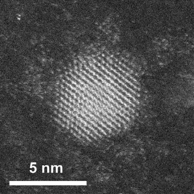
Extended Data Fig. 6 shows the energy-level scheme of the produced UCNPs. All materials and synthesis details of NPs, NP characterization, PCNS fabrication and characterization are in Supplementary Information sections 2–4 and Supplementary Figs. 5 and 6.
Optical characterization
Dispersion-band-diagram measurements, experimental interrogation and detection scheme of upconversion are provided, respectively, in Supplementary Information sections 5 and 6 and Supplementary Figs. 7–10. For upconversion, the pulsed (150-fs) Ti:Sa oscillator, with central wavelength λin = 810 nm and full-width at half-maximum of 6 nm, is tuned to the FW quasi-BIC and focused to a 6-µm spot on the PCNS. The power coupled with NPs was 5%, corresponding to 48 kW cm−2 at a pulse energy of 6.25 nJ (103 kW cm−2).
Photoluminescence, enhancement-factor and radiance-enhancement estimation
Enhancement-factor estimation, spectral emission datasets from samples and radiance-enhancement-factor estimation are provided, respectively, in Supplementary Information section 7 and Supplementary Figs. 11 and 12.
FDTD simulations
The radiation properties of the PCNS were evaluated using the FDTD method in Ansys Lumerical. A single dipole source was used to compute the isofrequency map using the Z-transform of the local optical field retrieved within the finite-structure domain with the 3D full-field monitor. The intensity of the Z-transform determines the strength of radiation in the momentum space and better represents the radiation properties associated with the PCNS. To validate the results found with this approach, we first simulated a literature case discussed in ref. 44, that is, supercollimation resulting from flat-band dispersion in the momentum space, which is shown in Supplementary Fig. 13. The isofrequency far-field intensity map in momentum space showed, in our case, non-trivial vanishing strips along orthogonal arms (cross of zeros; Fig. 3 and Extended Data Fig. 7). The near-field intensity map showed self-collimation as occurring when flat dispersion is involved. In Extended Data Fig. 7e, the experimental proof is reported using a rescaled geometry of the PCNS (using the fit in Extended Data Fig. 2e) to move the FW-BIC at 532 nm and make the beam easily visible. At this stage, the radiation properties were examined by placing an array of dipole sources (18 × 18) at the boundary of the finite PCNS with a uniform slab covering an area of several microns squared. The results are shown in Fig. 3c and Extended Data Fig. 8. The sources collectively add up their field and coherently emit radiation in the plane of the slab, as shown in Extended Data Fig. 8a, in which the field propagates along the direction (+1, 0) with intensity enhancement as large as 1.5 × 10⁴ (normalized to the number of emitters). The emission was always pointing towards the non-textured slab, thus—on the opposite edge—the propagation was along the direction (−1, 0). It was found that, at shorter wavelengths, other preferential directions of propagation were also possible, such as (1, ±1). The divergence was evaluated along 1 mm of propagation from the edge, as shown in Extended Data Fig. 8b, which showed a divergence of 0.02° (Extended Data Fig. 8c), which is even lower than the experimental values. Analysis of the whole visible and near-infrared spectrum revealed that the typical value of the divergence is less than 0.5° (Extended Data Fig. 8d), demonstrating that this regime of narrow radiation is expected to be common in this type of photonic structure. Indeed, as shown in Extended Data Fig. 8e, the full width at half maximum of the beam periodically contracts and expands along the propagation, which is because of a mechanism of self-healing that compensates for diffraction.
Directivity measurements
Extended Data Fig. 9a shows the microscopy inspection of light propagation near the edge. Extended Data Fig. 9b shows the experimental results on the divergence of the side beam (directed along the outer edge versor), with a polar plot of the edge emission in Extended Data Fig. 9c, in agreement with simulation in Extended Data Fig. 8 in the upconverted emission.