Tag: nanotechnology

  • Revolutionary Silicon Spikes Destroy 96% of Viruses on Contact

    Revolutionary Silicon Spikes Destroy 96% of Viruses on Contact

    [ad_1]

    Virus on Silicon Spikes

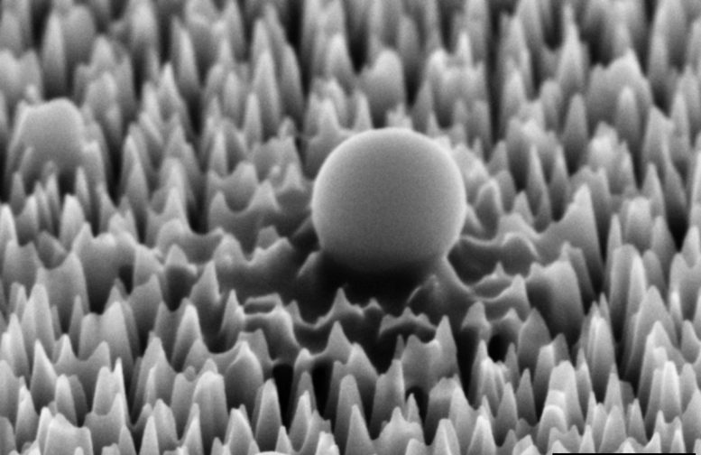
    A virus on the nano-spiked silicon surface, magnified 65,000 times. After 1 hour it has already begun to leak material. Credit: RMIT

    An international research team led by RMIT University has designed and manufactured a virus-killing surface that could help control disease spread in hospitals, labs, and other high-risk environments. The surface made of silicon is covered in tiny nanospikes that skewer viruses on contact.

    Lab tests with the hPIV-3 virus – which causes bronchitis, pneumonia, and croup – showed 96% of the viruses were either ripped apart or damaged to the point where they could no longer replicate to cause infection. These impressive results, featured on the cover of top nanoscience journal ACS Nano, show the material’s promise for helping control the transmission of potentially dangerous biological material in laboratories and healthcare environments.

    Virus Destroyed on Silicon Spikes

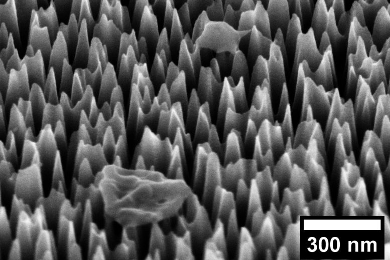
    A virus on the nano-spiked silicon surface, magnified 65,000 times. After 6 hours it has been completely destroyed. Credit: RMIT

    Spike the viruses to kill them

    Corresponding author Dr Natalie Borg, from RMIT’s School of Health and Biomedical Sciences, said this seemingly unsophisticated concept of skewering the virus required considerable technical expertise.

    “Our virus-killing surface looks like a flat black mirror to the naked eye but actually has tiny spikes designed specifically to kill viruses,” she said. “This material can be incorporated into commonly touched devices and surfaces to prevent viral spread and reduce the use of disinfectants.”

    Natalie Borg

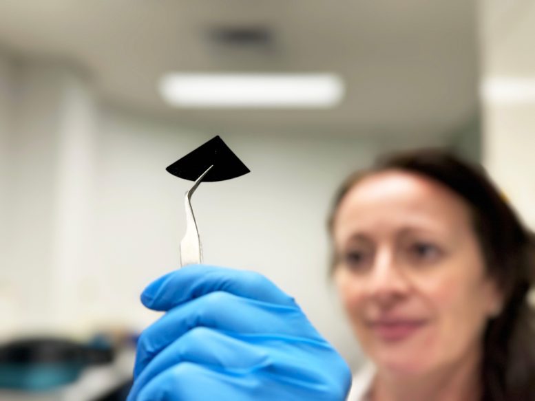
    Dr Natalie Borg inspects a sample of the nano-spiked silicon sheet. Credit: RMIT

    The nano-spiked surfaces were manufactured at the Melbourne Centre for Nanofabrication, starting with a smooth silicon wafer, which is bombarded with ions to strategically remove material. The result is a surface full of needles that are 2 nanometers thick – 30,000 times thinner than a human hair – and 290 nanometers high.

    Specialists in antimicrobial surfaces

    The team led by RMIT Distinguished Professor Elena Ivanova has years of experience studying mechanical methods for controlling pathogenic microorganisms inspired by the world of nature: the wings of insects such as dragonflies or cicadas have a nanoscale spiked structure that can pierce bacteria and fungi.

    In this case, however, viruses are an order of magnitude smaller than bacteria so the needles must be correspondingly smaller if they are to have any effect on them. The process by which viruses lose their infectious ability when they contact the nanostructured surface was analysed in theoretical and practical terms by the research team.

    Elena Ivanova Team

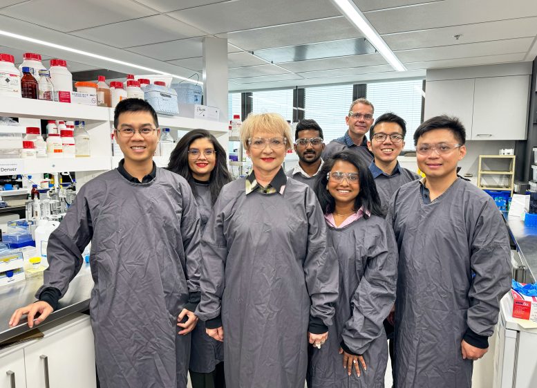
    Team Ivanova with study corresponding author Professor Elena Ivanova (3rd from left) and study lead author Samson Mah (2nd from right). Credit: RMIT

    Researchers at Spain’s Universitat Rovira i Virgili (URV), Dr. Vladimir Baulin, and Dr. Vassil Tzanov, computer-simulated the interactions between the viruses and the needles. RMIT researchers carried out a practical experimental analysis, exposing the virus to the nanostructured surface and observing the results at RMIT’s Microscopy and Microanalysis Facility.

    The findings show the spike design to be extremely effective at damaging the virus’ external structure and piercing its membranes, incapacitating 96% of viruses that came into contact with the surface within six hours. Study first author, Samson Mah, who completed the work under an RMIT-CSIRO Masters by Research Scholarship and has now progressed to working on his PhD research with the team, said he was inspired by the practical potential of the research.

    “Implementing this cutting-edge technology in high-risk environments like laboratories or healthcare facilities, where exposure to hazardous biological materials is a concern, could significantly bolster containment measures against infectious diseases,” he said. “By doing so, we aim to create safer environments for researchers, healthcare professionals, and patients alike.”

    Reference: “Piercing of the Human Parainfluenza Virus by Nanostructured Surfaces” by Samson W. L. Mah, Denver P. Linklater, Vassil Tzanov, Phuc H. Le, Chaitali Dekiwadia, Edwin Mayes, Ranya Simons, Daniel J. Eyckens, Graeme Moad, Soichiro Saita, Saulius Joudkazis, David A. Jans, Vladimir A. Baulin, Natalie A. Borg and Elena P. Ivanova, 21 December 2023, ACS Nano.
    DOI: 10.1021/acsnano.3c07099

    The project was a truly interdisciplinary and multi-institutional collaboration carried out over two years, involving researchers from RMIT, URV (Spain), CSIRO, Swinburne University, Monash University and the Kaiteki Institute (Japan).

    This study was supported by the ARC Research Hub for Australian Steel Manufacturing and by the ARC Industrial Transformational Training Centre in Surface Engineering for Advanced Materials.


    [ad_2]

    Source link

  • Nanoparticles turbocharge turmeric’s curcumin for enhanced health benefits

    Nanoparticles turbocharge turmeric’s curcumin for enhanced health benefits

    [ad_1]

    A review article published in the journal Antioxidants provides a detailed overview of nanoparticle-based strategies to improve the bioavailability and bioactivity of curcumin.

    Study: Enhancing the Bioavailability and Bioactivity of Curcumin for Disease Prevention and Treatment. Image Credit: Microgen / ShutterstockStudy: Enhancing the Bioavailability and Bioactivity of Curcumin for Disease Prevention and Treatment. Image Credit: Microgen / Shutterstock

    Background

    Curcumin, turmeric’s main bioactive compound, is a polyphenol found in Curcuma longa roots. This compound has numerous health benefits, including anticancer, antioxidant, anti-inflammatory, anti-obesity, anti-diabetic, anti-microbial, wound-healing, and lipid-lowering properties.

    Curcumin has low bioavailability in human organs and is rapidly converted to a number of bioactive metabolites after intestinal absorption. Dried turmeric powder prepared from Curcuma longa roots contains about 2-5% of curcumin.

    Curcumin consumed through dietary sources is sufficient to impact the gut microbiota. However, due to rapid metabolism, the concentration of intact curcumin in the circulation becomes very low (sub-micromolar concentrations), which is insufficient to trigger cellular signaling and gene expression, as observed in in vitro studies with cultured cells.   

    Examples of curcumin nano-delivery systems.Examples of curcumin nano-delivery systems.

    Strategies to increase curcumin bioavailability

    Dietary curcumin is inefficiently absorbed across the intestinal epithelium and undergoes rapid metabolism and systemic elimination. In an aqueous solution with a neutral pH, the enol state of curcumin is formed, which reduces the stability of curcumin.

    Several nanoformulations have been developed to increase curcumin concentration in the circulation as well as in specific cells, tissues, and organelles. These nanoformulations have been designed to increase curcumin solubility, improve stability during gastrointestinal absorption, alter absorption routes, and inhibit detoxification enzymes using adjuvants.

    The latest generation of curcumin nanoformulations can increase free curcumin bioavailability in plasma by more than 100-fold and improve absorption, cellular uptake, permeability through the blood-brain barrier, and tissue distribution.

    Factors that improve curcumin bioavailability include composition, size, and route of administration of nanoparticles. Curcumin preparations with smaller-size nanoparticles have been found to increase bioavailability when administered orally. In contrast, larger-size nanoparticles have been found to increase bioavailability when administered intravenously.

    Curcumin nanoformulations can induce senescence in malignant and normal cells, thus effectively treating various cancer types and age-related diseases, including cardiometabolic diseases, neurodegenerative diseases, and liver, lung, and gastrointestinal diseases.

    Regarding mode of action, existing evidence indicates that curcumin acts as an antioxidant and anti-inflammatory compound to reduce the production of reactive oxygen species (ROS) and modulate cellular signaling and gene expression related to inflammatory pathways. These activities work synergistically to maintain homeostasis of cellular macromolecules (proteins, DNA, and lipids).

    These activities can be increased by incorporating curcumin in nanoparticle-based formulations, such as polymeric curcumin–bioperine–PLGA. The isomerization of curcumin to cis-trans curcumin is known to increase its ability to bind adenosine receptors. Incorporation of cis-trans curcumin into nanoformulations is considered to be a valuable strategy to increase its therapeutic efficacy against inflammatory diseases.        

    Regarding safety profile, recent clinical trials indicate that the majority of curcumin nanoformulations are well-tolerated and safe for use in humans.

    Anti-microbial activities

    Curcumin is known to exert an anti-microbial effect against both Gram-positive and Gram-negative bacteria, and this activity is beneficial for topical applications against skin infection and oral and intestinal applications. Moreover, curcumin can indirectly prevent infection by inhibiting bacterial growth in foods. 

    The anti-microbial activities of curcumin can be enhanced by incorporating it into nanoformulations. Administration of curcumin with other compounds, such as antibiotics, honey, or other polyphenols, can also increase its anti-microbial and biofilm inhibitory activities.

    Effects of curcumin nanoformulations in the gastrointestinal tract  

    Several nanotechnology-based systems, such as micelles, liposomes, exosomes, phospholipid complexes, nanoemulsions, nanostructured lipid carriers, and biopolymer nanoparticles, have been found to increase oral curcumin bioavailability.

    Nanoparticle curcumin called ‘Theracurmin’ has been found to suppress colitis in mice by modulating gut microbiota. Improvement in gut microbiota composition has also been achieved using nanobubble curcumin extract. Curcumin loaded with nanostructured lipid carriers has been found to reduce colonic inflammation in animals.

    The incorporation of curcumin in liposomes has been found to increase its anticancer activity by improving gastrointestinal absorption. Moreover, the administration of curcumin with other bioactive compounds, such as piperine and salsalate, has been found to increase curcumin bioavailability and bioactivity.

    Effects of curcumin nanoformulations in liver and adipose tissue  

    Curcumin nanoformulations with adjuvants, such as piperine and quercetin, have been found to increase its bioavailability and bioactivity significantly. Various nanotechnology-based delivery systems, such as micelles, liposomes, polymeric, metal, and solid lipid nanoparticles, have been found to increase curcumin bioavailability.

    The anti-inflammatory, antioxidant, and antifibrotic properties of curcumin make it a potential therapeutic compound for liver diseases. In liver diseases, curcumin nanoformulations have been found to increase its therapeutic efficacy by increasing curcumin solubility, bioavailability, and membrane permeability and improving its pharmacokinetics, pharmacodynamics, and biodistribution.   

    Effects of curcumin nanoformulations on the cardiovascular system   

    Curcumin encapsulated in carboxymethyl chitosan nanoparticles conjugated to a myocyte-specific homing peptide has been found to increase the cardiac bioavailability of curcumin. The formulation has also been found to improve cardiac function by reducing the expression of hypertrophy marker genes and apoptotic mediators.

    Several curcumin nanoformulations, such as hyaluronic acid-based nanocapsules, nanoparticles encapsulated in PLGA or nanoemulsion systems, have been found to increase the aqueous solubility of curcumin and subsequently prevent hypertension in animals. Similar cardio-protective effects have been observed using nanocurcumin polymer-based nanoparticles and curcumin and nisin-based polylactic acid nanoparticles. These formulations have been found to prevent myocardial damage and improve cardiac muscle functions.

    Effects of curcumin nanoformulations on the brain   

    Curcumin complexed with galactomannans has been found to have better blood-brain barrier permeability and higher efficacy in preventing neuroinflammation, anxiety, fatigue, and memory loss in both humans and animals.

    Curcumin-laden liposomes have been found to exert anti-amyloidogenic and anti-inflammatory effects in animal and cellular models of Alzheimer’s disease. Curcumin’s preventive activities against Alzheimer’s disease are associated with its ability to reduce amyloid-beta production and tau aggregation, which are major hallmarks of Alzheimer’s disease.   

    However, clinical trials involving patients with mild to moderate Alzheimer’s disease could not find any beneficial effect of curcumin in reducing disease biomarkers and improving cognitive functions.

    A recent clinical trial involving non-demented adults, on the other hand, has shown that oral curcumin treatment can improve memory and reduce amyloid and tau accumulation in the amygdala and hypothalamus.   

    [ad_2]

    Source link

  • DNA origami vaccine DoriVac paves way for personalized cancer immunotherapy

    DNA origami vaccine DoriVac paves way for personalized cancer immunotherapy

    [ad_1]

    Therapeutic cancer vaccines are a form of immunotherapy in the making that could not only destroy cancer cells in patients, but keep a cancer from coming back and spreading. Multiple therapeutic cancer vaccines are being studied in clinical trials, but despite their promise, they are not routinely used yet by clinical oncologists to treat their patients. 

    The central ingredient of therapeutic cancer vaccines is antigens, which are preferentially produced or newly produced (neoantigens) by tumor cells and enable a patient’s immune system to search and destroy the cancerous cells. In most cases, those antigens cannot act alone and need the help of adjuvant molecules that trigger a general alarm signal in immune cells known as antigen-presenting cells (APCs). APCs internalize both antigen and adjuvant molecules and present the antigens to different types of T cells. Those T cells then launch an immediate attack against the tumor, or preserve a longer-lasting memory of the tumor for future defense.

    A cancer vaccine’s effectiveness depends on the level and duration of the “alarm” its adjuvants can ring in APCs. Previously, researchers found that delivering adjuvant and antigen molecules to APCs simultaneously using nanostructures like DNA origami can increase APC activation. However, none of these approaches systematically investigated how the number and nanoscale arrangement of adjuvant molecules affect downstream tumor-directed immunity. 

    Now, a research team at the Wyss Institute at Harvard University, Dana-Farber Cancer Institute (DFCI), Harvard Medical School (HMS), and Korea Institute of Science and Technology (KIST) has created a DNA origami platform called DoriVac, whose core component is a self-assembling square block-shaped nanostructure. To one face of the square block, defined numbers of adjuvant molecules can be attached in highly tunable, nanoprecise patterns, while the opposite face can bind tumor antigens. The study found that molecules of an adjuvant known as CpG spaced exactly 3.5 nanometers apart from each other resulted in the most beneficial stimulation of APCs that induced a highly-desirable profile of T cells, including those that kill cancer cells (cytotoxic T cells), those that cause beneficial inflammation (Th-1 polarized T cells), and those that provide a long-term immune memory of the tumor (memory T cells). DoriVac vaccines enabled tumor-bearing mice to better control the growth of tumors and to survive significantly longer than control mice. Importantly, the effects of DoriVac also synergized with those of immune checkpoint inhibitors, which are a highly successful immunotherapy that is already widely used in the clinic. The findings are published in Nature Nanotechnology.

    “DoriVac’s DNA origami vaccine technology merges different nanotechnological capabilities that we have developed over the years with an ever-deepening knowledge about cancer-suppressing immune processes,” said Wyss Core Faculty member William Shih, Ph.D., who led the Wyss Institute team together with first-author Yang (Claire) Zeng, M.D., Ph.D. “We envision that in the future, antigens identified in patients with different types of tumors could be quickly loaded onto prefabricated, adjuvant-containing DNA origami to enable highly effective personalized cancer vaccines that can be paired with FDA-approved checkpoint inhibitors in combination therapies.”

    Shih is also a Professor at HMS and DFCI’s Department of Cancer Biology and, as some of the other authors, a member of the NIH-funded cross-institutional “Immuno-engineering to Improve Immunotherapy” (i3) Center based at the Wyss. 

    DNA origami rationale

    The CpG adjuvant is a synthetic strand of DNA made up of repeated CpG nucleotide motifs that mimic the genetic material from immune cell-invading bacterial and viral pathogens. Like its natural counterparts, CpG adjuvants bind to a “danger receptor” called TLR9 in immune cells, which in turn induces an inflammatory (innate) immune response that works in concert with the antigen-induced (adaptive) immune response. 

    “We knew from previous work that to trigger strong inflammatory responses, TLR9 receptors need to dimerize and aggregate into multimeric complexes binding to multiple CpG molecules. The nanoscale distances between the CpG-binding domains in effective TLR9 assemblies revealed by structural analysis fell right into the range of what we hypothesized we could mirror with DNA origami structures presenting precisely spaced CpG molecules,” explained Zeng, who was an Instructor in Medicine at the time of the study and now is a senior scientist at DFCI and Harvard Medical School (HMS). In addition to Shih, Zeng was also mentored on the project by senior authors Ju Hee Ryu, Ph.D., a Principal Researcher at KIST, and Wyss Founding Core Faculty member David Mooney, Ph.D., who also is Professor at Harvard John A. Paulson School of Engineering and Applied Sciences (SEAS), and one of the i3 Center’s Principal Investigators. 

    Zeng and the team fabricated DoriVac vaccines in which different numbers of CpG strands were spaced at 2.5, 3.5, 5, or 7 nanometers apart from each other on one face of the square block, and a model antigen was attached to the opposite face. They protected their structures from being degraded in the body using a chemical modification method that Shih’s group had developed earlier. When internalized by different types of APCs, including dendritic cells (DCs), which orchestrate tumor-directed T cell responses, the DoriVac vaccines improved the uptake of antigens compared to controls consisting of free antigen molecules. A CpG spacing of 3.5 nanometers produced the strongest and most beneficial responses in APCs, and significantly outperformed a control vaccine containing only free CpG molecules. “We were excited to find that the DoriVac vaccine preferentially induced an immune activation state that supports anti-tumor immunity, which is what researchers generally want to see in a good vaccine,” said Zeng. 

    Besides spacing, the numbers of CpG molecules in DoriVac vaccines also mattered. The team tested vaccines containing between 12 to 63 optimally spaced CpG molecules and found that 18 CpG molecules provided the best APC activation. This meant that their approach can also help limit the dosage of CpG molecules and thus minimize commonly observed toxic side effects observed with adjuvants.

    Gained in (tumor) translation

    Importantly, these in vitro trends translated to in vivo mouse tumor models. When prophylactically injected under the skin of mice, DoriVac vaccines accumulated in the closest lymph nodes where they stimulated DCs. A vaccine loaded with a melanoma antigen prevented the growth of subsequently injected aggressive melanoma cells. While all control animals had succumbed to the cancer by day 42 of the experiment, DoriVac-protected animals all were alive. DoriVac vaccines also inhibited tumor growth in mice in which the formation of melanoma tumors was already underway, with a 3.5 nanometer spacing of 18 CpG molecules again providing maximum effects on DC and T cells, and the strongest reduction in tumor growth.

    Next, the team asked whether DoriVac vaccines could also boost immune responses produced by small “neoantigens” emerging in melanoma tumors. Neoantigens are ideal targets because they are exclusively made by tumor cells. However, they often are not very immunogenic themselves, which make highly effective adjuvants an important component in neoantigen vaccines. A DoriVac vaccine customized with four neoantigens enabled the researchers to significantly suppress growth of the tumor in mice that produced the neoantigens.

    Finally, the researchers asked whether DoriVac could synergize with immune checkpoint therapy, which reactivates T cells that have been silenced in tumors. In mice, the two therapies combined resulted in the total regression of melanoma tumors, and prevented them from growing back when the animals were exposed to the same tumor cells again four months later. The animals had built up an immune memory of the tumor. The team obtained a similar vaccination efficiency in a mouse lymphoma model.

    We think that DoriVac’s value for determining a sweet spot in adjuvant delivery and enhancing the delivery and effects of coupled antigens can pave the way to more effective clinical cancer vaccines for use in patients with a variety of cancers.”


    Yang (Claire) Zeng, M.D., Ph.D., First Author

    The team is currently translating the DoriVac platform toward its clinical application, which is supported by the study’s assessment of vaccine distribution and vaccine-directed antibodies in mice, as well as cytokines produced by immune cells in response to the vaccines in vivo. 

    “The DoriVac platform is our first example of how our pursuit of what we call Molecular Robotics – synthetic bioinspired molecules that have programmable shape and function – can lead to entirely new and powerful therapeutics. This technology opens an entirely new path for development of designer vaccines with properties tailored to meet specific clinical challenges. We hope to see its rapid translation into the clinic,” said Wyss Institute Founding Director Donald Ingber, M.D., Ph.D., who is also the Judah Folkman Professor of Vascular Biology at HMS and Boston Children’s Hospital, and the Hansjörg Wyss Professor of Bioinspired Engineering at SEAS.

    Other authors on the study are Olivia Young, Christopher Wintersinger, Frances Anastassacos, James MacDonald, Giorgia Isinelli, Maxence Dellacherie, Miguel Sobral, Haiqing Bai, Amanda Graveline, Andyna Vernet, Melinda Sanchez, Kathleen Mulligan, Youngjin Choi, Thomas Ferrante, Derin Keskin, Geoffrey Fell, Donna Neuberg, Cathrine Wu, and Ick Chan Kwon. The study was funded by the Wyss Institute’s Validation Project and Institute Project programs, Claudia Adams Barr Program at DFCI, Korean Fund for Regenerative Medicine (award #21A0504L1), Intramural Research Program of KIST (award #2E30840), and National Institutes of Health (under the i3 Center supporting U54 grant (award #CA244726-01).

    Source:

    Journal reference:

    Zeng, Y. C., et al. (2024). Fine tuning of CpG spatial distribution with DNA origami for improved cancer vaccination. Nature Nanotechnologydoi.org/10.1038/s41565-024-01615-3.

    [ad_2]

    Source link

  • Opposites Attract, Likes Repel? Scientists Overturn Fundamental Principle of Physics

    Opposites Attract, Likes Repel? Scientists Overturn Fundamental Principle of Physics

    [ad_1]

    Hexagonal Cluster Formation

    A new study has overturned a fundamental principle of physics by demonstrating that similarly charged particles can attract each other in a solution, with the effect varying between positive and negative charges depending on the solvent. This discovery has significant implications for various scientific processes, including self-assembly and crystallization. The research reveals the importance of solvent structure at the interface in determining interparticle interactions, challenging long-held beliefs and indicating a need for a re-evaluation of our understanding of electromagnetic forces. Credit: Zhang Kang

    “Opposites charges attract; like charges repel” is a fundamental principle of basic physics. However, a new study from Oxford University, recently published in the journal Nature Nanotechnology, has demonstrated that similarly charged particles in solution can, in fact, attract each other over long distances.

    Just as surprisingly, the team found that the effect is different for positively and negatively charged particles, depending on the solvent.

    Besides overturning long-held beliefs, these results have immediate implications for a range of processes that involve interparticle and intermolecular interactions across various length-scales, including self-assembly, crystallization, and phase separation.

    The team of researchers, based at Oxford’s Department of Chemistry, found that negatively charged particles attract each other at large separations whereas positively charged particles repel, while the reverse was the case for solvents such as alcohols.

    These findings are surprising because they seem to contradict the central electromagnetic principle that the force between charges of the same sign is repulsive at all separations.

    Experimental Observations

    Now, using bright-field microscopy, the team tracked negatively charged silica microparticles suspended in water and found that the particles attracted each other to form hexagonally arranged clusters. Positively charged aminated silica particles, however, did not form clusters in water.

    Using a theory of interparticle interactions that considers the structure of the solvent at the interface, the team established that for negatively charged particles in water, there is an attractive force that outweighs electrostatic repulsion at large separations, leading to cluster formation. For positively charged particles in water, this solvent-driven interaction is always repulsive, and no clusters form.

    This effect was found to be pH dependent: the team was able to control the formation (or not) of clusters for negatively charged particles by varying the pH. No matter the pH, the positively charged particles did not form clusters.

    Solvent-Specific Effects and Further Discoveries

    Naturally, the team wondered whether the effect on charged particles could be switched, such that the positively charged particles form clusters and the negatives do not. By changing the solvent to alcohols, such as ethanol, which has different interface behavior to water, this is exactly what they observed: positively charged aminated silica particles formed hexagonal clusters, whereas negatively charged silica did not.

    According to the researchers, this study implies a fundamental re-calibration in understanding that will influence the way we think about processes as different as the stability of pharmaceutical and fine chemical products or the pathological malfunction associated with molecular aggregation in human disease. The new findings also provide evidence for the ability to probe properties of the interfacial electrical potential due to the solvent, such as its sign and magnitude, which were previously thought immeasurable.

    Professor Madhavi Krishnan (Department of Chemistry, Oxford University), who led the study, says: “I am really very proud of my two graduate students, as well as the undergraduates, who have all worked together to move the needle on this fundamental discovery.”

    Sida Wang (Department of Chemistry, Oxford University), a first-author on the study, says: “I still find it fascinating to see these particles attract, even having seen this a thousand times.”

    Reference: “A charge-dependent long-ranged force drives tailored assembly of matter in solution” by Sida Wang, Rowan Walker-Gibbons, Bethany Watkins, Melissa Flynn and Madhavi Krishnan, 30 February 2024, Nature Nanotechnology.
    DOI: 10.1038/s41565-024-01621-5


    [ad_2]

    Source link

  • Innovative Subak tool offers affordable solution for detecting nuclease digestion

    Innovative Subak tool offers affordable solution for detecting nuclease digestion

    [ad_1]

    A new tool could reduce costs for diagnosing infectious diseases.

    Biomedical researchers from The University of Texas at Austin have developed a new, less expensive way to detect nuclease digestion – one of the critical steps in many nucleic acid sensing applications, such as those used to identify COVID-19 and other infectious diseases. 

    A new study published in the journal Nature Nanotechnology shows that this low-cost tool, called Subak, is effective at telling when nucleic acid cleavage occurs, which happens when an enzyme called nuclease breaks down nucleic acids, such as DNA or RNA, into smaller fragments. 

    The traditional way of identifying nuclease activity, Fluorescence Resonance Energy Transfer (FRET) probe, costs 62 times more to produce than the Subak reporter. 

    “To make diagnostics more accessible to the public, we have to reduce costs,” said Soonwoo Hong, a Ph.D. student in the lab of Tim Yeh, associate professor in the Cockrell School of Engineering’s Department of Biomedical Engineering, who led the work. “Any improvements in nucleic acid detection will strengthen our testing infrastructure and make it easier to widely detect diseases like COVID-19.”

    The research team – which also included Jennifer Brodbelt, professor of chemistry at UT Austin’s College of Natural Sciences, and MinJun Kim, professor of mechanical engineering in Southern Methodist University’s Lyle School of Engineering – replaced the traditional FRET probe with Subak reporter in a test called DETECTR (DNA endonuclease-targeted CRISPR trans reporter).

    Subak reporters are based on a special class of fluorescent nanomaterials known as silver nanoclusters. They are made up of 13 silver atoms wrapped inside a short DNA strand. This organic/inorganic composite nanomaterial is too small to be visible to the naked eye and ranging from 1 to 3 nanometers (one billionth of a meter) in size.

    Nanomaterials at this length scale, such as semiconductor quantum dots, can be highly luminescent and exhibit different colors. Fluorescent nanomaterials have found applications in TV displays and biosensing, such as the Subak reporters.

    We have very clear evidence from mass spectrometry that transformation from Ag13 to Ag10 underlines the green to red color conversion observed in the sample, after DNA template digestion.”


    Jennifer Brodbelt, professor of chemistry at UT Austin’s College of Natural Sciences

    Subak reporters, which can be synthesized at room temperature in a single-pot reaction, cost just $1 per nanomole to make. In contrast, FRET probe – which employs complex steps to label a donor dye and a quencher – costs $62 per nanomole to produce. 

    “These highly luminescent silver nanoclusters can be called quantum dots as they show strong size-tunable fluorescence emission due to quantum confinement effect,” Yeh said. “No one can precisely tune the cluster size (and the corresponding emission color) until our demonstration of Subak,” which highlights the innovation of this research. 

    In addition to further testing the Subak reporter for nuclease digestion, the team also wants to investigate whether it can be a probe for other biological targets. 

    The work is supported by a National Science Foundation grant to Yeh and Brodbelt.

    Source:

    Journal reference:

    Hong, S., et al. (2024). A non-FRET DNA reporter that changes fluorescence colour upon nuclease digestion. Nature Nanotechnology. doi.org/10.1038/s41565-024-01612-6.

    [ad_2]

    Source link

  • Researchers introduce a pioneering approach to combat neurodegenerative diseases

    Researchers introduce a pioneering approach to combat neurodegenerative diseases

    [ad_1]

    Researchers led by Northwestern University and the University of Wisconsin-Madison have introduced a pioneering approach aimed at combating neurodegenerative diseases such as Alzheimer’s disease, Parkinson’s disease and Amyotrophic lateral sclerosis (ALS).

    In a new study, researchers discovered a new way to enhance the body’s antioxidant response, which is crucial for cellular protection against the oxidative stress implicated in many neurodegenerative diseases.

    The study published today (Feb. 16) in the journal Advanced Materials. 

    Nathan Gianneschi, the Jacob & Rosaline Cohn Professor of Chemistry at Northwestern’s Weinberg College of Arts and Sciences and member of the International Institute for Nanotechnology, led the work with Jeffrey A. Johnson and Delinda A. Johnson of the University of Wisconsin-Madison School of Pharmacy.

    Targeting neurodegenerative diseases

    Alzheimer’s disease, characterized by the accumulation of beta-amyloid plaques and tau protein tangles; Parkinson’s disease, known for its loss of dopaminergic neurons and presence of Lewy bodies; and ALS, involving the degeneration of motor neurons, all share a common thread of oxidative stress contributing to disease pathology. 

    The study focuses on disrupting the Keap1/Nrf2 protein-protein interaction (PPI), which plays a role in the body’s antioxidant response. By preventing the degradation of Nrf2 through selective inhibition of its interaction with Keap1, the research holds promise for mitigating the cellular damage that underlies these debilitating conditions. 

    “We established Nrf2 as a principal target for the treatment of neurodegenerative diseases over the past two decades, but this novel approach for activating the pathway holds great promise to develop disease-modifying therapies,” Jeffrey Johnson said.

    Limitations of current therapeutics

    The research team embarked on addressing one of the most challenging aspects of neurodegenerative disease treatment: the precise targeting of PPIs within the cell. Traditional methods, including small molecule inhibitors and peptide-based therapies, have fallen short due to lack of specificity, stability and cellular uptake.

    The study introduces an innovative solution: protein-like polymers, or PLPs, are high-density brush macromolecular architectures synthesized via the ring-opening metathesis polymerization (ROMP) of norbornenyl-peptide-based monomers. These globular, proteomimetic structures display bioactive peptide side chains that can penetrate cell membranes, exhibit remarkable stability and resist proteolysis.

    This targeted approach to inhibit the Keap1/Nrf2 PPI represents a significant leap forward. By preventing Keap1 from marking Nrf2 for degradation, Nrf2 accumulates in the nucleus, activating the Antioxidant Response Element (ARE) and driving the expression of detoxifying and antioxidant genes. This mechanism effectively enhances the cellular antioxidant response, providing a potent therapeutic strategy against the oxidative stress implicated in many neurodegenerative diseases.

    The innovation behind protein-like polymers

    PLPs, developed by Gianneschi’s team, could represent a significant breakthrough in halting or reversing damage offering hope for improved treatments and outcomes.

    Focusing on the challenge of activating processes crucial for the body’s antioxidant response, the team’s research offers a novel solution. The team provides a robust, selective method enabling enhanced cellular protection and offering a promising therapeutic strategy for a range of diseases including neurodegenerative conditions. 

    Through modern polymer chemistry, we can begin to think about mimicking complex proteins. The promise lies in the development of a new modality for the design of therapeutics. This could be a way to address diseases like Alzheimer’s and Parkinson’s among others where traditional approaches have struggled.”


    Nathan Gianneschi, the Jacob & Rosaline Cohn Professor of Chemistry at Northwestern’s Weinberg College of Arts and Sciences 

    This approach not only represents a significant advance in targeting transcription factors and disordered proteins, but also showcases the PLP technology’s versatility and potential to revolutionize the development of therapeutics. The technology’s modularity and efficacy in inhibiting the Keap1/Nrf2 interaction underscore its potential for impact as a therapeutic, but also as a tool for studying the biochemistry of these processes. 

    A collaboration of minds 

    Highlighting the study’s collaborative nature, Gianneschi’s team worked closely with experts across disciplines, illustrating the rich potential of combining materials science with cellular biology to tackle complex medical challenges. 

    “We were contacted by Professor Gianneschi and colleagues proposing to use this novel PLP technology in neurodegenerative diseases due to our previous work on Nrf2 in models of Alzheimer’s disease, Parkinson’s disease, ALS and Huntington’s disease,” Jeffrey Johnson said. “We had never heard of this approach for Nrf2 activation and immediately agreed to initiate this collaborative effort that led to the generation of great data and this publication.” 

    This partnership underscores the importance of interdisciplinary research in developing new therapeutic modalities.

    Impact

    With the development of this innovative technology, Gianneschi, his colleagues at the International Institute for Nanotechnology and the Johnson Lab at the University of Wisconsin-Madison, are not just advancing the field of medicinal chemistry, they are opening new pathways to combat some of the most challenging and devastating neurodegenerative diseases faced by society today. As this research progresses towards clinical application, it may soon offer new hope to those suffering from diseases of oxidative stress such as Alzheimer’s and Parkinson’s diseases. 

    “By controlling materials at the scale of single nanometers, we’re opening new possibilities in the fight against diseases that are more prevalent than ever, yet remain untreatable,” Gianneschi said. “This study is just the beginning. We’re excited about the possibilities as we continue to explore and expand the development of macromolecular drugs, capable of mimicking some of the aspects of proteins using our PLP platform.”

    Source:

    Journal reference:

    Carrow, K. P., et al. (2024). Inhibiting the Keap1/Nrf2 Protein‐Protein Interaction with Protein‐Like Polymers. Advanced Materials. doi.org/10.1002/adma.202311467.

    [ad_2]

    Source link

  • Lasers smaller than a human hair emit doughnut-shaped light

    Lasers smaller than a human hair emit doughnut-shaped light

    [ad_1]

    Schematic image of GaN hollow nanowires on a sapphire substrate.

    An artist’s impression of a hollow nanowire emitting doughnut-shaped laser light

    Masato Takiguchi et al./ACS Photonics/American Chemical Society 2024

    Tiny, hollow wires can produce doughnut-shaped laser light that could be used to levitate small objects or transmit information.

    Conventional lasers typically make beams that appear as a single, small point of light when they hit a surface. But for some novel communication technologies that use light to transfer information, it can be better to use lasers that produce hollow beams like a drinking straw, which appear as a ring of light when they hit a surface.

    Such hollow laser…

    [ad_2]

    Source link

  • Injections of drug-filled nanoparticles may relieve arthritis pain

    Injections of drug-filled nanoparticles may relieve arthritis pain

    [ad_1]

    Arthritis at hip joint

    Osteoarthritis affects 530 million people worldwide

    Puwadol Jaturawutthichai/Shutterstock

    Drug-delivering nanoparticles might help treat osteoarthritis. In mice with signs of the condition, a single injection of the particles relieved pain for months.

    Osteoarthritis is the most common form of arthritis, affecting about 530 million people worldwide. It occurs when the cartilage cushioning bones breaks down, causing joint stiffness and pain. Treatments for the condition are limited, and none of them prevent cartilage degeneration.

    Previous research indicates a drug called pazopanib may help relieve osteoarthritis pain. However, its effects only last a few days. So Hee-Jeong Im Sampen at the University of Illinois Chicago and her colleagues developed a method of administering it that prolongs its release.

    The researchers encapsulated pazopanib inside nanoparticles already approved by the US Food and Drug Administration for delivering other medications. They then injected the nanoparticles into the knees of 16 mice, half of which had early signs of osteoarthritis and the other half of which had advanced signs. An equal number of animals received dummy injections of nanoparticles without pazopanib.

    It is difficult to assess joint pain in animals, so the researchers focused on the rodents’ sensitivity instead, using the widely accepted idea that when an individual is experiencing pain – such as joint soreness – they are also more sensitive to physical touch.

    Immediately after treatment, the scientists assessed how quickly the mice removed one of their paws from an uncomfortably hot plate. Mice with either early or advanced osteoarthritis took significantly longer to remove the paw if they had received pazopanib rather than a dummy injection, suggesting that the pazopanib nanoparticles quickly relieved their joint pain. When the researchers later repeated the hot plate test – after two months for the advanced osteoarthritis group and after three months for the early osteoarthritis group – they found a similar effect. This implies the pazopanib nanoparticles offer effective pain relief for several weeks. Animals treated with pazopanib also had less cartilage degeneration, indicating the nanoparticles may slow the progression of osteoarthritis.

    Yet Sampen says just because a treatment is effective in mice doesn’t mean it will be in humans. The researchers plan to explore other ways in which to assess the pain-relieving properties of pazopanib in animals. One approach would be to analyse gait and limb use to confirm the drug relieves joint pain, the researchers said.

    Topics:

    [ad_2]

    Source link

  • Cyborg locusts with brain nanoparticles could act as bomb sniffers

    Cyborg locusts with brain nanoparticles could act as bomb sniffers

    [ad_1]

    Researchers wanting to make use of locusts’ keen sense of smell to sniff out certain chemicals have found that injecting their brains with nanoparticles seems to make odour identification more reliable

    [ad_2]

    Source link

  • COPD: Inhalable nanoparticles could help treat chronic lung disease

    COPD: Inhalable nanoparticles could help treat chronic lung disease

    [ad_1]

    Human bronchi, illustration.

    Chronic obstructive pulmonary disease affects the lungs

    SEBASTIAN KAULITZKI/SCIENCE PHOTO LIBRARY

    Delivering medication to the lungs with inhalable nanoparticles may help treat chronic obstructive pulmonary disease (COPD). In mice with signs of the condition, the treatment improved lung function and reduced inflammation.

    COPD causes the lungs’ airways to become progressively narrower and more rigid, obstructing airflow and preventing the clearance of mucus. As a result, mucus accumulates in the lungs, attracting bacterial pathogens that further exacerbate the disease.

    This thick mucus layer also traps medications, making it challenging to treat infections. So, Junliang Zhu at Soochow University in China and his colleagues developed inhalable nanoparticles capable of penetrating mucus to deliver medicine deep within the lungs.

    The researchers constructed the hollow nanoparticles from porous silica, which they filled with an antibiotic called ceftazidime. A shell of negatively charged compounds surrounding the nanoparticles blocked off pores, preventing antibiotic leakage. This negative charge also helps the nanoparticles penetrate mucus. Then, the slight acidity of the mucus transforms the shells’ charge from negative to positive, opening up pores and releasing the medication.

    The researchers used an inhalable spray containing the nanoparticles to treat a bacterial lung infection in six mice with signs of COPD. An equal number of animals received only the antibiotic.

    On average, mice treated with the nanoparticles had about 98 per cent less pathogenic bacteria inside their lungs than those given just the antibiotic. They also had fewer inflammatory molecules in their lungs and lower carbon dioxide in their blood, indicating better lung function.

    These findings suggest the nanoparticles could improve drug delivery in people with COPD or other lung conditions like cystic fibrosis where thick mucus makes it difficult to treat infections, says Vincent Rotello at the University of Massachusetts Amherst, who wasn’t involved in the study. However, it is unclear if these nanoparticles are cleared by lungs. “If you have a delivery system that builds up over time, that would be problematic,” he says.

    Topics:

    [ad_2]

    Source link