Tag: Peptides

  • The impact of quinoa bioactive compounds on gut health

    The impact of quinoa bioactive compounds on gut health

    [ad_1]

    In a recent study published in Frontiers in Nutrition, researchers review the beneficial impacts of Chenopodium quinoa and its bioactive compounds, particularly its effects on intestinal microflora.

    Study: Progress in research on the effects of quinoa (Chenopodium quinoa) bioactive compounds and products on intestinal flora. Image Credit: Elena Schweitzer / Shutterstock.com

    What is quinoa?

    Quinoa is the common name of Chenopodium quinoa, a whole-grain crop belonging to the Amaranthaceae family. Quinoa is native to the South American Andes Mountains and can be found in three varieties differentiated by its white, black, or red color.

    Quinoa is becoming increasingly popular, especially among health- and fitness-conscious individuals, as it is a rich source of protein, fat, vitamins, minerals, fiber, and other bioactive compounds.

    Recent nutraceutical research using quinoa has discovered that its bioactive compounds can affect the body’s production of short-chain fatty acids (SCFAs) and alter intestinal pH, both of which are significant determinants of intestinal health. Intestinal microbiota health has profound effects on the risk and progression of chronic diseases, including cardiovascular diseases, neurological conditions, and cancers.

    Collating and discussing research on quinoa’s health and clinical benefits can better inform medical practitioners, health-conscious individuals, and future researchers of the optimal ways to utilize this natural, safe, and cost-effective plant.

    About the study

    The present study reviewed 85 scientific publications evaluating the biochemical composition of quinoa, its nutritional benefits, and the efficacy of its bioactives in improving intestinal health. The individual roles of quinoa-derived saponins, polyphenolic compounds, polysaccharides, and biopeptides in improving gut microbiota outcomes were also discussed.

    Saponins

    Saponins, which are also known as triterpene glycosides, are bitter plant-derived secondary metabolites with a broad spectrum of biologically relevant functions.

    Quinoa-derived saponins exhibit poor intestinal absorption and low bioavailability, thereby resulting in prolonged intestinal residence, which may allow these metabolites to be used by gut microbiota as a source of nutrition. Previous studies in rats have confirmed this interaction and shown that quinoa supplementation directly correlates with increasing gut microbial diversity.

    Metabolomic studies have found that saponins obtained from quinoa digestion can improve the metabolism of some vitamins and alter the ammonia cycle. However, caution must be taken when deciding upon supplementation dosages, as high concentrations of quinoa-derived saponins have been shown to be toxic in rat models.

    Polyphenolics

    Quinoa consists of many polyphenolic compounds. For example, as compared to placebo, red junglefowl treated with 1% quinoa quercetin exhibited reduced population sizes of opportunistic pathogens and increased populations of the beneficial bacterial phylum Firmicutes. When combined with supplementation of quinoa-derived cellulose, quercetin further increased the number of goblet cells, directly contributing to improved intestinal immunity.

    Quinoa polyphenols inhibit enzymes involved in the regulation of the digestive tract, thereby affecting the abundance of intestinal flora and improving the microenvironment of intestinal flora.”

    Polysaccharides

    Most quinoa-derived polysaccharides are prebiotics capable of increasing the proportion of beneficial probiotic Bifidobacteria and Collinsella bacteria. In combination with quinoa dietary fiber, quinia polysaccharides effectively modulate SCFA concentrations and reduce weight in high-fat diets (HFDs) characteristic in hyperlipidemia.

    Bioactive peptides

    In vivo studies using hypertensive rats (SHR) have shown that quinoa proteins contain numerous promising peptide precursors. While their mechanism of action remain unknown, these precursors have been shown to significantly reduce the blood pressure of SHR models, thus highlighting their application in cardiovascular research. These health benefits extend beyond blood pressure management, as some studies suggest the colorectal cancer applications of quinoa proteins.

    Quinoa proteins have also been found to be a naturally occurring source of angiotensin-converting enzyme (ACE) inhibitory peptides, an additional cardiovascular benefit.

    Flour containing quinoa protein can significantly enhance cecal microbial activity, the activities of α-glucosidase, β-glucosidase, and α-galactosidase, and the production of SCFAs in rats, while promoting a reduction in the pH of digesters, thereby indicating the favorable effects of these proteins on growth parameters and metabolism of intestinal flora.”

    Conclusions

    While research in the field remains in its infancy, a growing body of literature highlights the clinical and nutritional benefits of quinoa.

    The bioactive components of quinoa have been shown to promote the abundance of probiotic bacteria while simultaneously inhibiting pathogens. Furthermore, quinoa-derived bioactive compounds have been shown to reduce intestinal pH and increase the production of SCFAs.

    Journal reference:

    • Huang, H., Jia, C., Chen, X., et al. (2024). Progress in research on the effects of quinoa (Chenopodium quinoa) bioactive compounds and products on intestinal flora. Frontiers in Nutrition. doi:10.3389/fnut.2024.1308384

    [ad_2]

    Source link

  • Rare gene mutations in hereditary Alzheimer’s disease disrupt amyloid production, study shows

    Rare gene mutations in hereditary Alzheimer’s disease disrupt amyloid production, study shows

    [ad_1]

    A University of Kansas study of rare gene mutations that cause hereditary Alzheimer’s disease shows these mutations disrupt production of a small sticky protein called amyloid.

    Plaques composed of amyloid are notoriously found in the brain in Alzheimer’s disease and have long been considered responsible for the inexorable loss of neurons and cognitive decline. Using a model species of worm called C. elegans that’s often used in labs to study diseases at the molecular level, the research team came to the surprising conclusion that the stalled process of amyloid production -; not the amyloid itself -; can trigger loss of critical connections between nerve cells.

    The research, appearing in the journal Cell Reports, was headed by Michael Wolfe, Mathias P. Mertes Professor of Medicinal Chemistry at KU. 

    The research team focused on the rare inherited mutations because these mutations are found in genes that encode proteins that produce amyloid. 

    If we can understand what’s happening in this inherited form of the disease where a single mutation can trigger it. that might be a clue to what’s going on in all the other cases.” 


    Michael Wolfe, Mathias P. Mertes Professor of Medicinal Chemistry at KU

    The rare mutations are particularly devastating, as they fate the mutation carrier to Alzheimer’s disease in middle age, and children of a mutation carrier have a 50% chance of inheriting the disease-causing mutation.

    Wolfe said hereditary Alzheimer’s disease shows the same pathology, the same presentation clinically and the same progression of symptoms as the “common, garden-variety” of Alzheimer’s related to old age.

    “You see the same amyloid plaques in the hereditary disease,” he said. “We think that these inherited mutations, though rare, are key to what’s going on with all Alzheimer’s disease.”

    Wolfe, who earned his doctorate at KU and returned to the university seven years ago for collaborative research opportunities, joined forces with Brian Ackley, associate professor of molecular biology at KU, whose lab specializes in research with the C. elegans model worm. The research team also included other KU collaborators as well as investigators in Beijing, China, and at Harvard Medical School.

    Co-authors with KU’s Department of Medicinal Chemistry were Sujan Devkota, Vaishnavi Nagarajan, Arshad Noorani and Sanjay Bhattarai; co-authors at KU’s Department of Molecular Biosciences were Ackley and Yinglong Miao; and co-authors from KU’s Center for Computational Biology were Hung Do and Anita Saraf. Other KU co-authors were Caitlin Overmeyer of the Graduate Program in Neurosciences and Justin Douglas of KU’s Nuclear Magnetic Resonance Core Lab. The KU personnel collaborated with Rui Zhou of Tsinghua University in Beijing and Masato Maesako of Harvard Medical School.

    Wolfe said the discovery could point the way toward new approaches to Alzheimer’s therapies, and he hoped fellow researchers and developers of drug therapies would pay close attention to his team’s results. 

    “Our findings suggest what’s needed is a stimulator of the amyloid-producing enzyme, to restart stalled processes and address both problems: eliminating stalled protein complexes that lead to degeneration of nerve cell connections and producing more soluble forms of amyloid. This approach could address both contributing factors simultaneously.”

    Source:

    Journal reference:

    Devkota, S., et al (2024). Familial Alzheimer mutations stabilize synaptotoxic γ-secretase-substrate complexes. Cell Reports. doi.org/10.1016/j.celrep.2024.113761.

    [ad_2]

    Source link

  • Scientists uncover a new doorway for SARS-CoV-2 into human cells

    Scientists uncover a new doorway for SARS-CoV-2 into human cells

    [ad_1]

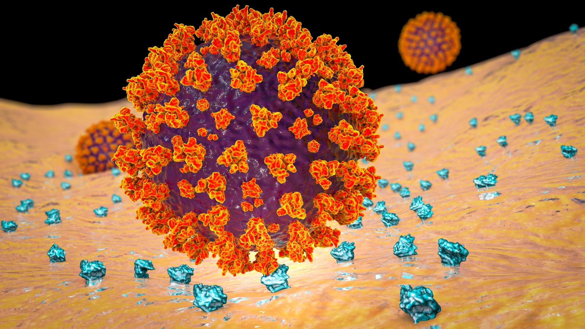
    In a recent study published in the journal Proceedings of the National Academy of Sciences, researchers demonstrated that human transferrin receptor (TfR) mediates severe acute respiratory syndrome coronavirus 2 (SARS-CoV-2) infection.

    Coronavirus disease 2019 (COVID-19), caused by SARS-CoV-2, presents influenza-like manifestations, including mild-to-severe pneumonia, acute respiratory distress syndrome, multiorgan failure, and fatal lung injury. Further, the etiology and pathogenesis of COVID-19 are not entirely understood and targeted therapies remain inadequate.

    The viral spike protein binds to the host receptor, angiotensin-converting enzyme 2 (ACE2), for cellular entry. Although SARS-CoV-2 preferentially infects cells in the respiratory tract, the virus has been detected in virtually all organs. Studies have revealed the presence of SARS-CoV-2 RNA in diverse cells lacking ACE2, suggesting that other receptors or co-receptors may mediate viral entry.

    Study: Human transferrin receptor can mediate SARS-CoV-2 infection. Image Credit: Kateryna Kon / ShutterstockStudy: Human transferrin receptor can mediate SARS-CoV-2 infection. Image Credit: Kateryna Kon / Shutterstock

    The study and findings

    In the present study, researchers identified TfR as an alternative receptor mediating the cellular entry of SARS-CoV-2. First, they used co-immunoprecipitation (Co-IP) to identify host proteins interacting with the viral spike in Calu-3 cells. This revealed 293 proteins, including 42 transmembrane proteins; two proteins were associated with entry (ACE2 and TfR). Next, the team evaluated TfR expression in the respiratory tract and liver in mice.

    TfR expression, both transcript and protein levels, was substantially higher in the lungs and trachea than in other tissues. Using immunohistochemical analysis, the researchers investigated the effects of SARS-CoV-2 on TfR expression in the lungs of humanized ACE2 (hACE2) mice and monkeys. This revealed a 1.5- and 1.8-fold increase in TfR expression in mice and monkeys, respectively.

    In addition, surface plasmon resonance revealed direct interactions between the viral spike and human TfR. Notably, the spike protein lacked interactions with Syrian hamster or mouse TfR. Docking analysis predicted two peptide sequences (QK8: QDSNWASK and SL8 SKVEKLTL) in TfR to be involved at the interface of TfR-spike interactions.

    Mutagenesis and Co-IP revealed that the A529 residue in TfR was essential for interactions with the spike. Further analysis indicated that physiological interactions between spike and TfR occurred at the cellular surface and during endocytosis. This was confirmed by electron microscopy using SARS-CoV-2 pseudoviral spike and HEK293/hACE2 and BHK-21/TfR cells.

    Next, the team evaluated the effects of soluble TfR, anti-TfR antibody, and SL8 and QK8 peptides on SARS-CoV-2 infection using reverse-transcription polymerase chain reaction (RT-PCR) and plaque assays. Results showed their inhibitory effects on SARS-CoV-2 in Vero E6 and Calu-3 cells. Cytotoxicity was not observed even at 1,000 nM.

    Confocal microscopy revealed that TfR was widespread on the surface of Calu-3 and Vero E6 cells, with the colocalization of TfR and SARS-CoV-2 at the surface and during endocytosis. Notably, treatment with the anti-TfR antibody inhibited the colocalization. Further, electron microscopy showed that viral particles were present in the cytosol and clathrin-coated pits in Vero E6 cells; likewise, treatment with anti-TfR antibody inhibited viral internalization.

    Next, ACE2 was knocked out (KO) from Calu-3 and Vero E6 cells and the cells were infected with SARS-CoV-2. This inhibited infection by 40% to 50%, suggesting that ACE2 might not be the only receptor mediating infection. In addition, TfR knockdown (KD) inhibited infection by 30%, whereas its overexpression (OE) promoted infection. TfR KO was not performed as it is lethal. TfR OE or KD did not impact ACE2 expression.

    Further, the team transfected C57 mice with adenovirus vector (Ad5) expressing hACE2 or humanized TfR (hTfR) and infected them with SARS-CoV-2. Viral load in the lungs in Ad5-hTfR and Ad5-hACE2 mice was significantly higher than in Ad5-empty mice. Finally, the researchers evaluated the effects of the anti-TfR antibody on infection in rhesus macaques. Anti-TfR antibody inhibited viral replication and reduced pneumonia.

    Viral load in the respiratory epithelium was also significantly lower between 3- and 7 days post-infection (dpi) compared to controls. Radiographs taken at 0 and 5 dpi revealed significantly less severe pulmonary infiltration in antibody-treated macaques relative to controls. Antibody-treated animals had no significant pulmonary lesions, while controls showed lung lesions of varying degrees.

    Conclusions

    Taken together, the study described the human TfR as a receptor for SARS-CoV-2. TfR can directly bind to the viral spike at an affinity comparable to that of ACE2. Notably, mouse TfR and the viral spike lacked interactions. Soluble TfR, SL8, and QK8 peptides and anti-TfR antibodies can inhibit infection. The team also illustrated the antiviral effects of the anti-TfR antibody in rhesus macaques. Overall, TfR could serve as an alternative infection pathway, facilitating viral entry through endocytosis.

    [ad_2]

    Source link

  • New research shows promise for urine-based test to detect ovarian cancer

    New research shows promise for urine-based test to detect ovarian cancer

    [ad_1]

    New research by Joseph Reiner and colleagues at Virginia Commonwealth University shows promise for a urine-based test for ovarian cancer. Reiner will present their research at the 68th Biophysical Society Annual Meeting, to be held February 10 – 14, 2024 in Philadelphia, Pennsylvania.

    Previous research showed that there are thousands of small molecules, called peptides, in the urine of people with ovarian cancer. While it is possible to detect those molecules using certain well-established techniques, those techniques aren’t straightforward or cost effective. Reiner sought a new approach to more easily detect those peptides. 

    He turned to nanopore sensing, which has the potential to simultaneously detect multiple peptides. The basic idea of nanopore sensing involves passing molecules through a tiny pore, or nanopore, and measuring the changes in electrical current or other properties as the molecules move through.

    To harness the nanopore technology to detect various peptides, Reiner used gold nanoparticles that can partially block the pore. Peptides, like those in the urine of people with ovarian cancer, will then “stick to the gold particle and basically dance around and show us a unique current signature,” Reiner explained.

    The method is capable of simultaneously identifying multiple peptides, and in their study they identified and analyzed 13 peptides, including those derived from LRG-1, a biomarker found in the urine of ovarian cancer patients. Of those 13 peptides, Reiner said, “we now know what those signatures look like, and how they might be able to be used for this detection scheme. It’s like a fingerprint that basically tells us what the peptide is.”

    Clinical data shows a 50-75% improvement in 5-year survival when cancers are detected at their earliest stages. This is true across numerous cancer types.”


    Joseph Reiner and colleagues, Virginia Commonwealth University

    Their ultimate goal is to develop a test that, combined with other information like CA-125 blood tests, transvaginal ultrasound, and family history, could improve early-stage ovarian cancer detection accuracy in the future.

    [ad_2]

    Source link

  • Monash-led study unveils potential long-term treatment for lupus

    Monash-led study unveils potential long-term treatment for lupus

    [ad_1]

    In a recent study published in Nature Communications, a group of researchers assessed Smith (Sm)-specific regulatory T cells (Tregs) efficacy in halting lupus nephritis (LN) by engineering and testing epitope-targeted Tregs for systemic lupus erythematosus (SLE) treatment.

    Study: Smith-specific regulatory T cells halt the progression of lupus nephritis. Image Credit: megaflopp/Shutterstock.com
    Study: Smith-specific regulatory T cells halt the progression of lupus nephritis. Image Credit: megaflopp/Shutterstock.com

    Background 

    Tregs play a crucial role in immune balance, with their dysfunction linked to autoimmune conditions like SLE. Treg therapies, especially those targeting specific antigens, have shown promise in controlling autoimmune responses. LN, a critical SLE manifestation, is often associated with the Sm autoantigen and specific human leukocyte antigen (HLA) haplotypes, suggesting a targeted therapeutic approach might be effective.

    Further research is needed to optimize Sm-specific Treg therapy for broader clinical application and to understand its long-term efficacy and safety in diverse SLE patient populations.

    About the study 

    The present study utilized a biophysical affinity binding assay. Researchers screened peptides derived from Sm proteins against HLA-DR15, identifying those with the highest affinity. This thorough approach enabled the calculation of binding affinities and half-life estimations, crucial for selecting epitopes with the potential to evoke a strong T-cell response.

    Ethical adherence was paramount, with all procedures conforming to the Declaration of Helsinki and receiving approval from relevant ethics committees. The study’s rigorous inclusion criteria ensured that only suitable participants, informed and consenting, contributed to the findings.

    Further investigations into the immunogenicity of top-ranking Sm epitopes utilized whole blood from an HLA-DR15 homozygous donor. This approach allowed for the differentiation of monocytes into dendritic cells and the subsequent activation of Cluster of Differentiation 4 (CD4)+ T cells, providing a robust platform for assessing T-cell responses to Sm epitopes.

    The process extended to detailed methodologies for expressing and purifying HLA-DR15, crucial for understanding the intricate interactions between Sm epitopes and the immune system. Protein crystallization and structural determination further illuminated the binding mechanisms at play, offering insights that could pave the way for novel therapeutic strategies.

    This comprehensive study not only identified potential targets for autoimmune therapy but also set a high standard for methodological rigor and ethical compliance, promising future research in this critical area of medicine.

    Study results 

    The researchers explored the potential of Tregs, particularly in SLE and its severe manifestation, LN. Tregs, known for their role in maintaining immune equilibrium, become a focus due to their decreased numbers or malfunction in autoimmune diseases. Targeted therapies using Tregs, especially those engineered to be antigen-specific, offer a promising approach to suppress the pathogenic autoactivity inherent in these conditions.

    The study embarked on identifying immunodominant Sm protein epitopes, given their association with LN and specific HLA haplotypes. Through a thorough screening process involving 145 overlapping peptides from Sm proteins, researchers identified a set of epitopes with strong binding affinity to HLA-DR15, with SmB/B’58-72 standing out for its binding stability and capacity to induce T-cell proliferation, marking it as a prime candidate for further exploration.

    Diving deeper, the team elucidated the crystal structure of the SmB/B’58-72 epitope in complex with HLA-DR15, revealing key amino acid residues pivotal for T-cell activation. This structural insight laid the groundwork for identifying high-affinity T-cell receptors (TCRs) specific to the SmB/B’58-72 epitope. Leveraging high-throughput sequencing and binding assays, they isolated TCRs with potent affinity, notably TCR1, which demonstrated significant clonal expansion and functional activity indicative of its therapeutic potential.

    The translational leap involved engineering Tregs with the identified Sm-specific TCRs using lentiviral vectors. These engineered Tregs not only retained their regulatory phenotype but also showcased enhanced specificity and suppressive capacity against Sm epitope-induced pro-inflammatory responses, both in vitro and in a humanized mouse model of LN. This specificity was further evidenced by their ability to form immune synapses upon encountering their target antigen, leading to effective T-cell activation and suppression of autoimmunity.

    Moreover, the study highlighted the therapeutic efficacy of these Sm-specific Tregs in suppressing SLE patient-derived autoreactivity, as demonstrated through in vitro cytokine profiling and in vivo models of LN. Mice treated with Sm-Tregs exhibited significantly less renal injury and proteinuria compared to those receiving polyclonal Tregs or no treatment, underscoring the potential of antigen-specific Tregs in restoring immune tolerance and halting disease progression.

    This research opens new avenues for targeted immunotherapy in autoimmune diseases, emphasizing the power of precision medicine in addressing complex disorders like SLE and LN. Harnessing the specificity and regulatory capabilities of Tregs presents a promising strategy for developing more effective and personalized treatments, marking a significant step forward in the battle against autoimmune diseases.

    [ad_2]

    Source link

  • Researchers uncover how deadly MRSA pneumonia inhibits body’s antimicrobial activity

    Researchers uncover how deadly MRSA pneumonia inhibits body’s antimicrobial activity

    [ad_1]

    In a recent study published in the American Journal of Physiology-Lung Cellular and Molecular Physiology, a group of researchers examined how heparan sulfate (HS)shedding impacts cathelicidin efficacy in Methicillin-Resistant Staphylococcus aureus (MRSA) pneumonia.

    Study: Bacterial pneumonia-induced shedding of epithelial heparan sulfate inhibits the bactericidal activity of cathelicidin in a murine model. Image Credit: Tatiana Shepeleva/Shutterstock.com
    Study: Bacterial pneumonia-induced shedding of epithelial heparan sulfate inhibits the bactericidal activity of cathelicidin in a murine model. Image Credit: Tatiana Shepeleva/Shutterstock.com

    Background 

    Pneumonia, particularly caused by MRSA, is a leading cause of infectious mortality. The mechanisms leading to Staphylococcal pneumonia are not fully understood. This study explores the interactions between MRSA, the pulmonary epithelial glycocalyx, and antimicrobial peptides (AMPs) in pneumonia.

    The focus is on the HS enriched glycocalyx, a sulfated layer lining the alveoli known to bind cationic proteins. We examine the shedding of HS into the airspace following lung injury and its potential impact on lung function and interactions with AMPs. Specifically, we investigate how shed HS oligosaccharides, especially during bacterial pneumonia, interact with AMPs like cathelicidins, impacting the host immune response and pathogen dynamics.

    Further research is needed to fully understand the mechanisms by which HS shedding impacts AMP function, offering potential for novel therapeutic strategies in pneumonia treatment.

    About the study 

    In this study, following the University of Colorado’s Institutional Animal Care and Use Committee (IACUC) and Animal Research: Reporting of In Vivo Experiments (ARRIVE) guidelines, male C57BL6 mice underwent intratracheal MRSA instillation, followed by bronchoalveolar lavage (BAL) for fluid analysis. Researchers used mass spectrometry to measure HS in BAL fluid, blind to treatment groups for objective results.

    Additionally, the team innovatively collected airspace fluid from pneumonia patients using heat and moisture exchanger (HME) filters at Vanderbilt University Medical Center under an approved Institutional Review Board (IRB) protocol. This aimed to detect lung changes due to respiratory failure.

    The study employed surface plasmon resonance (SPR) to examine the binding kinetics between AMP  and HS, providing real-time, label-free interaction insights. Concurrently, bacterial growth curves were studied under various conditions to assess the effect of different heparin types on MRSA strains.

    Detailed processes of bacterial ribonucleic acid (RNA) isolation and sequencing were conducted, involving MRSA culture in heparin or saline, followed by RNA extraction and sequencing. These steps were crucial in exploring transcriptomic changes and enhancing understanding of bacterial pneumonia dynamics.

    The research also included minimum inhibitory concentration (MIC) quantification for different pneumonia pathogens against AMP in varying HS concentrations. This was key in evaluating how HS influences AMP effectiveness against bacterial infections. Rigorous statistical analysis ensured the study’s findings were reliable and valid.

    Study results 

    In the present study, researchers utilized a murine model of MRSA pneumonia. Mass spectrometry analyses revealed a significant increase in HS in the airspace lining fluid of MRSA-infected mice compared to saline controls. Notably, this increase was characterized by a higher abundance of sulfated HS, particularly multi-sulfated disaccharides. Complementary analyses with HME filter samples indicated higher HS levels in patients with gram-negative pneumonia compared to those with gram-positive pneumonia, suggesting a nuanced relationship between bacterial etiology and HS shedding.

    Despite the observed increase in shed HS in the lung environment, the study found no direct impact of HS on MRSA growth or gene transcription. Experiments involving various sizes and sulfation patterns of HS showed no significant changes in MRSA growth or transcriptomic response. This finding suggested that HS, while a significant component of the lung milieu post-injury, did not directly inhibit bacterial growth or induce changes in bacterial gene expression.

    The study further delved into the interactions between HS and host immune mediators. Using surface plasmon resonance (SPR), the researchers quantified the binding of HS with murine cathelicidin-related antimicrobial peptide (mCRAMP). The strong binding observed indicated a likely interaction in vivo, which could potentially influence the host response to bacterial infection.

    Most critically, the study investigated the functional implications of HS binding to mCRAMP. Focusing on common nosocomial pneumonia pathogens including MRSA, Klebsiella pneumoniae, and Pseudomonas aeruginosa, the research employed a modified radial diffusion assay to assess the MIC of mCRAMP against these bacteria.

    Results showed significant increases in MIC with higher HS concentrations, indicating a diminished bactericidal effect of mCRAMP in the presence of HS. This finding was particularly noteworthy as it highlighted the complex interplay between HS and host defense mechanisms, where HS, despite not directly affecting MRSA growth, significantly altered the efficacy of an antimicrobial peptide.

    Conclusion

    Overall, the study underscored the intricate dynamics within the pulmonary environment following bacterial pneumonia. The acute shedding of epithelial HS, particularly when enriched in sulfated forms, presented a nuanced challenge to the host’s immune response, potentially influencing the effectiveness of innate immune mechanisms against bacterial pathogens.

    [ad_2]

    Source link

  • Interdisciplinary dream team receives $3 million grant to revolutionize Alzheimer’s diagnosis

    Interdisciplinary dream team receives $3 million grant to revolutionize Alzheimer’s diagnosis

    [ad_1]

    What do a synthetic chemist, a medical imaging expert, and a neurologist have in common? They’re coming together in the Biomedical Imaging Center at the Beckman Institute for Advanced Science and Technology to develop better diagnostic tools and imaging agents to detect early-stage Alzheimer’s disease and other neurodegenerative diseases.

    The dream team

    A team led by Liviu M. Mirica along with Wawryzneic “Wawosz” Dobrucki and Dr. Daniel A. Llano received a $3 million grant from the U.S. National Institute on Aging of the National Institutes of Health to develop and test multi-modal imaging agents for the detection of Alzheimer’s disease and related dementias. This grant is one of the first federal grants to bridge Beckman’s Magnetic Resonance Imaging Laboratory and Molecular Imaging Laboratory. They are both part of Beckman’s Biomedical Imaging Center.

    I’m really excited about the opportunity to collaborate with different scientists from different fields.” 


    Liviu M. Mirica, synthetic chemist and the William H. and Janet G. Lycan Professor of Chemistry, School of Chemical Sciences, University of Illinois Urbana-Champaign

    His research group specializes in building and characterizing synthetic inorganic molecules in vitro: outside of the body.

    Dobrucki, the Neil and Carol Ruzic Scholar for Biomedical and Translational Sciences, is an imaging expert who works extensively with PET scanning in Beckman’s Molecular Imaging Laboratory.

    “I’m looking forward to high-resolution imaging of the brain and its structures,” Dobrucki said.

    Llano, a professor of molecular and integrated physiology and a physician-surgeon, is a practicing neurologist who sees patients daily and specializes in in vivo brain studies: those inside the body.

    “The potential impact that this project will have on Alzheimer’s is what I’m most excited about,” Llano said.

    Understanding Alzheimer’s disease

    Alzheimer’s disease is a neurodegenerative disease that negatively affects brain function and cognitive abilities. Along with Parkinson’s disease, amyotrophic lateral sclerosis, and other disorders, Alzheimer’s falls under the category of amyloid diseases. Amyloids are small groups of abnormally fibrous or misfolded proteins that do not commonly serve a purpose in the body.

    A key marker of Alzheimer’s disease is the presence of amyloid plaques: large buildups of smaller beta-amyloid peptide aggregates. Peptides are short chains of amino acids that eventually create proteins. Neuroinflammation and oxidative stress in the brain are also major markers of Alzheimer’s.

    The detection and treatment of neurodegenerative diseases is especially difficult because of the blood-brain barrier, a semipermeable system of blood vessels and capillaries that controls the flow of ions, molecules, and cells between the blood and the brain. To be effective, imaging agents and drug therapies (which are made of molecules or antibodies) need to be able to pass through.

    Diagnosis and treatment

    Diagnosing Alzheimer’s disease with a high degree of accuracy requires identifying the amyloid aggregates and can only be completed during post-mortem investigation. This creates a need for diagnostic tools that can quickly locate soluble beta-amyloid peptide aggregates and larger amyloid plaques in a living patient.

    PET and MRI are two noninvasive imaging methods commonly used in clinical settings. However, no MRI contrast agents that target amyloid aggregates have been developed. The few FDA-approved PET imaging agents are insufficient at detecting small-scale amyloid abnormalities or in some cases, lead to false-positives test results when diagnosing Alzheimer’s.

    It’s important to develop diagnostic tools to target smaller beta-amyloid peptides and other signs of neuroinflammation and oxidative stress for a variety of reasons, Mirica said. Creating multi-modal tools that can be used for both PET and MRI scans will give researchers a better idea of who is at risk for developing Alzheimer’s, who truly has the disease, and at what stage.

    The $3M plan

    Mirica, Dobrucki, and Llano will receive the $3 million grant over the course of five years to generate novel dual-purpose imaging agents that can easily pass the blood-brain barrier and are compatible with both PET and MRI scanners.

    This will enable the detection of neurodegenerative diseases at earlier stages and “will help tremendously in developing better therapies,” Mirica said.

    Brad Sutton, a professor of bioengineering and the technical director of Beckman’s Biomedical Imaging Center, will assist the team by performing in vivo MRI studies. They will then evaluate the imaging agent’s ability as a dual modality diagnostic agent for Alzheimer’s disease and related dementias.

    Already, Mirica and his collaborators have developed a series of customized molecules that can cross the blood-brain barrier and help detect both smaller soluble beta-amyloid peptides and larger insoluble amyloids.

    They have also developed a copper-based PET imaging agent that led to the successful imaging of amyloid plaques in transgenic Alzheimer’s mice. Looking ahead, the team believes that these agents can be developed to pass through the blood-brain barrier in humans and image multiple markers of Alzheimer’s disease and other neurodegenerative diseases at earlier stages.

    [ad_2]

    Source link

  • SARS-CoV-2 fragments found to mimic immune system peptides, fueling inflammation

    SARS-CoV-2 fragments found to mimic immune system peptides, fueling inflammation

    [ad_1]

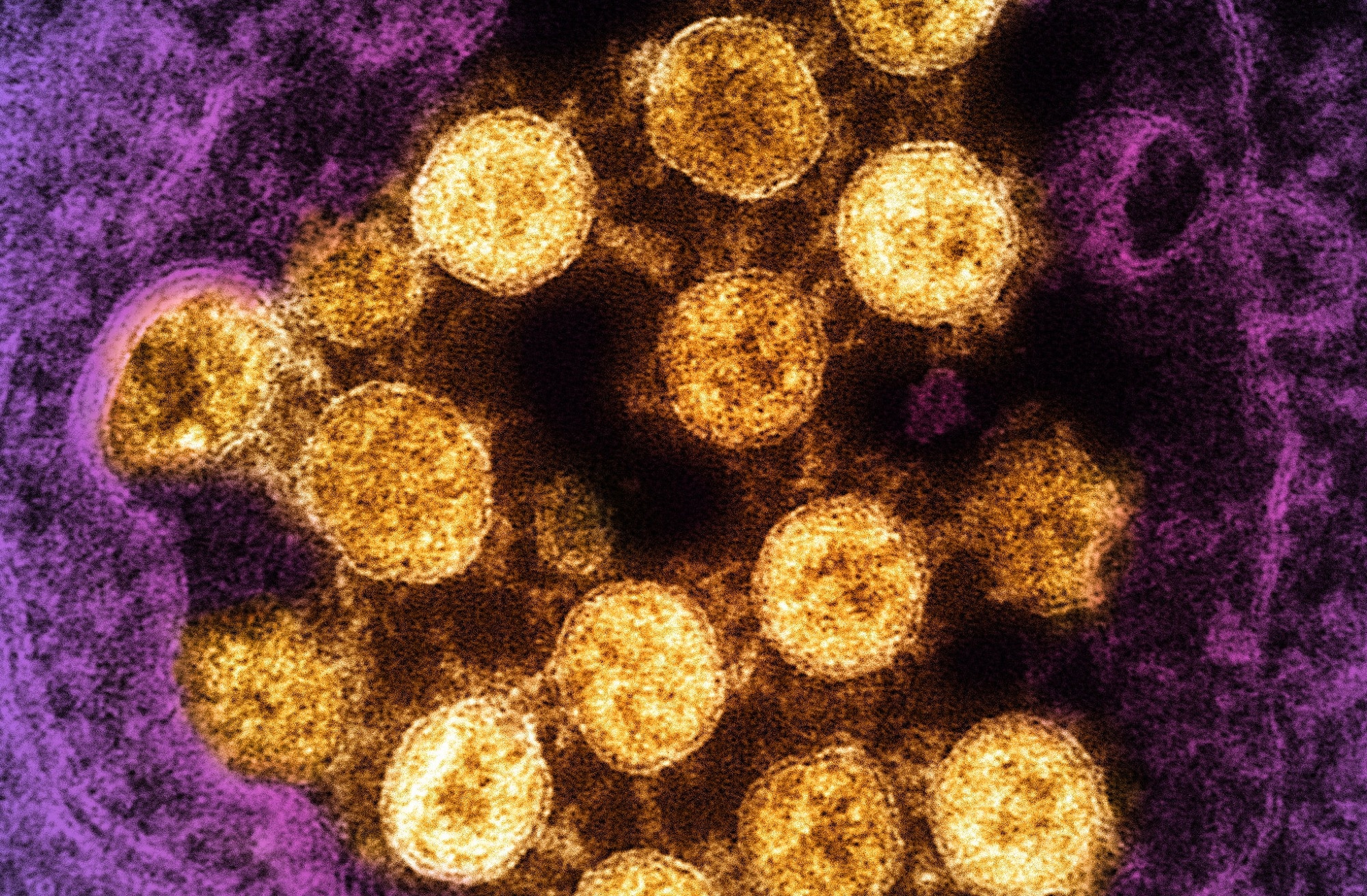
    In a recent study published in the journal Proceedings of the National Academy of Sciences, researchers analyzed the inflammatory capacity of fragmented components of severe acute respiratory syndrome coronavirus 2 (SARS-CoV-2).

    The intensive research during the coronavirus disease 2019 (COVID-19) pandemic has helped understand SARS-CoV-2 infection. Nevertheless, what makes the virus capable of causing a dangerous inflammatory response remains unclear. Research has suggested that amphiphilic, cationic peptides from the innate immune system undergo amyloid-like assembly with anionic nucleic acids and form proinflammatory complexes.

    Study: Viral afterlife: SARS-CoV-2 as a reservoir of immunomimetic peptides that reassemble into proinflammatory supramolecular complexes. Image Credit: NIAIDStudy: Viral afterlife: SARS-CoV-2 as a reservoir of immunomimetic peptides that reassemble into proinflammatory supramolecular complexes. ​​​​​​​Image Credit: NIAID

    The study and findings

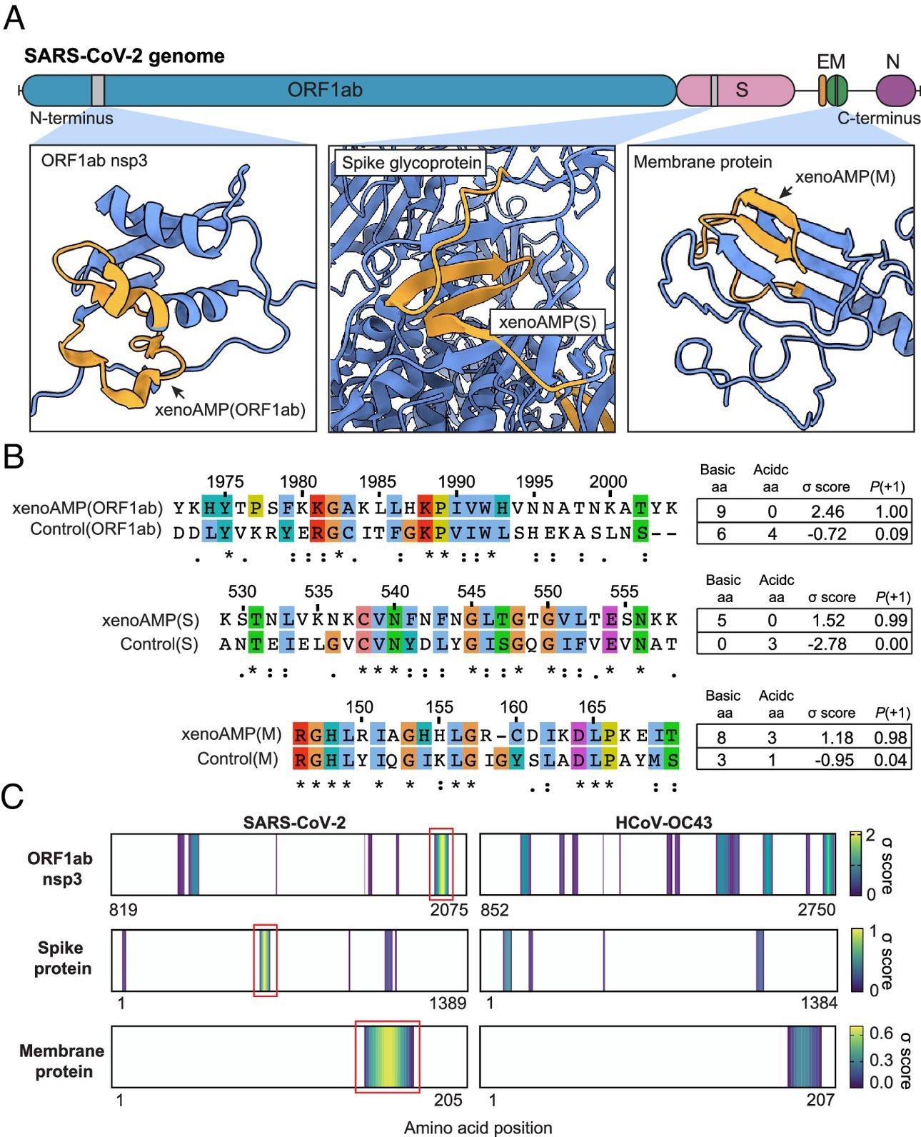
    The present study investigated whether fragmented SARS-CoV-2 peptides assemble with anionic double-stranded RNA (dsRNA) into supramolecular complexes. The viral proteome was considered a reservoir of peptide fragments liberating after the proteolytic destruction of virions. The researchers leveraged a support vector machine (SVM) classifier to recognize antimicrobial peptide (AMP)-like sequences (xenoAMPs) in the SARS-CoV-2 proteome.

    Viral protein sequences were scanned via a moving window of 24–34 amino acids to identify potential xenoAMPs and test whether they behave like AMPs if cleaved at different positions. Sequences were selected based on the output provided by the classifier as a sigma (σ) score, wherein a strongly positive score implied the sequence was highly likely to be an AMP.

    Existence of exogenous mimics of pro-inflammatory host antimicrobial peptides (xenoAMPs) in SARS-CoV-2 proteins. (A) SARS-CoV-2 proteins are scanned with a machine-learning AMP classifier. Each queried sequence is given a σ score that measures its AMP-ness. Three representative high-scoring sequences are studied: xenoAMP(ORF1ab), xenoAMP(S), and xenoAMP(M). The grey bars mark the location where the corresponding sequences are selected. (B) SARS-CoV-2 sequences are aligned and compared to their homologs in a common cold human coronavirus HCoV-OC43: Control (ORF1ab), Control(S), and Control(M). Asterisks, colons, and periods indicate positions that have fully conserved residues, those that have strongly similar properties, and those that have weakly similar properties, respectively. Color is assigned to each residue using the ClustalX scheme. (C) σ score heatmaps compare the distribution of high-scoring sequences in three proteins from SARS-CoV-2 and HCoV-OC43. The first amino acid in each sequence is colored according to its average σ score; regions with negative average σ scores (non-AMPs) are colored white. “Hot spot” clusters of high-scoring sequences for SARS-CoV-2 (bright yellow regions bracketed in red boxes) have systematically higher scores and span wider regions of sequence space compared to HCoV-OC43. This trend suggests that hot spots in SARS-CoV-2 can generate higher scoring sequences for a greater diversity of enzymatic cleavage sites than those in HCoV-OC43.

    Existence of exogenous mimics of pro-inflammatory host antimicrobial peptides (xenoAMPs) in SARS-CoV-2 proteins. (A) SARS-CoV-2 proteins are scanned with a machine-learning AMP classifier. Each queried sequence is given a σ score that measures its AMP-ness. Three representative high-scoring sequences are studied: xenoAMP(ORF1ab), xenoAMP(S), and xenoAMP(M). The grey bars mark the location where the corresponding sequences are selected. (B) SARS-CoV-2 sequences are aligned and compared to their homologs in a common cold human coronavirus HCoV-OC43: Control (ORF1ab), Control(S), and Control(M). Asterisks, colons, and periods indicate positions that have fully conserved residues, those that have strongly similar properties, and those that have weakly similar properties, respectively. Color is assigned to each residue using the ClustalX scheme. (C) σ score heatmaps compare the distribution of high-scoring sequences in three proteins from SARS-CoV-2 and HCoV-OC43. The first amino acid in each sequence is colored according to its average σ score; regions with negative average σ scores (non-AMPs) are colored white. “Hot spot” clusters of high-scoring sequences for SARS-CoV-2 (bright yellow regions bracketed in red boxes) have systematically higher scores and span wider regions of sequence space compared to HCoV-OC43. This trend suggests that hot spots in SARS-CoV-2 can generate higher scoring sequences for a greater diversity of enzymatic cleavage sites than those in HCoV-OC43.

    Further, the team selected specific sequences from this population of (high-scoring) sequences with a high cationic charge. Specifically, they focused on prototypical candidates from the membrane (M) protein, spike (S) protein, and open reading frame 1ab (ORF1ab) polyprotein. In silico analyses showed that these xenoAMPs could be generated during proteasomal degradation, with matrix metalloproteinase 9 (MMP9) and neutrophil elastase (NE) capable of generating them.

    Next, the team compared SARS-CoV-2 xenoAMPs with homologous sequences from SARS-CoV-1 and non-pandemic human CoVs. This showed that sequences were partially conserved. A comparison of σ score heat maps of ORF1ab, S, and M proteins between SARS-CoV-2 and HCoV-OC43 revealed that high-scoring sequences were clustered into hotspots, with SARS-CoV-2 hotspots having higher scores and spanning wider regions than those of HCoV-OC43.

    Further, mass spectrometry was performed on tracheal aspirate samples from patients with severe COVID-19. The team detected fragments of host AMP, cathelicidin LL-37, in 20 samples (out of 29). By contrast, 28 samples contained viral peptide fragments, some of which had sufficiently high σ scores to qualify as xenoAMPs.

    The three xenoAMPs, xenoAMP(S), xenoAMP(M), and xenoAMP(ORF1ab), were experimentally observed to chaperone and assemble with dsRNA into complexes similar to LL-37. Polyinosine: polycytidylic acid (Poly(I:C) was used as a synthetic analog to mimic the viral dsRNA generated during replication. The structures of xenoAMPs-poly(I:C) complexes were cognate to host AMPs-dsRNA complexes.

    Next, the team investigated the robustness of these self-assembled proinflammatory complexes under non-optimal conditions. They found that the nanocrystalline structures were preserved when participating xenoAMPs were shortened. Besides, SARS-CoV-2 xenoAMPs were found to co-crystallize with LL-37, suggesting that host AMPs and xenoAMPs could synergistically activate inflammatory responses.

    The immune activation capacity of xenoAMPs from SARS-CoV-2 was compared with that of homolog peptides from HCoV-OC43 using human monocytes. XenoAMP-poly(I:C)-treated monocytes released 1.7-fold more interleukin (IL)-8 than poly(I:C) treated controls. By contrast, complexes formed with homologous peptides from HCoV-OC43 induced much lower IL-8 levels.

    In addition, xenoAMP-poly(I:C) stimulation of primary human dermal microvascular endothelial cells (HDMVECs) triggered robust production of IL-6, which was not observed with complexes formed from HCoV-OC43 peptides. Notably, xenoAMP-poly(I:C)-treated HDMVECs showed significant upregulation of several proinflammatory chemokine and cytokine genes.

    Finally, the researchers measured the immune activation capacity in mice. C57BL/6 mice unexposed to infection were treated with xenoAMP(ORF1ab)-poly(I:C) complexes or poly(I:C)-alone (control). XenoAMP(ORF1ab)-poly(I:C) treatment increased plasma levels of IL-6 and C-X-C motif chemokine ligand 1 (CXCL1) by 1.6 and 2.2 times, respectively, compared to poly(I:C)-alone. Moreover, IL-6 and CXCL1 levels increased 1.2 times in the lung compared to the control treatment.

    Conclusions

    In sum, the study has illustrated an unexpected mechanism of inflammation propagating through uninfected cells in COVID-19, wherein viral fragments mimic AMPs like LL-37. This could be salient to understand why the host immune system in COVID-19 resembles that of individuals with autoimmune conditions like rheumatoid arthritis and lupus.

    The researchers found that host proteases could generate xenoAMPs, suggesting that protease inhibitors suppressing xenoAMP generation could have a clinical impact on viral-induced inflammation. The proteolytic degradation of SARS-CoV-2 could differ across host individuals, possibly explaining the heterogeneity of infection outcomes, e.g., asymptomatic and fatal.

    Journal reference:

    • Zhang Y, Bharathi V, Dokoshi T, et al. Viral afterlife: SARS-CoV-2 as a reservoir of immunomimetic peptides that reassemble into proinflammatory supramolecular complexes. Proc Natl Acad Sci USA, 2024, DOI: 10.1073/pnas.2300644120, https://www.pnas.org/doi/10.1073/pnas.2300644120

    [ad_2]

    Source link