Tag: Protein

  • Researchers discover mechanism that protects tissue after faulty gene expression

    Researchers discover mechanism that protects tissue after faulty gene expression

    [ad_1]

    The genetic material, in the form of DNA, contains the information that is crucial for the correct functioning of every human and animal cell. From this information repository, RNA, an intermediate between DNA and protein, the functional unit of the cell, is generated. During this process, the genetic information must be tailored for specific cell functions. Information that is not needed (introns) is cut out of the RNA and the important components for proteins (exons) are preserved. A team of researchers led by Professor Dr Mirka Uhlirova at the University of Cologne’s CECAD Cluster of Excellence in Aging Research has now discovered that if the processing of this information no longer works properly, a protein complex (C/EBP heterodimer) is activated and directs the cell towards a dormant state, known as cellular senescence. The results have appeared under the title ‘Xrp1 governs the stress response program to spliceosome dysfunction’ in Nucleic Acids Research.

    All eukaryotes (i.e. organisms in which DNA is enclosed within the cell nucleus) have a spliceosome. This is a machine that performs ‘splicing’, the removal of introns and linking exons to form messenger RNA (mRNA). Malfunctions in the spliceosome lead to diseases known as spliceosomopathies, which may affect many different tissues, and manifest as retinal degeneration or myelodysplastic syndrome, a group of bone marrow diseases affecting the blood. 

    In the study, the Uhlirova lab used the model organism Drosophila melanogaster, a fruit fly, to investigate how cells within a developing organism respond to spliceosome malfunction. The scientists used a combination of genomics and functional genetics to determine the role of individual genes and interactions among them. The study showed that cells suffering from a defective spliceosomal U5 snRNP (U5 small nuclear ribonucleoprotein particle) activate a stress signaling response and cellular behaviors that are characteristic of cellular senescence. The senescence program changes crucial functions of the cells. It prevents cells from dividing while stimulating their secretion. Senescence is triggered to preserve cells that are damaged, as their immediate elimination would cause more harm than good. However, senescent cell accumulation can have a negative impact on a tissue as well as the whole organism. Therefore, these cells are ultimately eliminated.

    Uhlirova’s team identified the C/EBP-heterodimer protein complex, Xrp1/Irbp18, as the critical driver of the stress response program caused by faulty splicing. Upregulation of Xrp1/Irbp18 in damaged cells led to increased protein production and induced a senescence-like state. “Senescence is a double-edged sword,” said Uhlirova. One advantage of senescent cells is that they are not all eliminated by cell death at the same time, thus maintaining the integrity of the tissue. After all, partially intact tissue is better than none at all. However, these cells create problems in the long term, as their accumulation promotes disease and aging. 

    “A functioning spliceosome is a basic prerequisite for healthy cells, tissue and the entire organism,” she concluded. “Additional investigation of the stress signaling program we have identified will be important to further unpack the complex responses triggered by defects in the essential machines controlling gene expression – and how we can influence them.” In future, the results could contribute to the development of therapeutic approaches to treat diseases that are caused by malfunctions of the spliceosome. 

    Source:

    Journal reference:

    Stanković, D., et al. (2024) Xrp1 governs the stress response program to spliceosome dysfunction. Nucleic Acids Research. doi.org/10.1093/nar/gkae055.

    [ad_2]

    Source link

  • Proteogenomics reveals new target for beating drug resistance in acute myeloid leukemia

    Proteogenomics reveals new target for beating drug resistance in acute myeloid leukemia

    [ad_1]

    Doctors have nearly a dozen new targeted drugs to treat patients with acute myeloid leukemia, or AML, yet three of four patients still die within five years. Some patients succumb within just a month or two, despite the battery of drugs used to treat the aggressive blood disease, where blood cells don’t develop properly.

    A new study draws on a field of science known as proteogenomics to try to improve the outlook. In a paper published Jan. 16 in Cell Reports Medicine, scientists report new findings about how drug resistance in some AML patients develops and how doctors might someday stop or slow the process.

    The research comes from a team of researchers from the Department of Energy’s Pacific Northwest National Laboratory and Oregon Health & Science University. For nearly a decade, OHSU and PNNL researchers have worked together to fill a critical gap in our knowledge of how cancer and other diseases happen. At one end of the spectrum, our body’s genes can go awry, creating mutations that can be harmful or deadly. At the other end of the spectrum is a real person whose life is affected or even ended as a result.

    What happens in the middle, between the genes and the person’s health?

    The answer: a dizzying number of complex molecular processes that scientists are grappling to understand. At the center are the body’s proteins and a field of study known as proteogenomics.

    Sorting the data with machine learning

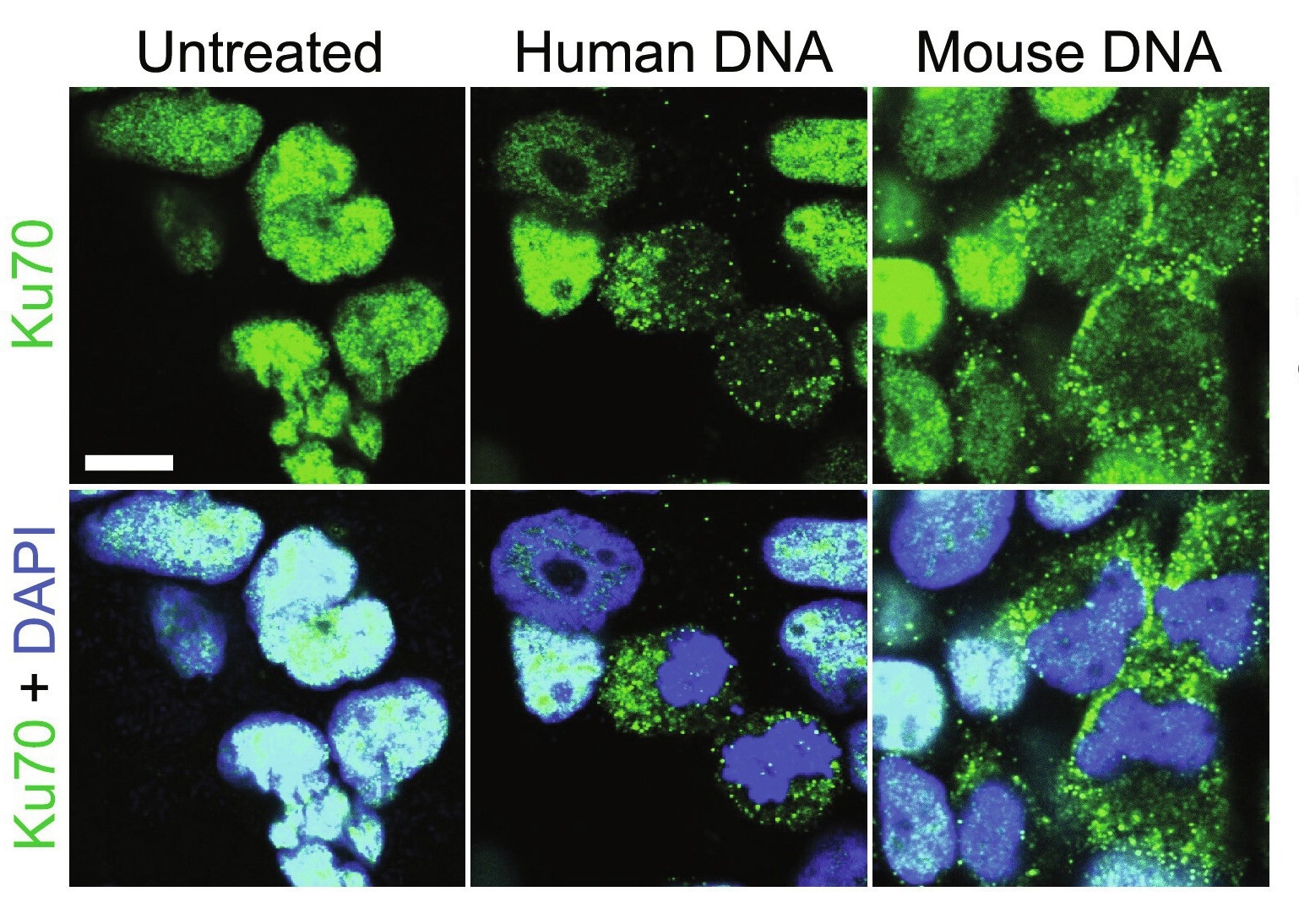
    The PNNL-OHSU team is studying thousands of proteins that could play a role in AML. Proteins are the body’s molecular workhorses, ferrying nutrients and other supplies back and forth between cells, turning genes on or off, and maintaining dozens of basic body processes. Even though genes get the glory, they do little directly to keep our bodies going. That’s the job of proteins. For nearly 20 years, study author Karin Rodland of OHSU, formerly of PNNL, has been a pioneer exploring the role of proteins in health and disease, building a program with OHSU and PNNL colleagues to study AML.

    In the latest study, a team led by Sara Gosline, a data scientist and computational biologist at PNNL, did an exhaustive study of the protein activity in 210 patients with AML. Altogether, the team measured levels of nearly half a million pieces of proteins from more than 9,000 proteins in patients’ blood samples. The team combined those findings with extensive data already known about the disease-;the genes and mutations involved, the molecular messengers that indicate which genes are active, and the effects of 46 drugs on AML patients, along with information about how the disease progressed in those patients.

    We were able to look at patterns of drug responses in hundreds of people by including protein and gene measurements together, and this gave us a level of detail that hasn’t been possible in prior studies. This is a great example where we are able to put our growing knowledge of protein signaling and machine learning models to benefit patients in the future.”


    Sara Gosline, data scientist and computational biologist at PNNL

    Gosline and colleagues, including first author James Pino of PNNL, deployed artificial intelligence, using several machine learning algorithms to make sense of the data.

    Beating drug resistance

    While the study yielded a load of data about what happens in the body of an AML patient, one finding stood out, pointing to a possible way to sidestep or delay drug resistance for some patients.

    The team showed that treatment with quizartinib, approved last year to treat AML, can shift how cancer cells respond to other drugs often used in combination to treat patients.

    Specifically, the team found that when patients on quizartinib stop responding to venetoclax, doctors might consider switching to another drug, panobinostat. It’s an example of how proteogenomic information could alter the roadmap that doctors use to navigate which medications patients receive at different stages of the disease.

    “The difficulty is that cancer keeps evolving,” said Gosline. “You hit the tumor with one drug and the tumor changes. This is what happens when patients experience drug resistance and the medicines stop working. Our study helps us understand exactly how this happens and what might be done in response. Which medication is best to turn to?”

    AML poses a particular challenge, said study author Cristina Tognon at OHSU.

    “When you treat a tumor with a drug, you are putting pressure on the tumor cells as they try to figure out a way to escape that pressure and survive. It’s a big problem in AML patients. What’s even more difficult is that in AML, there are many mutations at work; the disease doesn’t come in just one flavor,” said Tognon, who is an associate research professor and scientific director of the Druker Laboratory at OHSU.

    Ultimately, the team focused on 147 proteins and specific molecular locations known as phosphosites that play a key role in determining which proteins are turned on and which are off.

    Using just the protein data, the team sorted the samples into four distinct groups that predicted how the patients fared. Patients whose samples placed them in one of the groups had a better prognosis than the others, surviving far more than five years. Doctors hope that this type of information will eventually become available in the clinic. That would allow some patients who do not need aggressive therapies with severe side effects to avoid them while assuring that patients who have the worst prognosis are treated as aggressively as possible.

    “There is potential for clinical applications to be derived from this work, for example, diagnostics, such as protein biomarkers to predict responses to therapies, and the design of new drug combinations that might outperform current ones,” said OHSU’s Jeff Tyner, professor of medicine at the OHSU School of Medicine and Knight Cancer Institute.

    The work is the latest of more than 200 studies that have looked at protein activity in many forms of cancer, including colon, brain, endometrial, brain, blood and ovarian cancers. An OHSU-PNNL team

    discussed the emerging role of proteins for treating patients with precision medicine in a recent article in the Annual Reviews of Pharmacology and Toxicology. More and more, scientists are using proteomics-;the study of proteins-;to bridge the gap between genomics (the study of genes) to phenomics (phenotypes or observable characteristics).

    PMedIC: an OHSU-PNNL collaboration

    OHSU and PNNL scientists work collaboratively on many projects. OHSU brings outstanding clinical expertise about disease as well as extensive laboratory knowledge and is a world-class center for new treatments of leukemia. PNNL offers an unparalleled ability to measure tiny amounts of important molecules in great detail. Much of this work happens through the Pacific Northwest Biomedical Innovation Co-Laboratory, or PMedIC, a joint research collaboration between the two organizations. Through PMedIC and other collaborations, the institutions have made discoveries about several diseases, including Alzheimer’s disease, COVID-19, and the Zika virus.

    At PNNL, additional authors include Camilo Posso, Michael Nestor, Jamie Moon, Joshua Hansen, Chelsea Hutchinson-Bunch, Marina Gritsenko, Karl Weitz, Jason McDermott, Tao Liu and Paul Piehowski. Other authors from OHSU include Sunil Joshi, Kevin Watanabe-Smith, Nicola Long, Brian Druker, Anupriya Agarwal and Elie Traer.

    This work was supported by the National Cancer Institute’s Office of Cancer Clinical Proteomics Research (CPTAC U01CA271412), the ARCS Scholar Foundation, a Paul & Daisy Soros Fellowship, the National Cancer Institute (F30CA239335, R01 CA229875-01A1), the American Cancer Society (RSG-17-187-01-LIB), the National Heart, Lung, and Blood Institute (R01 HL155426-01), the Alex’s Lemonade Stand Foundation/RUNX1 Research Program, and the EvansMDS Foundation.

    Source:

    Journal reference:

    Pino, J. C., et al. (2024). Mapping the proteogenomic landscape enables prediction of drug response in acute myeloid leukemia. Cell Reports Medicine. doi.org/10.1016/j.xcrm.2023.101359.

    [ad_2]

    Source link

  • Engrailed-1 protein promotes pancreatic cancer progression and metastasis

    Engrailed-1 protein promotes pancreatic cancer progression and metastasis

    [ad_1]

    Pancreatic cancer is the No. 3 cause of cancer-related deaths in the United States, and only 12% of patients survive five years after being diagnosed. Severe pancreatic cancer is associated with metastasis, and it is this spread of secondary tumors that usually causes death, but little is known about the molecular mechanisms that drive metastasis.

    In a study published Dec. 18 in Advanced Science, researchers from the University of California, Davis showed that abnormal expression of the protein Engrailed-1 (EN1) promotes pancreatic cancer progression and metastasis in vitro and in mouse models. The team also found that elevated EN1 was associated with severe, metastatic pancreatic cancer in human patients, which suggests that EN1 might make a good target for pancreatic cancer therapies.

    We identified a novel epigenetic factor that can contribute to metastasis in pancreatic cancer, which is one of the most challenging cancers to treat. A better understanding of these mechanisms would allow us to identify potential targets and improve patient survival.”


    Chang-Il Hwang, assistant professor in the UC Davis Department of Microbiology and Molecular Genetics and a senior author on the paper

    Uncovering a main actor in pancreatic metastasis

    Metastasis is an important component of pancreatic cancer progression, but researchers have not been able to identify genetic mutations responsible for it. For this reason, Hwang thought that nongenetic factors, such as epigenetic changes or altered protein production, might be at play. His team previously identified several transcription factors -; proteins that control the production of other proteins -; that are elevated in pancreatic cancers that have undergone metastasis compared to primary tumors.

    One of these proteins, EN1, is essential for the survival of neurons during development and is not usually produced in adult pancreatic cells. EN1 has been shown to promote aggressive forms of breast cancer, and it is also associated with poor prognosis in other cancers, including glioblastoma and salivary gland adenoid cystic carcinoma, but its role in pancreatic cancer had not previously been described.

    The researchers tested whether inhibiting EN1 or ramping up its expression impacted the growth and survival of pancreatic cancer “organoids” -; three-dimensional clumps of lab-grown tissue. They found that, without EN1, pancreatic cancer cells were less likely to survive and divide, but adding extra EN1 increased the tumors’ survival. Furthermore, when the researchers genetically modified mouse pancreatic cancer cell lines so that they produced more EN1 than usual, the cells showed increased rates of cell invasion and migration, key features of metastasis.

    “It’s very clear that EN1 is a really important factor behind the aggressiveness of pancreatic cancer,” said first author Jihao (Reno) Xu, a doctoral candidate in the Biochemistry, Molecular, Cellular and Development Biology graduate group. “When we take the tumor cells and make them overexpress EN1, they become more metastatic and aggressive, and when we knock it down, they become less metastatic.”

    By analyzing publicly available patient databases, the researchers also showed that EN1 is important for prognosis in human pancreatic cancer. They found that EN1 levels were elevated in a subset of patients with advanced pancreatic cancer, and that patients with elevated EN1 tended to have worse prognoses.

    “Patients with high levels of EN1 have shorter survival times, which suggests that it is contributing to the aggressiveness of pancreatic cancer,” said Hwang.

    Now, Hwang, Xu and their colleagues are working on ways to translate their findings into the clinic by testing different ways to target EN1. They also plan to continue investigating other nongenetic factors that might contribute to pancreatic cancer progression.

    “Ultimately, we want to identify new therapeutic strategies to tackle this disease,” Xu said.

    Additional authors on the paper are: at UC Davis, EunJung Lee, Keely Y. Ji, Omar W. Younis and Alexander D. Borowsky; Jae-Seok Roe, Yonsei University; Claudia Tonelli, Tim D.D. Somerville, Melissa Yao, Joseph P. Milazzo, Herve Tiriac, Youngkyu Park, Christopher R. Vakoc and David A. Tuveson, Cold Spring Harbor Laboratory; Ania M. Kolarzyk and Esak Lee, Cornell University; Jean L. Grem, Audrey J. Lazenby, James A. Grunkemeyer and Michael A. Hollingsworth, University of Nebraska Medical Center.

    The work was supported by the UC Davis Comprehensive Cancer Center Pilot Grant and the National Institutes of Health.

    Source:

    Journal reference:

    Xu, J., et al. (2023). Engrailed‐1 Promotes Pancreatic Cancer Metastasis. Advanced Science. doi.org/10.1002/advs.202308537.

    [ad_2]

    Source link

  • New microscopy technology unveils detailed images of brain cancer tissue

    New microscopy technology unveils detailed images of brain cancer tissue

    [ad_1]

    Brigham and MIT researchers uncovered never-before-seen details in human brain tissue with new, inexpensive microscopy technology.

    Key takeaways:

    • Researchers have developed a new microscopy technology called decrowding expansion pathology (dExPath) to analyze brain tissue.
    • By pulling proteins apart with dExPath, researchers can stain proteins in tissue that could not be accessed before, highlighting nanometer sized structures or even cell populations that were previously hidden.
    • This “super-resolution imaging” technology has the potential to provide insights that could improve diagnostic strategies and patient outcomes.

    Researchers from Brigham and Women’s Hospital, a founding member of Mass General Brigham, and the Massachusetts Institute of Technology (MIT) have unveiled unprecedentedly detailed images of brain cancer tissue through the use of a new microscopy technology called decrowding expansion pathology (dExPath).Their findings, published in Science Translational Medicine, provide novel insights into brain cancer development, with potential implications for advancing the diagnosis and treatment of aggressive neurological diseases.

    “In the past, we have relied on expensive, super-resolution microscopes that only very well-funded labs could afford, required specialized training to use, and are often impractical for high-throughput analyses of brain tissues at the molecular level,” said Pablo Valdes, MD, PhD, a neurosurgery resident alumnus at the Brigham and lead author of the study. “This technology brings reliable, super-resolution imaging to the clinic, enabling scientists to study neurological diseases at a never-before-achieved nanoscale level on conventional clinical samples with conventional microscopes.”

    Researchers previously relied on costly, super-high-resolution microscopes to image nanoscale structures in cells and brain tissue, and, even with the most advanced technology, they often struggled to effectively capture these structures at the nanoscale level.

    Ed Boyden, PhD, the Y. Eva Tan Professor in Neurotechnology at MIT and co-senior author on this study, began addressing this problem by labeling tissues, and then chemically modifying them to enable uniform physical expansion of tissues. However, this expansion technology was far from perfect. Relying on enzymes known as proteases to break up tissue, scientists found that this chemical treatment with enzymes destroyed proteins before they could analyze them, leaving behind only a skeleton of the original structure, retaining only the labels.

    Working together, Boyden and E. Antonio Chiocca, MD, PhD, Neurosurgery Chair at Brigham and Women’s Hospital and co-senior author on this study, mentored Valdes during his training as a neurosurgeon-scientist, to develop novel chemistries with dExPath to address the limitations of the original expansion technology.

    Their new technology chemically modifies tissues by embedding them in a gel and ‘softening’ the tissues with a special chemical treatment that separates protein structures without destroying them and which allows tissues to expand. This provided exciting findings to the MIT and Brigham researchers, who routinely use commercially available antibodies to bind to and illuminate biomarkers in a sample. Antibodies, however, are large and many times cannot easily penetrate cell structures to reach their target. Now, by pulling proteins apart with dExPath, these same antibodies used for staining can penetrate spaces to bind proteins in tissue that could not be accessed before expansion, highlighting nanometer sized structures or even cell populations that were previously hidden.

    The human brain has several stop guards in place to protect itself from pathogens and environmental toxins. But these elements make studying brain activity challenging. It can be a bit like driving a car through mud and ditches. We cannot access certain cell structures in the brain because of barriers that stand in the way. That is just is one of the reasons that this new technology could be so practice changing. If we can take more detailed and accurate images of brain tissue, we can identify more biomarkers and be better equipped to diagnose and treat aggressive brain diseases.”


    E. Antonio Chiocca, MD, PhD, Chair of the Department of Neurosurgery, Brigham and Women’s Hospital

    To validate the effectiveness of dExPath, Boyden and Chiocca’s team applied the technology to healthy human brain tissue, high and low-grade brain cancer tissues, and brain tissues affected by neurodegenerative diseases including Alzheimer’s and Parkinson’s diseases. Investigators stained tissue for brain and disease specific biomarkers and captured images before and after expanding samples with dExPath.

    The results revealed uniform and consistent expansion of the tissue without distortion, enabling accurate analysis of protein structures. Additionally, dExPath effectively eliminated fluorescent signals in brain tissue called lipofuscin, which makes imaging of subcellular structures in brain tissues very difficult, further enhancing image quality. Further, dExPath provided stronger fluorescent signals for improved labeling as well as simultaneous labeling of up to 16 biomarkers in the same tissue specimen. Notably, dExPath imaging revealed that tumors previously classified as “low-grade” contained more aggressive features and cell populations, suggesting the tumor could become far more dangerous than anticipated.

    While promising, dExPath requires validation on larger sample sizes before it can contribute to the diagnosis of neurological conditions such as brain cancer. Valdes underscores that, although still in its early stages, his team aspires for this technology to eventually serve as a diagnostic tool, ultimately enhancing patient outcomes.

    “We hope that with this technology, we can better understand at the nanoscale levels the intricate workings of brain tumors and their interactions with the nervous system without depending on exorbitantly expensive lab equipment,” said Valdes who is now an assistant professor of neurosurgery and Jennie Sealy Distinguished Chair in Neuroscience at the University of Texas Medical Branch. “The accessibility of dExPath will bring enable super-resolution imaging to understand biological processing at the nanometer level in human tissue in neuro-oncology and in neurological disease such as Alzheimer’s and Parkinson’s, and one day, could even improve diagnostic strategies and patient outcomes.”

    Source:

    Journal reference:

    Valdes, P. A., et al. (2024) Improved immunostaining of nanostructures and cells in human brain specimens through expansion-mediated protein decrowding. Science Translational Medicine. doi.org/10.1126/scitranslmed.abo0049.

    [ad_2]

    Source link

  • Is there an association between vitamin D, immunocompetence, and aging?

    Is there an association between vitamin D, immunocompetence, and aging?

    [ad_1]

    In a recent review published in the journal Nutrients, researchers explored the role of an individual’s immunocompetence in the responsiveness to vitamin D.

    They discussed the modulation of immunocompetence via the epigenetic programming function of the vitamin D receptor (VDR) and its ligand and highlighted the impact of aging on immunocompetence.

    Study: Vitamin D and Aging: Central Role of Immunocompetence. Image Credit: Iryna Imago/Shutterstock.comStudy: Vitamin D and Aging: Central Role of Immunocompetence. Image Credit: Iryna Imago/Shutterstock.com

    Background

    Vitamin D plays a crucial role in bone health by regulating calcium homeostasis and preventing conditions like rickets and osteomalacia. However, its influence on immunity extends beyond this function.

    Vitamin D deficiency, linked to modern lifestyle factors like limited sun exposure, affects the endogenous production of active vitamin D metabolites.

    The inactive vitamin D3 is converted to active 1,25(OH)2D3 in the liver and kidneys, which acts as a hormone and affects various tissues. Notably, various cells, including those of the innate immune system, can produce 1,25(OH)2D3 locally, contributing to auto- and paracrine effects. This compound acts as a ligand of high affinity for VDR, regulating the expression of numerous genes.

    The vitamin D status, indicated by serum 25(OH)D3 levels, categorizes individuals into deficient, insufficient, or sufficient groups. Vitamin D responsiveness varies among people due to genetic and epigenetic factors influencing molecular responses.

    Low responders, constituting about 25% of the population, may have increased susceptibility to diseases related to compromised immunity. The VDR-based modulation of immunocompetence may contribute to aging and reduce the risk of age-related diseases.

    The present review offers insights into the immunomodulatory functions of vitamin D and its impact on various health aspects beyond bone metabolism.

    Vitamin D signaling

    VDR binds specifically to genomic DNA, recognizing the motif RGKTSA. In complex with retinoid X receptor (RXR), VDR preferentially binds to direct repeat sequences in the euchromatin. Various “pioneer factors” facilitate VDR in opening chromatin, which is crucial for efficient binding.

    Chromatin accessibility and VDR binding can be assessed using next-generation sequencing technologies, including ChIP-seq (chromatin immunoprecipitation sequencing) and ATAC-seq (assay for transposase-accessible chromatin using sequencing), especially in peripheral blood mononuclear cells.

    Genomic regions of vitamin D target genes demonstrate changes in chromatin accessibility and VDR binding after vitamin D3 supplementation.

    Enhancers and transcription start site regions, even at a considerable linear distance, can interact via DNA looping within the same topologically associating domain, influencing gene expression.

    VDR’s genomic actions involve protein-protein interactions with the Mediator complex and RNA polymerase II, influencing transcription. Vitamin D also exerts epigenomic effects, altering DNA methylation, histone modifications, and chromatin organization, dynamically shaping the cell’s epigenetic landscape.

    These genomic and epigenomic effects contribute to vitamin D’s modulatory role in hematopoiesis and immunocompetence, affecting human immune cells both in vitro and in vivo.

    Epigenetic programming of immune cells

    Throughout embryogenesis and adult cellular differentiation, stem and progenitor cells undergo epigenetic programming, determining the function of terminally differentiated cells. 1,25(OH)2D3 plays a crucial role in this process, influencing hematopoiesis and the differentiation of immune cells.

    Hematopoietic stem cells (HSCs) differentiate into various blood and immune cell types, and 1,25(OH)2D3 regulates embryonic HSC numbers.

    Various transcription factors influenced by vitamin D drive the differentiation of myeloid progenitor cells into granulocytes and monocytes. Vitamin D is also the differentiation of monocytes into dendritic cells and macrophages.

    Epigenetic programming by vitamin D contributes to innate immune cell adaptation, modulating responses to infections, inflammation, and diseases.

    Variability in vitamin D status and response index among individuals affects the epigenetic programming of monocytes and derived cells, emphasizing the potential of optimized vitamin D3 supplementation for supporting proper immune cell epigenetics and overall immunocompetence. However, further research is needed to validate this concept fully.

    Decline in immunocompetence during aging

    Aging involves accumulating molecular damage, resulting in cellular dysfunction and weakened organs. Immunocompetence, crucial for appropriate immune responses, declines with age, leading to increased susceptibility to infections and diseases.

    The thymus atrophies, diminishing the production of T-cells, and “inflammaging” ensues. However, interindividual differences exist, and some individuals may display relatively higher immunocompetence.

    Lower immunocompetence correlates with accelerated aging and heightened disease risks. Vitamin D sufficiency may protect against cancers by preserving immunocompetence.

    Adequate vitamin D levels could stabilize immune resilience, safeguard against diseases, and contribute to healthy aging by mitigating various hallmarks of aging, including inflammation and cellular stress.

    Conclusion

    In conclusion, the active form of vitamin D plays a crucial role in modulating the epigenome of immune cells, particularly in monocytes.

    The observed associations between vitamin D deficiency, increased disease risk, and accelerated aging may be attributed to diminished immunocompetence.

    Considering individual responsiveness, a precautionary daily vitamin D3 dose of 1 µg (40 IU)/kg body mass is suggested, exceeding general recommendations but staying within safe limits to strengthen immunocompetence. The researchers emphasize personalized vitamin D supplementation to safeguard against prevalent diseases and promote healthy aging.

    [ad_2]

    Source link

  • Unveiling the mystery behind rapid memory loss in cancer patients

    Unveiling the mystery behind rapid memory loss in cancer patients

    [ad_1]

    In a rare but serious complication of cancer, the body’s own immune system can start attacking the brain, causing rapid-onset memory loss and cognitive deficits. What triggers this sudden biological civil war was largely unknown.

    Now, researchers at University of Utah Health have found that some tumors can release a protein that looks like a virus, kickstarting an out-of-control immune reaction that may damage brain cells.

    Their findings published in Cell on Jan. 31, 2024.

    A rapid immune attack

    Jason Shepherd, Ph.D., associate professor of neurobiology at University of Utah Health and last author on the study, explains that the swift escalation of symptoms-;which can include memory and behavioral changes, loss of coordination, and even seizures-;is a hallmark of the disease, called anti-Ma2 paraneoplastic neurological syndrome. The disease is one of a group of cancer-related neurological syndromes that occur in less than one in 10,000 people with cancer. The precise symptoms of these diseases vary, but all involve rapid immune reactions against the nervous system. “The symptoms come in quickly and can be quite debilitating,” Shepherd says.

    This fascinating research illustrates how tumor cells can manipulate their environment. We hope that this innovative transdisciplinary research will positively impact both the lives of cancer patients and of those who experience neurological symptoms.”


    Neli Ulrich, Ph.D., executive director of the Comprehensive Cancer Center at Huntsman Cancer Institute at the University of Utah and a Jon M. and Karen Huntsman Presidential Professor in Cancer Research at the U

    Stacey L. Clardy, M.D., Ph.D., a neurologist at U of U Health and a coauthor on the study, adds, “Most patients begin to experience these unusual neurologic symptoms before they even know they have a cancer.”

    These rapid-onset symptoms are the result of the immune system suddenly starting to target specific proteins that are found in the brain. Scientists knew that this flare of immunity often targets a protein called PNMA2. But nobody knew why PNMA2 provokes such a strong immune response, which left researchers at a loss for ways to prevent it. “We do not understand what is happening at the cellular or molecular level to actually cause the syndrome,” Clardy says, “and understanding the mechanism of disease is crucial to developing better treatments.”

    A virus lookalike

    To figure out how PNMA2 kickstarts an immune reaction, Junjie Xu, a graduate researcher in neurobiology at U of U Health and the lead author on the study, examined the protein’s structure using advanced microscopy. When he saw the first clear image of the protein, he was “so, so excited,” Xu says. Multiple PNMA2 proteins had spontaneously self-organized into 12-sided complexes that bore a striking resemblance to the geometric protein shells of some viruses.

    One of the immune system’s healthy functions is to attack viruses, and PNMA2’s virus-like structure makes it particularly prone to being targeted as well, the researchers found. In fact, in experiments in mice, the immune system only attacked PNMA2 protein when it was assembled into virus-like complexes.

    Wrong place, wrong time

    The location of PNMA2 in the body is also a crucial piece of the puzzle, the scientists found. “This protein normally is only expressed in the brain, in neurons,” Xu says, “but some cancer cells can express it, which can trigger an immune response.”

    As long as PNMA2 stays in the brain, the immune system won’t react to it. But rarely, a tumor elsewhere in the body will start producing PNMA2 protein. And when the immune system detects PNMA2 protein outside the brain, it reacts like it would to any foreign invader. The immune system makes antibodies that bind to the unfamiliar substance, and those antibodies direct immune cells to attack.

    But, once activated, the immune system doesn’t just attack the PNMA2 produced by the cancer. It also targets the parts of the brain that produce PNMA2 normally, including regions involved in memory, learning, and movement. The brain normally has a degree of protection from the immune system, but cancer weakens that barrier, leaving the brain especially vulnerable to this immune onslaught.

    In future work, the researchers aim to figure out which aspect of the immune response leads to patients’ rapid cognitive decline-;the antibodies themselves, immune cells making their way into the brain, or some combination of the two.

    Understanding how the immune system causes neurological symptoms may help scientists design targeted treatments, Shepherd says. “If we show that PNMA2 antibodies are the culprit that really drives the neurological symptoms, you could devise a way to block those antibodies from getting into the brain or mop them up with something as a treatment… If you can alleviate some of those neurological symptoms, it really would be huge.”

    Source:

    Journal reference:

    Xu, J., et al. (2024) PNMA2 forms immunogenic non-enveloped virus-like capsids associated with paraneoplastic neurological syndrome. Cell. doi.org/10.1016/j.cell.2024.01.009.

    [ad_2]

    Source link

  • Study reveals key mechanism behind obesity-related metabolic dysfunction

    Study reveals key mechanism behind obesity-related metabolic dysfunction

    [ad_1]

    In a recent study published in Nature Metabolism, researchers found that feeding a high-fat diet (HFD) causes mitochondrial dysfunction and fragmentation in white adipocytes in mice.

    Study: Obesity causes mitochondrial fragmentation and dysfunction in white adipocytes due to RalA activation. Image Credit: Kateryna Kon/Shutterstock.com
    Study: Obesity causes mitochondrial fragmentation and dysfunction in white adipocytes due to RalA activation. Image Credit: Kateryna Kon/Shutterstock.com

    Background

    Obesity has become a global epidemic, increasing the incidence of non-alcoholic steatohepatitis, diabetes, and other cardiometabolic disorders. White adipose tissue (WAT) expands chronically during the development of obesity, with metabolic changes characterized by fibrosis, inflammation, hormone insensitivity, and apoptosis. Obese individuals have impaired mitochondrial function, and the underlying mechanisms and their contribution to obesity remain unclear.

    The study and findings

    In the present study, researchers demonstrated increased expression and activity of Ras-like proto-oncogene A (RalA) in adipocytes from obese mice and attenuation of HFD-induced obesity upon targeted Rala deletion in white adipocytes. First, they noted upregulation of Rala expression in epididymal (eWAT) and inguinal WAT (iWAT) adipocytes during obesity development in HFD-fed mice relative to controls.

    Further, RalA protein levels were elevated in iWAT adipocytes from obese mice. No changes in RalA were observed in brown adipose tissue (BAT) after HFD feeding. Next, RalA-floxed (Ralaf/f) mice and adiponectin-promoter-driven Cre transgenic mice were crossed to generate adipocyte-specific Rala knockout (KO) mice (RalaAKO). RalaAKO mice showed over 90% reduced RalA protein in primary adipocytes from BAT and WAT compared to Ralaf/f littermates.

    RalA depletion reduced insulin-stimulated glucose uptake in BAT and iWAT. Additionally, brown adipocyte-specific KO mice (RalaBKO) were produced by crossing Ralaf/f mice and uncoupling protein 1 (Ucp1)-promoter-driven Cre transgenic mice. This reduced glucose uptake in the BAT of RalaBKO mice, and insulin-stimulated glucose uptake was mainly limited to brown fat.

    Adipocyte-specific Rala deletion did not affect the body weight of chow-diet (CD)-fed mice, albeit they had reduced fat mass and depot weight. RalaAKO mice had smaller iWAT adipocytes than CD-fed controls. RalaAKO mice gained less weight than controls when fed 60% HFD. HFD-fed RalaAKO mice had smaller adipocytes in iWAT but not in BAT or eWAT compared to controls.

    HFD-fed RalaAKO mice also showed improved glucose tolerance, without changes in insulin tolerance; they also had reduced insulin levels and improved homeostasis model assessment of insulin resistance (HOMA-IR) than controls. RalaAKO mice showed lower glucose excursions in a pyruvate tolerance test than controls, with downregulation of hepatic gluconeogenic genes.

    HFD-fed RalaAKO mice had lower triglyceride levels and liver weight and less lipid accumulation in the liver than controls. Moreover, the expression of lipogenic, fibrosis-related, and inflammatory genes was reduced in the livers of RalaAKO mice. The team found that adipocyte Rala ablation did not affect food intake and energy metabolism in CD-fed mice.

    However, HFD-fed RalaAKO mice had increased energy expenditure. In contrast, energy expenditure and food intake were identical in HFD-fed RalaBKO mice and controls, suggesting that WAT-specific Rala deficiency increased energy expenditure. Further, oxidative phosphorylation proteins were upregulated in the iWAT of RalaAKO mice but not in eWAT.

    Next, the team explored mechanisms underlying increased energy metabolism in RalaAKO mice and mitochondrial activity in adipocytes. They observed an elevated oxygen consumption rate in iWAT mitochondria from KO mice relative to controls. Moreover, fatty acid oxidation was higher in KO adipocytes. The expression of mitochondrial biogenesis-related genes in WAT was comparable between HFD-fed RalaAKO and Ralaf/f mice.

    Electron microscopy showed that HFD feeding of wild-type mice induced smaller, spherical iWAT mitochondria. iWAT mitochondria in CD-fed mice had an elongated shape, while those in HFD-fed mice had smaller mitochondria. Besides, adipocyte Rala ablation did not grossly impact mitochondrial morphology in the iWAT of CD-fed mice; in contrast, the HFD-induced morphological change in mitochondria was prevented in Rala KO iWAT.

    Mitochondrial morphology in BAT was unaltered upon Rala deletion in HFD- or CD-fed mice. HFD feeding downregulated protein levels of long and short forms of optic atrophy 1 (Opa1), a mitochondrial fusion regulator, in iWAT. However, only the short form (S-Opa1) was downregulated in eWAT. Further, they focused on dynamin-related protein 1 (Drp1), which regulates mitochondrial fission, and found increased phosphorylation at the anti-fission site (S637) in Rala KO iWAT.

    The researchers analyzed microarray data of WAT from non-obese and obese females to examine the relevance of Drp1 in human obesity. They found that the human Drp1 homolog, dynamin 1 like (DNM1L), was positively correlated with HOMA-IR and body mass index. DNM1L expression was upregulated in obese subjects.

    Conclusions

    Taken together, the study demonstrated that RalA was induced and activated in white adipocytes of HFD-fed mice. Targeted RalA deletion in white adipocytes prevented obesity-related mitochondrial fragmentation and resulted in resistance to HFD-induced weight gain through heightened energy expenditure.

    HFD-fed RalaAKO mice showed improved liver function and pyruvate tolerance and reduced gluconeogenesis and hepatic lipids. Overall, chronically increased RalA activity plays a role in repressing energy expenditure in obese adipose tissue by shifting mitochondrial dynamics towards excessive fission and contributing to weight gain and metabolic dysfunction.

    [ad_2]

    Source link

  • Immune protein Ku70 key in fighting bowel cancer

    Immune protein Ku70 key in fighting bowel cancer

    [ad_1]

    In a recent study published in the journal ScienceAdvances, researchers conducted a plethora of experiments on human cell lines and transgenic murine models aimed at investigating the relationship between the Ku70 DNA repair protein and intestinal cancer. Their results highlight the potent tumor-suppressive function of the protein. Mutations in the gene encoding the protein or downregulations in expression substantially increased the risk of subsequent spontaneous intestinal cancer. Colitis and colitis-associated colorectal cancer risks were similarly heightened.

    This study furthers progress in understanding the Ku70-mediated Ras-ERK signaling pathway and Ku70 activation’s molecular mechanisms. These, in turn, could form the basis for the future development of DNA-based therapeutics.

    Study: Ku70 senses cytosolic DNA and assembles a tumor-suppressive signalosomeStudy: Ku70 senses cytosolic DNA and assembles a tumor-suppressive signalosome

    What is Ku70?

    Ku70 is a DNA repair subunit protein that helps repair DNA via the non-homologous end-joining (NHEJ) pathway. In humans, the protein is encoded by the XRCC6 gene, which studies have revealed is evolutionarily conserved. Ku70 can be found both in the cell nucleus and cytoplasm and, until recently, was thought to be restricted to its primary (DNA repair) role. Over the last decade, however, a growing body of literature suggests secondary protein functions, including antimicrobial and anti-tumor.

    Cytoplasmic Ku70 has been shown to produce type III interferons (IFNs) in response to bacterial or viral DNA in both human and murine model systems. It has additionally been shown to bind to Rickettsia conorii, a human bacterial pathogen, thereby facilitating its neutralization by nonphagocytic mammalian cells.

    Recently, research has suggested that the protein may also have a tumor-suppressive role. In an unrelated experiment, genetically altered mice lacking the Ku70 gene were found remarkably susceptible to hepatocellular carcinoma and spontaneous T-cell lymphoma development. Another study revealed genetic deletion of Ku70 to enhance colorectal cancer risk. Unfortunately, the Ku70 gene is implicated in growth, with its deletion resulting in stunted murine growth. Since smaller body size and poor growth would have confounded interpretation of these results, the association between Ku70 and cancer remains speculative and hitherto unconfirmed.

    About the study

    The present study aims to elucidate any association between Ku70 protein expression and intestinal cancer risk. Once the association is identified, the mechanism underlining Ku70’s protective anti-tumor function is explored. The experimental sample group consisted of wild-type (WT), Ku70+/−(heterozygous for Ku70), Ku70−/−(homozygous recessive), and C57BL/6NcrlAnu transgenic mice. All four mice types were equally divided into case (AOM-DSS) and control (untreated) cohorts.

    The study began with the experimental induction of colitis and colitis-associated colorectal tumorigenesis in the case-cohort via the intraperitoneal injection of 10 mg of azoxymethane (AOM). This was followed five days later by administering 1.5% Dextran Sodium Sulfate (DSS) for six days. Fourteen days after AOM administration, mice were euthanized, and their intestines and colon tissues were harvested for methodological validation and downstream analysis.

    Induced cancers were identified and characterized using histology, immunohistochemistry, and microscopy techniques. Ku70 and related proteins (e.g., cytokines) were identified and quantified using immunoblotting and enzyme-linked immunosorbent assays (ELISAs), respectively. Quantitative Real Time-Polymerase Chain Reaction (qRT-PCR) was used to isolate, amplify, and identify RNA of interest within the colon tissue. RNA was further purified using lithium chloride (LiCl) precipitation. Genomic DNA obtained from mouse feces was used to identify and characterize gut microbiome assemblages using 16S ribosomal RNA (rRNA) gene sequencing.

    Separately, cell line-based lines of evidence were obtained by constructing a plasmid containing the Ku70 gene, which was then transformed into NEB 5-alpha competent Escherichia coli. Finally, the recombinant E. coli was used to transfect the study subjects, namely HEK293T human cell lines and colon cells cultured from harvested colon tissue.

    Cell line analyses incorporated genomics, immunoblotting, and immunofluorescence techniques used for the murine models and included coimmunoprecipitation, proliferation, and organoid culture analyses. Active RAS was screened as a confounding variable using the Active Ras Detection Kit. RAS is a gene family whose mutations are estimated to account for 95% of pancreatic and 45% of colorectal cancers.

    Study findings

    This study’s highlight is validating that the cytosolic DNA sensor Ku70 has the secondary role of tumor suppression. Reductions in Ku70 expression or mutation in its gene were rapidly followed by tumorigenesis in both murine models and cell cultures. This study further unravels the mechanism of action of Ku70, which depicts an unexpectedly high mutation co-occurrence with genes encoding ARAF, RAF1, HRAS, NRAS, and BRAF, RAS family genes previously implicated in intestinal cancers.

    “Our study suggests that the Ku70-ERK signaling pathway is tumor suppressive, which is in contrast to the observation that Ras/Raf mutations, which are common in colorectal cancer, drive aberrant activation of downstream ERK-MAPK signaling.”

    Study findings further suggest that Ku70 may function in a cell-specific manner – epithelial and stromal cells from patients with Crohn’s disease or colorectal cancer display decreased Ku70 gene expression even in homozygous dominant conditions. Parallelly, Ku70 was found to form a cytosolic signalosome consisting of Ras, Raf, and Ku70, which docks at the endosome’s membrane and mediates the MEK-ERK-Cdc25A-CDK1 signaling axis activation, thereby resulting in an antitumorigenic effect.

    “…activation of the Ras-ERK pathway protects mice against colitis (83) and inhibits mammalian cell proliferation. Further studies are required to elucidate which cell types undergo Ras-ERK signaling for the progression of colorectal cancer and which cell types undergo Ku70 signaling for the attenuating of colorectal cancer.”

    “We speculate that the activation of the Ku70-mediated Ras-ERK signaling might be initiated by the cytoplasmic DNA arising from the gut microbiome introduced into the host cells following a rupture of the intestinal barrier. However, it is also possible that damaged nucleus and/or mitochondria may be a source of cytoplasmic DNA that triggers Ku70-mediated Ras-ERK signaling.”

    Journal reference:

    • Pandey, A., Shen, C., Feng, S., Tuipulotu, D. E., Ngo, C., Liu, C., Kurera, M., Mathur, A., Venkataraman, S., Zhang, J., Talaulikar, D., Song, R., Wong, L., Teoh, N., Kaakoush, N. O., & Man, S. M. (2024). Ku70 senses cytosolic DNA and assembles a tumor-suppressive signalosome. Science Advances, DOI – 10.1126/sciadv.adh3409, https://www.science.org/doi/10.1126/sciadv.adh3409

    [ad_2]

    Source link