[ad_1]
In a recent study published in The Lancet Microbe, researchers investigated the effects of molnupiravir on the evolution of severe acute respiratory syndrome coronavirus 2 (SARS-CoV-2) in immunocompromised patients.
Persistent SARS-CoV-2 infection in individuals who are immunocompromised offers genomic variation and has been linked to viral evolution. Antiviral therapy is recommended in immunocompromised patients with acute infection to prevent severe disease. Molnupiravir is the only alternative when first-line therapies (remdesivir and ritonavir-boosted nirmatrelvir) are not feasible, available, or appropriate.
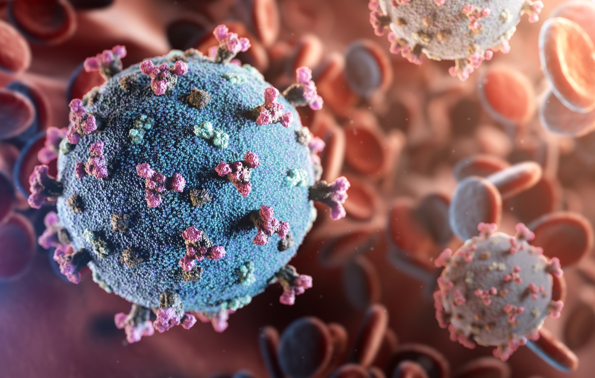 Study: Effect of molnupiravir on SARS-CoV-2 evolution in immunocompromised patients: a retrospective observational study. Image Credit: creativeneko / Shutterstock
Molnupiravir has been used worldwide in hospital and community settings as well as for immunocompromised patients. Nevertheless, it has been ineffective at reducing coronavirus disease 2019 (COVID-19) hospitalization and mortality rates in high-risk groups, and consequently, it has been designated a third-line therapeutic option. The drug triggers mutagenesis by introducing β-D-N4-hydroxycytidine, the prodrug, into the viral ribonucleic acid (RNA).
The viral RNA polymerase uses this modified RNA as the template, and an error catastrophe occurs, inhibiting the viral replication. During RNA synthesis, molnupiravir behaves like a cytosine (C) and pairs with guanine (G); however, once incorporated, it transforms into a tautomer analogous to uracil (U), leading to G-to-A mutations in the subsequent round of replication. Likewise, it can induce C-to-U (or -thymine [T]) mutations during the synthesis of the positive-sense genome.
Reverse T-to-C and A-to-G mutations are also possible but are less frequent. G-to-A mutations indicate molnupiravir treatment; distinctive mutational profiles with extensive G-to-A mutations have been found in global sequences and phylogenetic trees. This is linked to the use of molnupiravir as countries showing long G-to-A branches had increased use of the drug. Contrastingly, countries with infrequent G-to-A branches have not authorized molnupiravir.
About the study
In the present study, researchers analyzed the sequencing data from immunocompromised patients with SARS-CoV-2 infection to assess the effects of molnupiravir on viral evolution. The team sequenced around 100 genomes weekly from December 2021 to September 2022, specifically focusing on samples from reinfections, hospitalized patients, overseas travelers, and suspected residential care- and healthcare-related infections.
Immunocompromised patients with protracted infection were also covered. The team selected nine patients with the same variant with multiple samples (from distinct time points). Four patients (controls) were tested before molnupiravir was available, and five were sampled pre- and post-molnupiravir treatment. All molnupiravir recipients and two controls were immunocompromised. Seven patients received ≥ two vaccine doses, and two were non-vaccinated.
Patients’ prior infection status was unknown. Patients infected with similar variants and high-quality genomes were selected for group comparisons across time points. Accumulated mutations were compared between groups. The ultrafast sample placement on existing trees (UShER) pipeline and the University of California Santa Cruz genome viewer were leveraged to compare variants from patients with global reference sequences and visualize the locations of mutations.
Findings
The team noted that SARS-CoV-2 genomes acquired an average of 30 new low/mid frequency variants by 10 days post-molnupiravir treatment. These changes in viral diversity were not observed in patients who did not receive molnupiravir. On average, 3.3 mutations were acquired per day in the molnupiravir group.
The probability of observing no mutations among controls during the study period was extremely low. Non-synonymous mutations were common in the spike protein, and subsequent samples indicated that some mutations were fixed. In one patient, 10 non-synonymous mutations were fixed by 35 days post-treatment.
Accrued mutations were scattered throughout the genome, including those not detected in global Omicron genomes. Mutations acquired in the spike protein clustered at two locations, and their functional relevance was unclear. No known drug-resistance mutations were observed; however, non-synonymous mutations in the open reading frame 1b (ORF1b) were noted.
The UShER analysis revealed potentially rare/novel mutations in the sequences following treatment. Some samples could not be placed on the global SARS-CoV-2 phylogeny as many mutations were phylogenetically distinct. Mutational profiles post-treatment revealed dominant G-to-A and C-to-T mutations, representing 70% of mutations, which persisted up to 44 days post-treatment.
Conclusions
In sum, the findings showed that molnupiravir use in immunocompromised patients modified the patterns of viral evolution, with effects lasting beyond the five-day treatment period. This highlights the risks of treating this subgroup of patients with an error-generating antiviral. The evolution rate in molnupiravir recipients exceeded that observed in non-recipients in this study and globally. Overall, the researchers provided more evidence of the causal link between molnupiravir and the altered mutational landscape of SARS-CoV-2.
[ad_2]
Source link