In a recent study published in Nature Neuroscience, researchers investigated whether the neurological response to coronavirus disease 2019 (COVID-19) may be due to brain-brain barrier (BBB) disruption and subsequent extravasation of serum components.
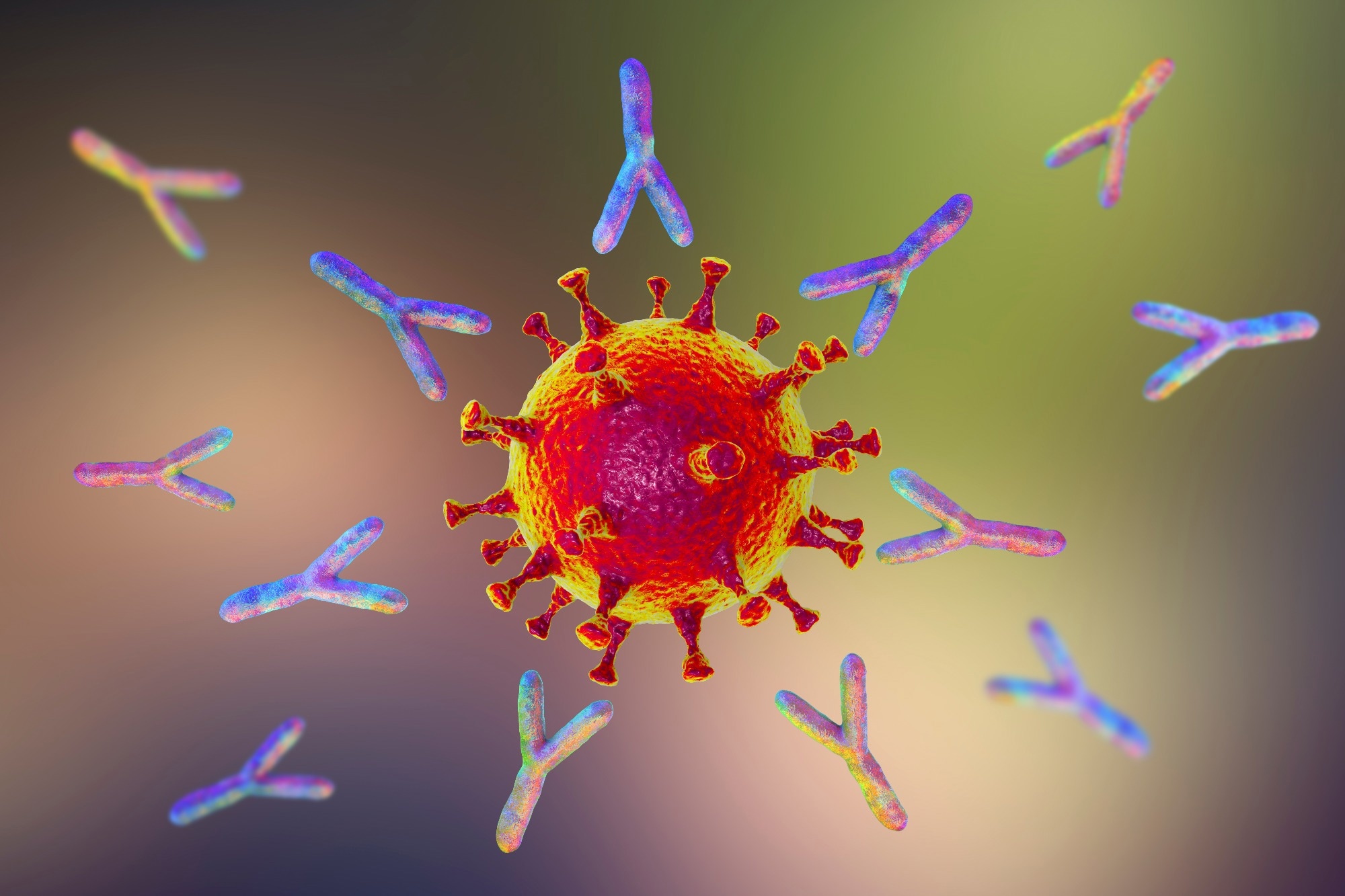
 Study: Blood–brain barrier disruption and sustained systemic inflammation in individuals with long COVID-associated cognitive impairment. Image Credit: fran_kie/Shutterstock.com
Background
COVID-19, caused by severe acute respiratory syndrome coronavirus 2 (SARS-CoV-2), is a respiratory viral infection resulting in severe acute respiratory distress syndrome (ARDS) and long-term neurological consequences such as headache, lethargy, malaise, and altered consciousness.
BBB breakdown allows serum components and cytokines to penetrate the brain. The cerebrovascular pathophysiology and processes remain unknown, warranting further research.
About the study
The present study determined the association between COVID-19-related cognitive impairment and BBB breakdown in COVID-19 patients.
The researchers collected blood and plasma samples from 76 acute COVID-19 patients, evaluated them for inflammatory, coagulation, and BBB dysfunction indicators, and rated their severity using World Health Organization (WHO) severity guidelines.
They next examined brain fog status to identify changes in patients’ inflammatory profiles. During the first round of COVID-19 in March and April 2020, they recruited participants from St. James’ Hospital at Trinity College in Dublin.
To investigate BBB function, the researchers selected ten recovered individuals, 11 suffering from long COVID or post-acute COVID-19 (PASC) and 11 with PASC-related brain fog, diagnosed with SARS-CoV-2 infection during the disease’s April 2020 outbreak in Ireland.
All patients had polymerase chain reaction (PCR)-verified moderate-intensity COVID-19, requiring no antiviral therapy or hospitalization.
The researchers used the quick smell identification test (Q-SIT) to evaluate anosmia status, grouping them based on self-reported cognition difficulties known as brain fog.
They classified subjects as recovered in the case of no symptom recurrence after recovering from acute COVID-19. They used the Montreal Cognitive Assessment (MOCA) to measure cognitive impairment.
The researchers used dynamic contrast-enhanced magnetic resonance imaging (DCE-MRI) volume and thickness measurements on recovered individuals with PASC and 60 age-matched healthy controls from the IXI dataset to investigate structural brain alterations associated with increased BBB permeability.
They used multiplexed assays to assess 50 BBB integrity and inflammation markers and correlation analysis adjusting for age and sex to identify associations between neuroinflammatory and BBB dysfunction markers in the recovered and PASC cohort.
RNA-seq was also used to evaluate gene expression alterations in peripheral blood mononuclear cells (PBMCs) and the human brain endothelial cell line hCMEC/d3 isolated from unaffected, recovered, and patients with protracted COVID in the absence or presence of brain fog.
They used gene ontology (GO) to examine the transcriptome profiles of those with and without brain fog in the group with extended COVID.
The study included COVID-19 convalescents aged ≥18 years without neurological symptoms and individuals suffering from long-term COVID with symptoms lasting over 12 weeks after infection.
Results
COVID-19-induced brain fog was associated with BBB impairment. This disturbance is visible during acute COVID-19 and in individuals with long-term COVID-related cognitive impairment, popularly termed brain fog.
In addition, the team found that PBMCs showed coagulation system instability and a suppressed adaptive immunological response in those with brain fogging.
In vitro, PBMCs showed enhanced adherence to cells of the human brain endothelium, whereas the endothelial cells were exposed to sera from individuals with prolonged COVID-19-induced inflammation.
The findings indicated that assessing BBB integrity might be a clinically helpful indicator of neurological sequelae linked with COVID-19 in a few individuals. Furthermore, targeted control of BBB integrity may provide a novel strategy for therapeutically treating patients with chronic COVID.
Common symptoms of brain fog include dyspnea, loss of smell and taste, coughing, weariness, and fever. Patients with brain fogging had a higher average age, were more likely to be hospitalized, and needed oxygen treatment.
There was a strong association between COVID-19 severity and age, hospitalization length, and comorbidities.
BBB failure was linked to long-term COVID-induced cognitive impairment, indicating that both active and acute SARS-CoV-2 infections may cause BBB dysfunction in individuals with neurological disabilities.
MRI imaging indicated significantly higher brain leakage in individuals with PASC and brain fog, with volumetric deficits mostly in the frontal and temporal lobes and increases in the occipital lobes and lateral ventricles.
White blood cells from COVID-19 patients stimulated brain endothelial cells. Compared to unaffected individuals, there were 950 differentially expressed genes (DEGs) in recovered individuals, 481 in individuals with protracted COVID, and 126 in those with brain fogging.
Upregulated genes were associated with T cell development and activation pathways, immune response negative control, and gene expression circadian regulation.
Conclusion
Overall, the study findings showed that PASC-related brain fog is associated with systemic inflammation and persistent localized BBB malfunction, with disruption apparent up to a year after infection.
Dysregulation of the coagulation system is a primary cause of prolonged COVID-19. BBB disruption is associated with neurological impairment during acute COVID-19, and high serum levels in neurological diseases such as epilepsy, traumatic brain injury, and schizophrenia.
Understanding the long-term effects of COVID-19 is critical to developing new treatments.