Tag: Severe Acute Respiratory Syndrome

  • Man takes 217 COVID vaccines with no ill effects, shows immune boost

    Man takes 217 COVID vaccines with no ill effects, shows immune boost

    [ad_1]

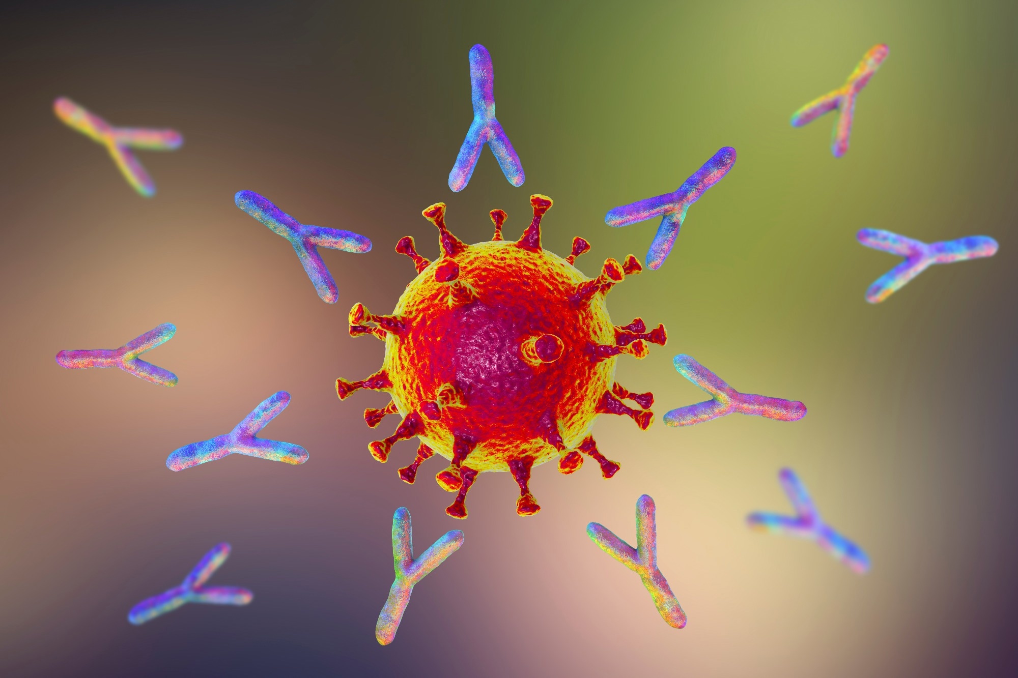
    In a recent case report published in The Lancet Infectious Diseases, researchers described a case of a 62-year-old male who received 217 vaccinations against severe acute respiratory syndrome coronavirus 2 (SARS-CoV-2) in 29 months and examined his immunological responses. They found that hyper-vaccination did not cause adverse events or significantly affect the quality of adaptive immune responses while resulting in increased T-cells and spike-specific antibodies.
    Study: Adaptive immune responses are larger and functionally preserved in a hypervaccinated individual. Image Credit: Douglas Sacha / ShutterstockStudy: Adaptive immune responses are larger and functionally preserved in a hypervaccinated individual. Image Credit: Douglas Sacha / Shutterstock

    Background

    Booster vaccinations may potentially amplify immune responses, while persistent antigen exposure may induce immune tolerance. However, the advantages, constraints, and risks of recurrent vaccination in humans remain to be thoroughly investigated. In the present study, researchers investigated the immunological responses in an older man hyper-vaccinated against SARS-CoV-2.

    The case

    In this case study, a 62-year-old male from Magdeburg, Germany (referred to as HIM), engaged in deliberate hyper-vaccination against SARS-CoV-2, receiving 217 vaccinations over 29 months for personal reasons. This occurred outside a clinical study context and contrary to national recommendations. Despite an investigation by a public prosecutor for potential fraud, no criminal charges were filed. Notably, HIM’s immunological evaluation, initiated during the public prosecutor’s investigation, received active and voluntary cooperation from HIM and was ethically approved. Throughout the extensive hyper-vaccination, HIM reported no vaccine-related side effects, and routine clinical chemistry parameters displayed no abnormalities between November 2019 and October 2023. In the repeated negative SARS-CoV-2 tests, including antigen tests, polymerase chain reaction (PCR) test, and nucleocapsid serology, HIM showed no signs of past SARS-CoV-2 infection.

    Starting from the 214th vaccination, HIM’s anti-spike SARS-CoV-2 immunoglobulin G (IgG) levels were measured before and after vaccinations. The antibody peak occurred at the 214th vaccination, and there was a slight increase after the 217th vaccination. Additionally, HIM showed IgG4 subclass switching after the 215th vaccination, which is uncommon in regimens with adenoviral-based vaccines as the first dose.

    A total of 29 individuals who received three doses of a messenger ribonucleic acid (mRNA) vaccine formed the control group. As compared to controls, HIM exhibited mildly elevated levels of anti-spike IgM and IgA in the serum. However, in saliva samples, HIM showed detectable levels of anti-spike IgG, contrary to the control participants. HIM’s serum neutralization capacity was higher (5.4-fold for wildtype and 11.5-fold for Omicron B1.1.529 spike proteins) than the controls, indicating elevated quantities of spike-specific IgG. This observed difference was not attributed to antibody avidity as it remained comparable among the groups.

    HIM showed a slightly increased number of spike-specific B-cells, with the same phenotype as seen in single-cell RNA sequencing (scRNA-seq). No significant differences were observed in the rates of somatic hypermutation or clonal expansion. CD8+ T-cells specific to the spike epitope were about six-fold more frequent in HIM, with a preference for effector memory T-cells. Further, scRNA-seq of LTD-specific T-cells showed a more differentiated phenotype and increased clonal expansion compared to controls. Flow-cytometric analysis and metabolic profiling showed no significant abnormalities in 14 protein markers.

    LTD-specific CD8+ T-cells in HIM showed a proliferative capacity similar to control individuals, aligned with conserved numbers of T-cells with a phenotype like early differentiated stem cells. After epitope-specific stimulation, HIM displayed higher cytokine-positive cells, but the cytokine release per cell remained roughly equal. Cytokine analysis in the supernatant revealed the typical pattern of virus-specific CD8+ T-cells. Additionally, HIM’s CD8+ T-cells showed higher peptide sensitivity than the control group. Examination of spike-reactive CD4+ T-cells revealed a dearth of nucleocapsid-specific immunity, with similar cytokine-producing CD4+ T-cell amounts in HIM compared to the control group while retaining peptide sensitivity.

    Conclusion

    In conclusion, the present case report showed that hyper-vaccination against SARS-CoV-2 yielded no adverse events and elevated T-cell levels and spike-specific antibodies. Notably, the implicit quality of adaptive immune responses showed no significant effects. Although breakthrough SARS-CoV-2 infections were not observed in the individual, any causal link with the hyper-vaccination regimen remains unclear. The researchers emphasize that they do not advocate for hyper-vaccination as an approach to improve adaptive immunity.

    [ad_2]

    Source link

  • COVID-19 exposes deep-rooted structural inequities affecting vaccine uptake among ACB groups

    COVID-19 exposes deep-rooted structural inequities affecting vaccine uptake among ACB groups

    [ad_1]

    In a recent review published in Vaccines, researchers explored the influence of poor vaccination uptake among African, Caribbean, and Black (ACB) communities on public health in high-income nations.

    Study: Understanding Low Vaccine Uptake in the Context of Public Health in High-Income Countries: A Scoping Review. Image Credit: SeventyFour/Shutterstock.comStudy: Understanding Low Vaccine Uptake in the Context of Public Health in High-Income Countries: A Scoping Review. Image Credit: SeventyFour/Shutterstock.com

    Background

    The severe acute respiratory syndrome coronavirus 2 (SARS-CoV-2) pandemic has resulted in a massive vaccination drive; however, ACB communities have experienced significant adverse consequences and are unwilling to get the vaccine.

    These communities are at risk due to health disparities, such as the greater prevalence of SARS-CoV-2 infections and hospitalizations. These imbalances significantly impact the social determinants of health (SDOH), and vaccine hesitancy can lead to delayed or uncertain immunization.

    Global vaccination uptake has fallen; therefore, public health initiatives must adapt to present conditions and plan for future epidemics.

    About the review

    In the present review, researchers explored the variables contributing to poor vaccination uptake by ACB individuals, emphasizing healthcare in developed nations.

    They sought to find current data sources, map the evidence, identify research gaps, and identify existing treatments for increasing vaccination uptake in the study population.

    The team searched the Cochrane Central Register of Controlled Trials, Embase, MEDLINE, APA PsycInfo, CINAHL, Web of Science, Open Science Framework, and the Allied and Complimentary Medicine databases.

    They followed the Joanna Briggs Institute (JBI) model, supplemented by the Preferred Reporting Items for Systematic Reviews and Meta-Analyses extension for scoping literature reviews (PRISMA-ScR). They included articles published in English or French between 2020 and July 19, 2022.

    The researchers performed the data search using the population, concept, and context (PCC) model to identify records discussing vaccine uptake among ACB residents of high-income nations, as defined by the World Bank.

    Evidence sources included primary studies, secondary research, abstracts, posters, conference proceedings, reports, and commentaries.

    Two researchers screened the data independently, and a third researcher resolved disagreements. The team used the social determinants of health (SDOH) method, the socioeconomic model (SEM), and thematic mapping (TM) to analyze and interpret the data.

    TM phases included individual-level, intra- and inter-group analyses to generate descriptive, analytical, and primary themes.

    Results

    Initially, the team identified 9,378 records, from which they removed 4246 duplicates. After title and abstract screening, they excluded 2,746 records.

    Of the remaining 2,386 records undergoing full-text screening, 60 eligible records were analyzed, including 24 quantitative, ten qualitative, 19 commentary records, and seven mixed-method records. Most records were from the United Kingdom, Canada, and the United States.

    Analysis performed via thematic mapping highlighted four primary themes: (i) inequities and racism, (ii) behaviors and sentiments, (iii) communication and knowledge, and (iv) influence and engagement.

    Inequities and racism in the healthcare system originate from mistrust, racial burden, and institutional impediments to access. Vaccine hesitancy (VH) is associated with increased rates of unwillingness to receive vaccinations, thereby perpetuating health inequities in black communities.

    The demographics of individuals exhibiting vaccine hesitancy reflect societal determinants of health, such as age, housing instability, poor income, and low education.

    Black vaccination starting in the United States is lower among immigrants from other countries, with the Americas and Caribbean Islands having a lower incidence than Africa.

    Factors like willingness, vaccination views, life events, and vaccine confidence determine vaccine uptake. Key causes include a lack of vaccination requirements, religious and political opinions, abuse, mortality exposure, and prior diseases.

    Mistreatment, exposure to mortality, and past diseases exemplify lived experiences. Vaccine confidence encompasses skepticism, timeliness, novelty, side effects, effectiveness, and safety.

    Black individuals are more likely to be vaccinated because of their existing health or their view that immunizations are the incorrect strategy.

    Vaccine hesitation might be due to a desire to protect oneself, a need for school or a job, or a desire to avoid infection. To lower vaccination uptake and COVID-19 infections, the government and healthcare institutions must address these variables.

    The lack of knowledge, disinformation, and misunderstandings in black communities all contribute to vaccination hesitation. Black parents are actively looking for information about kid vaccines, but the highest barrier is a lack of research on the short- and long-term impacts.

    Education, confidence in vaccine information, and open communication are critical for increasing immunization. Addressing distrust can boost vaccine confidence and intentions, while customizing messages to target populations can encourage immunization. Racism and prejudice, which serve as structural impediments to fair health care, must be addressed through culturally responsive techniques.

    Conclusion

    The review findings showed that ACB populations had lower vaccination uptake than high-income nations. Complacency, discomfort, and a lack of confidence are factors that contribute to vaccination reluctance, which past and contemporary racism and prejudice cannot entirely explain.

    The issue is complicated, encompassing knowledge and psychological, economic, and organizational constraints contributing to structural injustices. High-income nations should collect race-specific data for targeted treatments and increase the number of ACB participants in vaccine studies to boost vaccination trust.

    [ad_2]

    Source link

  • COVID-19 linked to long-lasting cognitive deficits, study finds

    COVID-19 linked to long-lasting cognitive deficits, study finds

    [ad_1]

    In a recent study published in the New England Journal of Medicine, researchers assessed cognitive functioning among adults with varying levels of persistence of coronavirus disease 2019 (COVID-19) caused by the severe acute respiratory syndrome coronavirus 2 (SARS-CoV-2) in England.

    Their results suggest that COVID-19 is associated with measurable cognitive deficits, which may persist in the long term.

    Study: Cognition and Memory after Covid-19 in a Large Community Sample. Image Credit: Berit Kessler / ShutterstockStudy: Cognition and Memory after Covid-19 in a Large Community Sample. Image Credit: Berit Kessler / Shutterstock

    Background

    The first documented cases of ‘brain fog,’ with symptoms such as poor memory, impaired concentration, and difficulty thinking, emerged as early as 2020, indicating that COVID-19 could have long-term cognitive impacts.

    Though the phenomenon is well-known, what is lacking is information on how it may persist and which aspects of cognitive functioning are most affected.

    About the study

    In this study, researchers hypothesized that cognitive deficits after the onset of COVID-19 should be quantifiable and associated with covariates related to illness severity and duration.

    Their second hypothesis was that individuals with prolonged COVID-19 symptoms should show more observable memory and executive function impairment, including brain fog and poor memory.

    They conducted a cohort-based study tracking the prevalence of SARS-CoV-2 infection among 3,099,386 individuals aged over 18 years. Of these, 800,000 people were invited to complete a cognitive assessment and follow-up survey.

    To be included, they should have received a positive result on a SARS-CoV-2 diagnostic test or suspected that they had COVID-19 and experienced symptoms for 12 weeks or more. Additionally, unvaccinated people with SARS-CoV-2 immunoglobulin-G antibodies and other randomly selected people from the full sample were included.

    The cognitive assessment tested immediate and spatial working memory, verbal analogical reasoning, two-dimensional mental manipulation, spatial planning, word definitions, delayed memory, and information sampling. For each domain, participants were scored on accuracy; secondary information was collected on types of errors and response times.

    Individuals were categorized into six groups based on SARS-CoV-2 duration. The first category included those who had never experienced an infection or had an unconfirmed one; all other categories required a positive test result.

    People in the second category had asymptomatic infections, those in the third had short COVID-19 that resolved in four weeks or less, and those in the fourth had symptoms that resolved in less than 12 weeks. To be in the fifth category, individuals had symptoms that persisted for more than 12 weeks; those in the sixth had persistent symptoms continuing until the cognitive assessments.

    Researchers assessed nonresponse bias to examine which factors were associated with accessing and completing the cognitive assessment. Linear regressions, factor analysis, and propensity-score matching (PSM) were used to analyze the data. Sensitivity analyses were also conducted to test the validity and robustness of the results.

    Findings

    Of the 800,000 people invited to participate, 34.6% completed the questionnaire, with 141,583 completing at least one cognitive testing task and 112,964 completing all eight.

    Among individuals infected with SARS-CoV-2 once, being infected earlier during the pandemic was associated with greater decreases in the overall cognitive score compared to those infected later. However, the gap narrowed after adjusting for the severity of the illness.

    On average, people who were ill for longer, were hospitalized, or were infected early on in the pandemic had lower overall cognitive scores than those who had never had COVID-19.

    Multivariate regression results indicated that people infected during the initial stages (when the original virus or alpha variant dominated) showed higher cognitive functioning decreases than those infected with the alpha or omicron variants.

    Similarly, greater decreases were seen in people with persistent and unresolved symptoms compared to those who never had COVID-19 and among people who were hospitalized compared to those who were not.

    The PSM analysis showed similar trends; cognitive advantages were observed based on vaccination status, with people who received two or more doses performing best. There was, however, no significant difference based on which vaccine was taken.

    Conclusions

    This large-sample community-based study suggests that COVID-19 may be associated with long-term and quantifiable cognitive deficits. However, people infected with more recent variants may experience more negligible effects on cognitive functioning.

    This could be because earlier strains of SARS-CoV-2 were dominant at a time when effective treatments were not available, and the health system faced heavy burdens. Repeated infections do not appear to have any effect, but vaccination (particularly two or more doses) may provide small cognitive advantages.

    Limitations of this study include the possibility of participant self-selection bias and reliance on self-reported data. Certain groups were overrepresented in the sample, including White persons and women; younger people and certain underprivileged groups were underrepresented.

    Further studies are required to provide information on the longer-term implications of these findings.

    Journal reference:

    • Cognition and memory after Covid-19 in a large community sample. Hampshire, A., Azor, A., Atchison, C., Trender, W., Hellyer, P.J., Giunchiglia, V., Husain, M., Cooke, G.S., Cooper, E., Lound, A., Donnelly, C.A., Chadeau-Hyam, M., Ward, H., Elliott, P. New England Journal of Medicine (2024). DOI: 10.1056/NEJMoa2311330, https://www.nejm.org/doi/10.1056/NEJMoa2311330

    [ad_2]

    Source link

  • How long do cognitive and memory dysfunctions persist after SARS-CoV-2 infection?

    How long do cognitive and memory dysfunctions persist after SARS-CoV-2 infection?

    [ad_1]

    A recent New England Journal of Medicine study assessed whether cognitive deficits can be measured after severe acute respiratory syndrome (SARS-CoV-2) infection, the causal agent of the coronavirus disease 2019 (COVID-19) pandemic.

    This study further explored the period for which these symptoms persist.

    Study: Cognition and Memory after Covid-19 in a Large Community Sample. Image Credit: Orawan Pattarawimonchai/Shutterstock.comStudy: Cognition and Memory after Covid-19 in a Large Community Sample. Image Credit: Orawan Pattarawimonchai/Shutterstock.com

    Background

    Brain fog is the common name for problems associated with thinking, concentrating, or memory. It has been observed frequently in patients recovered from COVID-19, but there is a dearth of objective data on cognitive performance.

    Furthermore, it is not clear which cognitive functions are most vulnerable and how long such deficits persist.

    About the study

    The primary hypothesis in this observational study was that cognitive deficits after COVID-19 were measurable and scalable regarding illness severity and duration.

    The secondary hypothesis was that objective impairments in memory and executive functions would be noted in patients showing prolonged symptoms.

    These hypotheses were tested using data from the Real-Time Assessment of Community Transmission (REACT) cohort in England.

    Eight hundred thousand adults were invited to complete an online assessment of cognitive function.

    Across eight tasks, a global cognitive score was estimated. These tasks helped test immediate memory, verbal analogical reasoning, two-dimensional mental manipulation, verbal analogical reasoning, spatial working memory, word definitions, delayed memory, spatial planning, and information sampling.

    Individuals who were invited to participate in this study were SARS-CoV-2 test positive or with persistent COVID-19 symptoms for at least 12 weeks.

    The eligible participants were categorized into six groups based on the duration of SARS-CoV-2 infection.

    The categories were defined as follows:

    • Category 1: No infection with SARS-CoV-2 detected.
    • Category 2: Infected with SARS-CoV-2, but no symptoms developed (asymptomatic infection).
    • Category 3: Experienced a brief infection period, with symptoms lasting less than four weeks.
    • Category 4: The period of infection lasted between 4 and 12 weeks.
    • Category 5: Symptoms were resolved at least 12 weeks after the onset of the infection.
    • Category 6: Symptoms persisted for more than 12 weeks after the infection began and were present during the cognitive assessment.

    Study findings

    This community-based study noted that COVID-19 and long-term objectively measurable cognitive deficits were associated with each other.

    The difference in the cognitive score between the no-COVID group and the group of participants whose symptoms had resolved was small. The difference was, however, large for participants who experienced unresolved symptoms.

    The probability of task performance was below the cut-off point in the case of moderate impairment.

    Relative to the no-COVID group, larger cognitive differences were noted in the group where ICU admission was needed. This finding is consistent with the ones documented in previous studies where medium-to-large-scale cognitive deficits were observed in patients who were hospitalized in a critical care unit.

    It was noted that with the progression of the pandemic, the association between cognitive deficits and COVID-19 attenuated.

    Smaller cognitive declines were noted among those infected with newer variants, compared to the original strain or the Alpha strain. Individuals who had received two or more vaccine doses were seen to have a small cognitive advantage.

    A progressive decline in cognitive deficits was observed among those infected during the pandemic’s first wave.

    The observation that patients whose persistent symptoms had resolved had experienced global cognitive deficits similar in nature to patients with shorter-duration symptoms is indicative of the fact that individuals with persistent symptoms could experience some degree of cognitive improvement once symptoms resolve.

    The executive, memory, and reasoning tasks were the most sensitive to COVID-19. The performance was dependent on hospitalization and illness duration.

    The scores were also weakly correlated with recent brain fog or poor memory. The results documented here validated the associations between mood swings, fatigue, and cognitive deficits, besides a variety of other symptoms.

    Limitations of the study

    A key limitation centers around subjective reporting to identify patients with persistent symptoms.

    Linking these results to the literature is difficult owing to the lack of definitive criteria for post–COVID-19 syndromes. Furthermore, causality could not be inferred owing to the observational nature of the data.

    The global cognitive score calculation adjusted for demographic characteristics and pre-existing health conditions. However, some residual confounding could have remained due to the observational nature of the data.

    To mitigate this bias, the propensity score matching method was used to show a highly consistent pattern of results.

    As with any study requiring active participant engagement, self-selection bias could not be ruled out.

    Patients with the most severe cognitive impairment may not have been willing or able to take a cognitive assessment.

    Additionally, some groups were less representative compared to the base population. Individuals from deprived backgrounds and younger persons were underrepresented.

    [ad_2]

    Source link

  • Long-term smell and taste loss

    Long-term smell and taste loss

    [ad_1]

    A recent study published in Life investigates the mechanisms responsible for severe olfactory dysfunction (OD) and gustatory dysfunction (GD) following recovery from the severe acute respiratory syndrome coronavirus 2 (SARS-CoV-2) infection.

    Study: Persistent Olfactory and Taste Dysfunction after COVID-19. Image Credit: DimaBerlin / Shutterstock.com Study: Persistent Olfactory and Taste Dysfunction after COVID-19. Image Credit: DimaBerlin / Shutterstock.com

    Background

    SARS-CoV-2 is the causal agent of the coronavirus disease 2019 (COVID-19). A sudden loss of smell or taste is one of the most common symptoms of SARS-CoV-2 infection.

    Typically, OD and GD occur in the early phase of the infection, which has led these symptoms to be used as a screening tool for COVID-19. Both OD and GD occur primarily among young patients and are independent of gender.

    COVID-19 patients with OD often experience a spontaneous return of their sense of smell to pre-symptomatic levels within one month of infection. However, between 7-20% of COVID-19 patients report prolonged OD, even after other symptoms have resolved.

    Mechanistically, patients with severe nasal and sinus symptoms undergo swelling of the mucous membranes of the olfactory cleft, which increases mucus secretion. This increase in mucus causes a mechanical blockage to odor molecules, manifesting as conductive OD. Infiltration of viral particles into the non-neural cells of the olfactory epithelium also induces significant immune responses.

    In the context of immune responses to viral infiltration into olfactory epithelium, pro-inflammatory cytokines such as interleukin 1 β (IL-1β) and tumor necrosis factor α (TNF- α) are released. The impaired non-neural cells subsequently disrupt the connection between nerve cells and sensory OD, which inhibits the transfer of olfactory stimuli to the brain.

    Importantly, the precise mechanism of action on the penetration of SARS-CoV-2 into the central nervous system remains unclear. One possible mechanism could be the direct penetration of SARS-CoV-2 into the cerebrospinal fluid from non-neural cells of the olfactory epithelium.

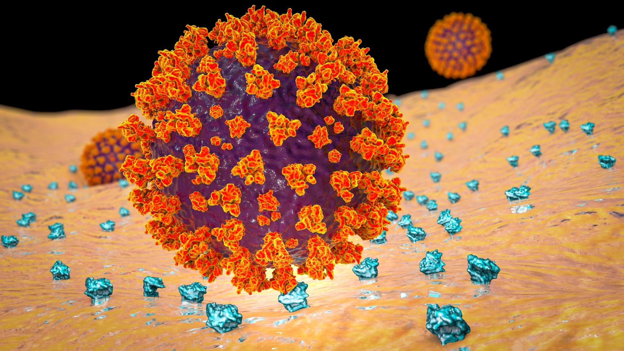
    The penetration of SARS-CoV-2 into the olfactory neuroepithelium causing sensorineural OD has been well documented. In this context, the spike (S) protein of SARS-CoV-2 binds to the angiotensin-converting-enzyme-2 (ACE2) receptor, which triggers the synthesis of the transmembrane serine protease 2 (TMPRSS2), thereby causing membrane fusion.

    To date, few studies have elucidated the mechanism that underlies the manifestation of GD due to COVID-19.

    About the study

    The current prospective and single-center study involved a subjective and physical examination of patients. Study participants also underwent multiple olfactory and gustatory tests, such as the Sniffin’ Sticks Test (SST) and Taste Strips Test (TS).

    A total of 81 participants were recruited, including 16 men and 65 women between 12 and 73 years of age. All participants experienced OD due to COVID-19, which persisted for at least one month after the resolution of other acute symptoms.

    Study findings

    The study participants experienced persistent OD or GD (OGD) with durations ranging between one and 25 months.

    Few clinical facilities offer help to patients with persistent OGD following recovery from COVID-19. This could be because clinicians are still developing their knowledge regarding long-term COVID and formulating strategies to combat the condition. Additionally, patients often seek treatment for OD only after resolving more serious symptoms.

    The degree of damage in the olfactory cells by SARS-CoV-2 penetration dictates the duration of OD. For conductive disorders that occur in most infected individuals, the sense of smell improves after a reduction in acute inflammation of the nasal mucosa. Patients with damaged olfactory cells exhibit persistent OD, as neurons take longer to recover and re-establish adequate intercellular connections.

    About 64% of the study participants had hyposmia, and 22% had functional anosmia. SST scores revealed that the participants scored lowest on the threshold part of the test. The SST score of other parts concerning discrimination and identification of odor revealed that patients could sense intense smell correctly.

    SST scores significantly depend on the function of the olfactory epithelium, which suggests that post-COVID-19 OD is predominantly associated with damage in peripheral olfactory due to acute immune responses. More specifically, the cytokine storm leads to leukocytic infiltration and disrupts olfactory epithelial cells, including stem cells.

    There was no difference in recognizing odorants that stimulate only the olfactory nerve and those that trigger the trigeminal nerve, such as mint and lemon. Interestingly, some participants with subjective ODs scored within a normal range on the SST test. 

    Around 17% of participants reported abnormal taste. The best-recognized taste was sweet, followed by salty. Only bitter taste was marginally correlated with TTS and TS scores.

    Conclusions

    The findings provide important insights into the relationship between OD and SARS-CoV-2 infection. It is possible that the relationship between OD and COVID-19 is more peripheral than central; therefore, clinicians must pay close attention to the condition of the nasal mucosa and provide better information to patients about good nasal hygiene that can help alleviate the condition.

    Journal reference:

    • Buksinska, M., Skarzynski, P. H., Raj-Koziak, D., et al. (2024) Persistent Olfactory and Taste Dysfunction after COVID-19. Life 14(3); 317. doi:10.3390/life14030317.

    [ad_2]

    Source link

  • Scientists uncover a new doorway for SARS-CoV-2 into human cells

    Scientists uncover a new doorway for SARS-CoV-2 into human cells

    [ad_1]

    In a recent study published in the journal Proceedings of the National Academy of Sciences, researchers demonstrated that human transferrin receptor (TfR) mediates severe acute respiratory syndrome coronavirus 2 (SARS-CoV-2) infection.

    Coronavirus disease 2019 (COVID-19), caused by SARS-CoV-2, presents influenza-like manifestations, including mild-to-severe pneumonia, acute respiratory distress syndrome, multiorgan failure, and fatal lung injury. Further, the etiology and pathogenesis of COVID-19 are not entirely understood and targeted therapies remain inadequate.

    The viral spike protein binds to the host receptor, angiotensin-converting enzyme 2 (ACE2), for cellular entry. Although SARS-CoV-2 preferentially infects cells in the respiratory tract, the virus has been detected in virtually all organs. Studies have revealed the presence of SARS-CoV-2 RNA in diverse cells lacking ACE2, suggesting that other receptors or co-receptors may mediate viral entry.

    Study: Human transferrin receptor can mediate SARS-CoV-2 infection. Image Credit: Kateryna Kon / ShutterstockStudy: Human transferrin receptor can mediate SARS-CoV-2 infection. Image Credit: Kateryna Kon / Shutterstock

    The study and findings

    In the present study, researchers identified TfR as an alternative receptor mediating the cellular entry of SARS-CoV-2. First, they used co-immunoprecipitation (Co-IP) to identify host proteins interacting with the viral spike in Calu-3 cells. This revealed 293 proteins, including 42 transmembrane proteins; two proteins were associated with entry (ACE2 and TfR). Next, the team evaluated TfR expression in the respiratory tract and liver in mice.

    TfR expression, both transcript and protein levels, was substantially higher in the lungs and trachea than in other tissues. Using immunohistochemical analysis, the researchers investigated the effects of SARS-CoV-2 on TfR expression in the lungs of humanized ACE2 (hACE2) mice and monkeys. This revealed a 1.5- and 1.8-fold increase in TfR expression in mice and monkeys, respectively.

    In addition, surface plasmon resonance revealed direct interactions between the viral spike and human TfR. Notably, the spike protein lacked interactions with Syrian hamster or mouse TfR. Docking analysis predicted two peptide sequences (QK8: QDSNWASK and SL8 SKVEKLTL) in TfR to be involved at the interface of TfR-spike interactions.

    Mutagenesis and Co-IP revealed that the A529 residue in TfR was essential for interactions with the spike. Further analysis indicated that physiological interactions between spike and TfR occurred at the cellular surface and during endocytosis. This was confirmed by electron microscopy using SARS-CoV-2 pseudoviral spike and HEK293/hACE2 and BHK-21/TfR cells.

    Next, the team evaluated the effects of soluble TfR, anti-TfR antibody, and SL8 and QK8 peptides on SARS-CoV-2 infection using reverse-transcription polymerase chain reaction (RT-PCR) and plaque assays. Results showed their inhibitory effects on SARS-CoV-2 in Vero E6 and Calu-3 cells. Cytotoxicity was not observed even at 1,000 nM.

    Confocal microscopy revealed that TfR was widespread on the surface of Calu-3 and Vero E6 cells, with the colocalization of TfR and SARS-CoV-2 at the surface and during endocytosis. Notably, treatment with the anti-TfR antibody inhibited the colocalization. Further, electron microscopy showed that viral particles were present in the cytosol and clathrin-coated pits in Vero E6 cells; likewise, treatment with anti-TfR antibody inhibited viral internalization.

    Next, ACE2 was knocked out (KO) from Calu-3 and Vero E6 cells and the cells were infected with SARS-CoV-2. This inhibited infection by 40% to 50%, suggesting that ACE2 might not be the only receptor mediating infection. In addition, TfR knockdown (KD) inhibited infection by 30%, whereas its overexpression (OE) promoted infection. TfR KO was not performed as it is lethal. TfR OE or KD did not impact ACE2 expression.

    Further, the team transfected C57 mice with adenovirus vector (Ad5) expressing hACE2 or humanized TfR (hTfR) and infected them with SARS-CoV-2. Viral load in the lungs in Ad5-hTfR and Ad5-hACE2 mice was significantly higher than in Ad5-empty mice. Finally, the researchers evaluated the effects of the anti-TfR antibody on infection in rhesus macaques. Anti-TfR antibody inhibited viral replication and reduced pneumonia.

    Viral load in the respiratory epithelium was also significantly lower between 3- and 7 days post-infection (dpi) compared to controls. Radiographs taken at 0 and 5 dpi revealed significantly less severe pulmonary infiltration in antibody-treated macaques relative to controls. Antibody-treated animals had no significant pulmonary lesions, while controls showed lung lesions of varying degrees.

    Conclusions

    Taken together, the study described the human TfR as a receptor for SARS-CoV-2. TfR can directly bind to the viral spike at an affinity comparable to that of ACE2. Notably, mouse TfR and the viral spike lacked interactions. Soluble TfR, SL8, and QK8 peptides and anti-TfR antibodies can inhibit infection. The team also illustrated the antiviral effects of the anti-TfR antibody in rhesus macaques. Overall, TfR could serve as an alternative infection pathway, facilitating viral entry through endocytosis.

    [ad_2]

    Source link

  • Study reveals how long COVID can affect brain function through vascular disruption

    Study reveals how long COVID can affect brain function through vascular disruption

    [ad_1]

    In a recent study published in Nature Neuroscience, researchers investigated whether the neurological response to coronavirus disease 2019 (COVID-19) may be due to brain-brain barrier (BBB) disruption and subsequent extravasation of serum components.

    Study: Blood–brain barrier disruption and sustained systemic inflammation in individuals with long COVID-associated cognitive impairment. Image Credit: fran_kie/Shutterstock.comStudy: Blood–brain barrier disruption and sustained systemic inflammation in individuals with long COVID-associated cognitive impairment. Image Credit: fran_kie/Shutterstock.com

    Background

    COVID-19, caused by severe acute respiratory syndrome coronavirus 2 (SARS-CoV-2), is a respiratory viral infection resulting in severe acute respiratory distress syndrome (ARDS) and long-term neurological consequences such as headache, lethargy, malaise, and altered consciousness.

    BBB breakdown allows serum components and cytokines to penetrate the brain. The cerebrovascular pathophysiology and processes remain unknown, warranting further research.

    About the study

    The present study determined the association between COVID-19-related cognitive impairment and BBB breakdown in COVID-19 patients.

    The researchers collected blood and plasma samples from 76 acute COVID-19 patients, evaluated them for inflammatory, coagulation, and BBB dysfunction indicators, and rated their severity using World Health Organization (WHO) severity guidelines.

    They next examined brain fog status to identify changes in patients’ inflammatory profiles. During the first round of COVID-19 in March and April 2020, they recruited participants from St. James’ Hospital at Trinity College in Dublin.
    To investigate BBB function, the researchers selected ten recovered individuals, 11 suffering from long COVID or post-acute COVID-19 (PASC) and 11 with PASC-related brain fog, diagnosed with SARS-CoV-2 infection during the disease’s April 2020 outbreak in Ireland.

    All patients had polymerase chain reaction (PCR)-verified moderate-intensity COVID-19, requiring no antiviral therapy or hospitalization.

    The researchers used the quick smell identification test (Q-SIT) to evaluate anosmia status, grouping them based on self-reported cognition difficulties known as brain fog.

    They classified subjects as recovered in the case of no symptom recurrence after recovering from acute COVID-19. They used the Montreal Cognitive Assessment (MOCA) to measure cognitive impairment.

    The researchers used dynamic contrast-enhanced magnetic resonance imaging (DCE-MRI) volume and thickness measurements on recovered individuals with PASC and 60 age-matched healthy controls from the IXI dataset to investigate structural brain alterations associated with increased BBB permeability.

    They used multiplexed assays to assess 50 BBB integrity and inflammation markers and correlation analysis adjusting for age and sex to identify associations between neuroinflammatory and BBB dysfunction markers in the recovered and PASC cohort.

    RNA-seq was also used to evaluate gene expression alterations in peripheral blood mononuclear cells (PBMCs) and the human brain endothelial cell line hCMEC/d3 isolated from unaffected, recovered, and patients with protracted COVID in the absence or presence of brain fog.

    They used gene ontology (GO) to examine the transcriptome profiles of those with and without brain fog in the group with extended COVID.

    The study included COVID-19 convalescents aged ≥18 years without neurological symptoms and individuals suffering from long-term COVID with symptoms lasting over 12 weeks after infection.

    Results

    COVID-19-induced brain fog was associated with BBB impairment. This disturbance is visible during acute COVID-19 and in individuals with long-term COVID-related cognitive impairment, popularly termed brain fog.

    In addition, the team found that PBMCs showed coagulation system instability and a suppressed adaptive immunological response in those with brain fogging.

    In vitro, PBMCs showed enhanced adherence to cells of the human brain endothelium, whereas the endothelial cells were exposed to sera from individuals with prolonged COVID-19-induced inflammation.

    The findings indicated that assessing BBB integrity might be a clinically helpful indicator of neurological sequelae linked with COVID-19 in a few individuals. Furthermore, targeted control of BBB integrity may provide a novel strategy for therapeutically treating patients with chronic COVID.

    Common symptoms of brain fog include dyspnea, loss of smell and taste, coughing, weariness, and fever. Patients with brain fogging had a higher average age, were more likely to be hospitalized, and needed oxygen treatment.

    There was a strong association between COVID-19 severity and age, hospitalization length, and comorbidities.

    BBB failure was linked to long-term COVID-induced cognitive impairment, indicating that both active and acute SARS-CoV-2 infections may cause BBB dysfunction in individuals with neurological disabilities.

    MRI imaging indicated significantly higher brain leakage in individuals with PASC and brain fog, with volumetric deficits mostly in the frontal and temporal lobes and increases in the occipital lobes and lateral ventricles.

    White blood cells from COVID-19 patients stimulated brain endothelial cells. Compared to unaffected individuals, there were 950 differentially expressed genes (DEGs) in recovered individuals, 481 in individuals with protracted COVID, and 126 in those with brain fogging.

    Upregulated genes were associated with T cell development and activation pathways, immune response negative control, and gene expression circadian regulation.

    Conclusion

    Overall, the study findings showed that PASC-related brain fog is associated with systemic inflammation and persistent localized BBB malfunction, with disruption apparent up to a year after infection.

    Dysregulation of the coagulation system is a primary cause of prolonged COVID-19. BBB disruption is associated with neurological impairment during acute COVID-19, and high serum levels in neurological diseases such as epilepsy, traumatic brain injury, and schizophrenia.

    Understanding the long-term effects of COVID-19 is critical to developing new treatments.

    [ad_2]

    Source link

  • SARS-CoV-2-infection and vaccine-induced antibodies wane initially but stabilize for lasting protection

    SARS-CoV-2-infection and vaccine-induced antibodies wane initially but stabilize for lasting protection

    [ad_1]

    In a recent observational study published in the journal Immunity, researchers from the United States of America investigated the longevity of antibody responses to severe acute respiratory syndrome coronavirus 2 (SARS-CoV-2) infection and vaccination. They found that the humoral responses to SARS-CoV-2 infection and vaccination were long-lasting and biphasic, with an initial decline followed by stabilization after seven to nine months.

    Study: SARS-CoV-2-infection- and vaccine-induced antibody responses are long lasting with an initial waning phase followed by a stabilization phase. Kateryna Kon / ShutterstockStudy: SARS-CoV-2-infection- and vaccine-induced antibody responses are long lasting with an initial waning phase followed by a stabilization phase. Kateryna Kon / Shutterstock

    Background

    The COVID-19 pandemic, which began four years ago, prompted the rapid development of messenger RNA (mRNA) vaccines, including the BNT162b2 and mRNA-1273, helping save millions of lives. However, emerging variants of SARS-CoV-2 and the waning immunity against them pose challenges. Although mRNA-based vaccine-induced immunity is perceived to decline rapidly, this perception is based on limited data, primarily from short-term studies.

    Amidst the exponential rise of SARS-CoV-2 cases in March 2020, the New York metropolitan area faced a crisis, with essential healthcare workers at a high infection risk. In response, a specific and sensitive SARS-CoV-2 enzyme-linked immunosorbent assay (ELISA) was developed, and the Protection Associated with Rapid Immunity to SARS-CoV-2 (PARIS) study was launched. This initiative tracked antibody responses, reinfection rates, and immunity factors in healthcare workers, offering vital insights into pandemic dynamics. Researchers in the present study utilized data from the PARIS study, one of the most comprehensive investigations on SARS-CoV-2 immunity longevity, and analyzed the humoral responses to SARS-CoV-2 infection and vaccination.

    About the study

    The PARIS study was an observational, longitudinal study conducted from April 2020 to March 2023 and enrolled 501 healthcare workers. Their mean age was 41 years, and 67% of them were female. Weekly saliva samples and bi-weekly blood samples were collected for the first two months. Nasopharyngeal/ante-near swabs were taken for respiratory symptoms or after vaccination. About 38% of participants showed baseline SARS-CoV-2-spike-binding immunoglobulin G (IgG) antibodies. A total of 93% of participants were vaccinated– 0.2% received four mRNA boosters, 2.6% had three boosters, 16.6% had two boosters, and 53.7% had one booster. Approximately 21.3% of the participants chose not to receive boosters.

    The study utilized REDCap for monthly surveys on general health and SARS-CoV-2 risk, focusing on side effects after mRNA vaccinations and booster doses. Data from 228 participants were analyzed, and severity scoring was conducted, revealing reported incidence and severity trends across doses and subgroups.

    Antibody titers in serum were assessed using enzyme-linked immunosorbent assay (ELISA) and optical density at 490 nm (OD490). Statistical and quantitative analysis involved the use of the Wilcoxon test, Mann-Whitney U test, log-rank test, unweighted pair group method with arithmetic mean (UPGMA) clustering, antibody kinetic modeling including nonlinear mixed-effects (NLME) models, and demographic factor assessment in post-vaccine and post-boost models.

    Results and discussion

    While 38% of the participants had detectable spike-binding IgG antibodies at baseline, 62% were seronegative at the first visit. Vaccination-naïve individuals exhibited low antibody titers after the first mRNA vaccine dose, with a substantial increase after the second dose. However, individuals with pre-existing immunity reached higher and faster peak titers, maintaining over threefold higher responses after primary immunization.

    Seven to nine months post-primary vaccination, antibody titers were found to achieve a steady state. Individuals with hybrid immunity maintained higher and more stable titers compared to naïve recipients, indicating the induction of long-lasting serum antibodies. Furthermore, vaccine type and age were found to affect the antibody titers in participants without hybrid immunity modestly. As per the study, the administration of booster doses elevated the threshold at which long-term serum antibody responses reached a stable state.

    A total of 225 SARS-CoV-2 infections were observed in the study period, predominantly occurring after immunization, with breakthrough infections more prevalent during the Omicron wave. In individuals with vaccine-only immunity, breakthrough infections acted as equivalent boosts to antibody responses, while in those with hybrid immunity, vaccination had a more robust boosting effect compared to a second infection.

    Participants with pre-existing immunity experienced more side effects after the first vaccine dose, with overall reactogenicity decreasing after subsequent doses. Booster doses induced fewer systemic side effects than the second dose in naïve participants, while those with hybrid immunity had a different pattern, showing slightly increased side effects with booster doses.

    However, the study is limited by the inability to analyze mucosal immune responses, the lack of measuring neutralizing antibodies or antibodies to specific epitopes, and the lack of assessment of later variant spikes or nucleoprotein.

    Conclusion

    In conclusion, the present study provides evidence that antibody responses to SARS-CoV-2 mRNA vaccination exhibit a classical biphasic decay, transitioning from rapid waning to stabilization. The findings emphasize the prolonged protection provided by hybrid immunity against several variants and the potential booster-like effect of breakthrough infections in enhancing immunity.

    [ad_2]

    Source link

  • Survey of US adults reveals common cognitive symptoms in post-COVID-19 patients, linked to impaired daily functioning and depression

    Survey of US adults reveals common cognitive symptoms in post-COVID-19 patients, linked to impaired daily functioning and depression

    [ad_1]

    In a recent study published in the journal JAMA Network Open, a team of scientists examined how prevalent self-reported cognitive symptoms were in individuals with post-coronavirus disease 2019 (COVID-19) condition as compared to individuals who had prior severe acute respiratory syndrome coronavirus 2 (SARS-CoV-2) infections but had not developed post-COVID-19 condition. They also evaluated the impact of these cognitive symptoms on mood, function, and employment status.

    Study: Cognitive Symptoms of Post–COVID-19 Condition and Daily Functioning. Image Credit: PeopleImages.com - Yuri A/Shutterstock.com
    Study: Cognitive Symptoms of Post–COVID-19 Condition and Daily Functioning. Image Credit: PeopleImages.com – Yuri A/Shutterstock.com

    Background

    One of the long-term impacts of the COVID-19 pandemic has been post-COVID-19 condition, commonly referred to as long coronavirus disease (long COVID), where the symptoms of acute SARS-CoV-2 infections persist or remerge months after recovering from the initial infection. The condition consists of wide-ranging symptoms affecting numerous organ systems, with fatigue, shortness of breath, and post-exertional malaise being the most common symptoms.

    Changes in mood and cognitive impairments have also been reported, with studies confirming the long-lasting impact of SARS-CoV-2 infections on neurological health. These persistent physical and neurological symptoms continue to have a significant impact on the functioning and quality of life of the patients long after they have recovered from the initial infection. Understanding how this condition impacts the individual’s productivity or employment status is essential to forming effective treatment mechanisms and public health strategies.

    About the study

    In the present study, the researchers used data from a survey conducted across the United States (U.S.) during two COVID-19 waves among individuals who had reported post-COVID-19 condition symptoms and those who reported complete recovery after a SARS-CoV-2 infection. The data was collected between December 2022 and January 2023 and then again from April to May 2023 across 50 U.S. states.

    The participants were above 18 years of age, and the study population was balanced for demographic factors such as gender, age, race, and ethnicity. A validated measure for patient-reported outcomes was used to design the questions on cognitive symptoms, which largely included questions on how often patients experienced specific symptoms over the previous week with replies on a five-point scale.

    The questions addressed the prevalence of symptoms such as trouble remembering, trouble starting tasks, slowed thinking, finding multitasking difficult, decision-making problems, and needing to pay extra attention to avoid errors. The number of symptoms and presence of these symptoms based on an occurrence rate of at least once a day were recorded for each patient.

    A nine-item questionnaire was also used to assess depressive symptoms in patients. Additionally, the patients were asked to describe how these cognitive post-COVID-19 symptoms interfered with their daily activities. The employment status of the participants was also recorded and categorized as full-time, contract, part-time, self-employed, homemaker, student, retired, or unemployed.

    Sociodemographic information collected from the participants included self-reported race and ethnicity data. The initial SARS-CoV-2 infection and post-COVID-19 condition were defined based on self-reported symptoms from the participants, such as reports of positive test results for COVID-19.

    Results

    The results showed that cognitive symptoms were prevalent in individuals experiencing post-COVID-19 conditions, and these symptoms were associated with functional impairments and a lower likelihood of holding full-time employment. The severity of depressive symptoms was also greater for individuals with cognitive post-COVID-19 symptoms.

    The number of individuals with post-COVID-19 condition who reported experiencing cognitive impairments was significantly higher than those who reported cognitive symptoms but did not have post-COVID-19 condition. Furthermore, women, younger individuals, and people with lower income levels showed a higher prevalence of cognitive symptoms than those in other sociodemographic groups.

    The researchers believe that the higher prevalence of cognitive impairments reported among younger individuals could be due to the notable change from the baseline measurements before the COVID-19 pandemic. Among older individuals, who might already be experiencing cognitive decline associated with age, the cognitive impairments due to post-COVID-19 condition might not be as apparent as in younger individuals.

    The study also suggested that the association between increased prevalence of cognitive impairments among individuals from lower-income households could reflect the influence of economic stress on the vulnerability to cognitive symptoms of post-COVID-19 conditions.

    Conclusions

    Overall, the study found that cognitive decline was highly prevalent among individuals with long COVID or post-COVID-19 conditions, especially among younger individuals, women, and those from low-income households.

    Furthermore, the probability of full-time employment was found to be lower among individuals experiencing cognitive impairments due to long COVID, highlighting the need for public health strategies and treatment measures to improve the quality of life and functional abilities of individuals suffering from post-COVID-19 condition.

    Journal reference:

    [ad_2]

    Source link

  • New study pinpoints key markers for Long COVID diagnosis

    New study pinpoints key markers for Long COVID diagnosis

    [ad_1]

    In a recent preprint* uploaded to the medRxiv server, an international team of researchers conducted a large-scale systems-level immunological screening of more than 1,000 confirmed COVID-19 patients to identify diagnostic markers of Long-term COVID-19. The analyses using multiple orthogonal detection methods reveal elevated serologic responses as a highlight of Long COVID and that its correlated memory CD8+ T cell clonal expansion is a more reliable and sensitive marker of the condition than conventional antigen (SARS-CoV-2 RNA and protein) detection approaches.

    Study: Restrained memory CD8+ T cell responses favors viral persistence and elevated IgG responses in patients with severe Long COVID. Image Credit: Lightspring / ShutterstockStudy: Restrained memory CD8+ T cell responses favors viral persistence and elevated IgG responses in patients with severe Long COVID. Image Credit: Lightspring / Shutterstock

    *Important notice: medRxiv publishes preliminary scientific reports that are not peer-reviewed and, therefore, should not be regarded as conclusive, guide clinical practice/health-related behavior, or treated as established information.

    COVID-19 and the need for Long COVID diagnosis

    The ongoing Severe Acute Respiratory Syndrome Coronavirus 2 (SARS-CoV-2) caused Coronavirus disease 2019 (COVID-19) viral pandemic is one of the worst in human memory, estimated to have infected more than 700 million individuals since its discovery in Wuhan, China, in late 2019. While global legislative policy and the widespread development and dissemination of anti-COVID-19 vaccines have substantially reduced the disease burden, with reports of vaccination efforts saving 70% of patients or more, survivors of the pandemic are plagued by a hitherto unknown condition – Long COVID.

    Also called the ‘Post COVID-19 condition’, ‘chronic COVID syndrome,’ and clinically, ‘post-acute sequelae of COVID-19 (PASC)’, Long COVID presents itself as perhaps the worst legacy of the pandemic. The now well-established yet poorly understood condition is characterized by the persistence or development of COVID-19-associated symptoms that may persist for months or even years following initial infection recovery. These symptoms include severe cognitive decline (brain fog), chronic fatigue, and multiple organ damage, resulting in significant economic and quality of life (QoL) losses in patients.

    Alarmingly, research has revealed that despite vaccination efforts substantially reducing adverse Long COVID outcomes, between 30 and 60% of all COVID-19 infections result in Long COVID, with an estimated 350+ million individuals suffering from the condition. Unfortunately, extensive global scientific efforts remain unable to elucidate the mechanisms underpinning Long COVID, hampering the development of diagnostic assays and clinical interventions for patients.

    About the study

    In the present study, researchers screened more than 1000 prospective patients enrolled at Long COVID clinics in Belgium and Sweden to elucidate the shared mechanisms of Long COVID pathology and subsequently develop a sensitive and reliable diagnostic test for the condition. Only subjects with a clinically confirmed mild or moderate COVID-19 infection were included. Severe cases were excluded due to overlapping symptoms with those of the post-intensive care syndrome.

    Patients without objective measures of disease-associated organ damage (e.g., magnetic resonance imaging [MRI], pulsatile arterial tonometry [EndoPAT], and postural orthostatic tachycardia syndrome [POTS]) were excluded. Inclusion and exclusion criteria resulted in a final sample cohort of 121 patients from Belgium (n = 31) and Sweden (n = 90).

    Experimental procedures included the enzyme-linked immunosorbent assay (ELISA) for detecting and measuring patients’ antibody responses against SARS-CoV-2. Since these standard ELISAs were not observed to elucidate differences in immunoglobulin A (IgA) and IgM despite clear case-convalescent control differences in IgG titers, single-molecule array (SIMOA) assays were employed. The SPEAR immunoassay was used to detect the presence of persistent SARS-CoV-2 spike proteins in patients’ plasma samples.

    Since these assays revealed that antigen responses were only depicted by about 10% of the study cohort, suggesting its unreliability and poor sensitivity as a diagnostic tool, researchers used a 51-parameter-panel mass cytometry assay to investigate possible immunological correlates. The Olinks assay was further conducted to measure levels of cytokines and other plasma proteins in patients’ plasma samples.

    “Autoantibodies to type-I IFN have been associated with life-threatening COVID-19 pneumonia due to impaired IFN-I-mediated inhibition of viral replication. Such autoantibodies increase in frequency with age, are more common in males than females for unknown reason, and could explain up to 20% of COVID-19 deaths. The reasons for the development of anti-cytokine autoantibodies are unknown in most cases, but most, if not all, patients with inborn errors of central tolerance due to AIRE deficiency in cis (APECED or APS1) or in trans (mutations of the alternative NF-kB pathway) all carry these autoantibodies and are highly susceptible to severe SARSCoV-2 infections.”

    To investigate the above, single-cell T-cell receptor (TCR) and message RNA (mRNA) sequencing of peripheral blood mononuclear cells (PBMCs) was carried out. Memory CD8 T cell TCR sequences were then clustered using the GLIPH methodology.

    Study findings

    The present study reveals that, while IgG response to SARS-CoV-2 spike (receptor binding domain [RDB]) proteins as measured by the SIMAO assay can be used as a sensitive Long COVID marker, IgA and IgM cannot due to their detection in ~10% of afflicted patients. This suggests that memory CD8+ T cells were restrained, and their clonal expansion is restricted by SARS-CoV-2, inconsistent with the previously hypothesized exhausted phenotype pathology.

    Strong and persistent Long COVID symptoms despite high IgG tirtes suggest differences between the initial and long-term adaptive responses of patients’ immunity to SARS-CoV-2.

    “A strong initial adaptive response might increase the chance of viral clearance and reduce the risk of Long COVID, while a sustained and elevated long-term response to SARSCoV-2 with elevated titers occur once a viral reservoir has been established leading to chronic antigen stimulation.”

    Results highlight that in Long COVID cases, the elevated serologic response was inversely correlated to expanding CD8+ T cell populations, elucidating the role of the restrained antiviral T cell response as a crucial component of Long COVID pathology. Current and future work aimed at understanding the genetic basis of this revelation may allow for the development of clinical therapeutics capable of treating this hitherto incurable condition.

    Conclusions

    The present study uses a combination of ELISA, SIMOA, and sequencing assays to investigate the associations between circulating immunoglobulin titers and Long COVID pathology, with the dual aim of elucidating Long COVID’s mechanism of action and progressing the discovery of a universal Long COVID diagnostic test. Their findings reveal that contrary to expectation, IgG titers in Long COVID patients increase following initial infection recovery, suggesting chronic antigen stimulation.

    IgA and IgM titers, in contrast, were extremely low and detectable in only 10% of cases, making them unreliable in Long COVID diagnosis.

    *Important notice: medRxiv publishes preliminary scientific reports that are not peer-reviewed and, therefore, should not be regarded as conclusive, guide clinical practice/health-related behavior, or treated as established information.

    Journal reference:

    • Preliminary scientific report.
      Lucie Rodriguez, Ziyang Tan, Lakshmi Kanth Tadepally, Jun Wang, Hugo Barcenilla, Zoe Swank, Fanglei Zuo, Hassan Abolhassani, Ana Jimena Pavlovitch-Bedzyk, Chunlin Wang, Laura Gonzalez, Constantin Habimana Mugabo, Anette Johnsson, Yang Chen, Anna James, Jaromir Mikes, Linn Kleberg, Christopher Sundling, Mikael Bjornson, Malin Nygren-Bonnier, Marcus Stahlberg, MIchael Runold, Sofia Bjorkander, Erik Melen, Isabelle Meyts, Johan Van Weyenbergh, Qiang Pan Hammarstrom, Mark M Davis, David R. Walt, Nils Landegren, COVID Human Genetic Effort, Alessandro Aiuti, Giorgio Casari, Jean-Laurent Casanova, MARC JAMOULLE, Judith Bruchfeld, Petter Brodin. Restrained memory CD8+ T cell responses favors viral persistence and elevated IgG responses in patients with severe Long COVID. medRxiv (2024), DOI – 10.1101/2024.02.11.24302636, https://www.medrxiv.org/content/10.1101/2024.02.11.24302636v1

    [ad_2]

    Source link