Tag: stress

  • Ketamine’s rapid impact on depression linked to immune system pathways

    Ketamine’s rapid impact on depression linked to immune system pathways

    [ad_1]

    In a recent study published in Molecular Psychiatry, researchers review the anti-inflammatory effects of ketamine in the peripheral and central nervous systems. To this end, all relevant articles were obtained from PubMed and Web of Science databases, with both animal and human studies published until September 2023 considered for the analysis.

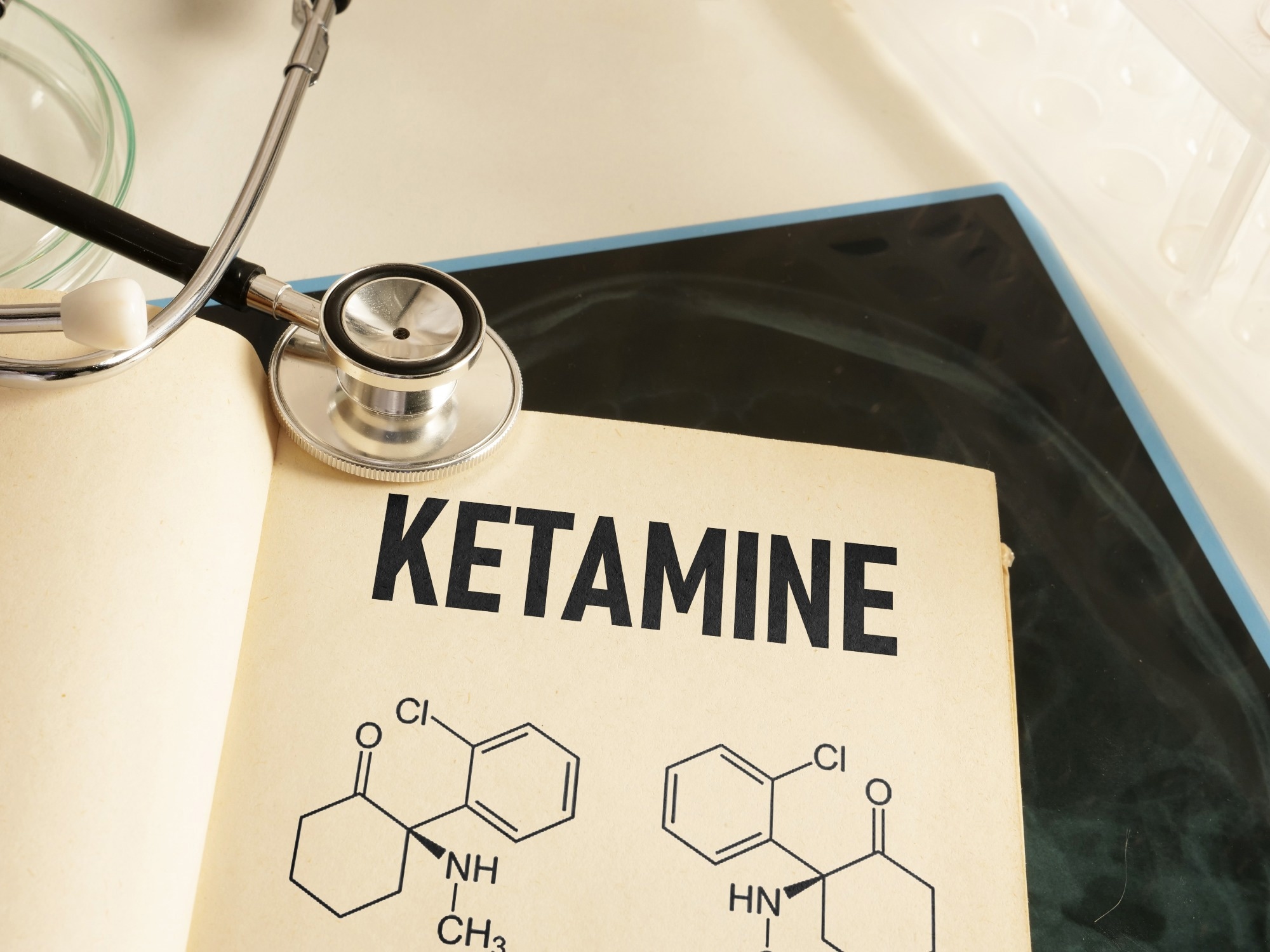
    Study: Ketamine’s mechanism of action with an emphasis on neuroimmune regulation: Can the complement system complement ketamine’s antidepressant effects? Image Credit: Jack_the_sparrow / Shutterstock.com

    Treating major depressive disorder

    Major depressive disorder (MDD) is a mood disorder associated with persistent feelings of loss of interest and sadness. Current estimates indicate that over 300 million individuals are affected by MDD globally, about 700,000 of whom commit suicide each year. Altered neurotrophin levels and monoamine dysregulation are both mechanisms that have been attributed to manifestations of MDD.

    Monoamines associated with noradrenergic, serotoninergic, and dopaminergic activities can be regulated through certain pharmaceutical agents to improve the cognition, sleep, and mood of MDD patients. However, conventional monoamine antidepressant therapy has only been shown to be effective in 30-40% of patients with MDD. 

    According to the Sequenced Treatment Alternatives to Relieve Depression (STAR*D) study, a significant number of MDD patients do not respond to standard treatment. Patients who fail to respond to two antidepressants of suitable dosage are known to suffer treatment-resistant depression (TRD). 

    Racemic (R, S)-ketamine, which is more commonly referred to as ketamine, and (S)-ketamine (esketamine) have shown significant positive effects on MDD. As compared to conventional treatments, ketamine has been shown to exert antidepressant effects within a few hours. Many TRD patients have also responded positively to a single ketamine infusion.

    Mechanism of action of ketamine for MDD treatment

    The mechanisms that underlie the antidepressant effects of ketamine are associated with the N-methyl-D-aspartate (NMDA) receptor, opioid pathway, α-amino-3-hydroxy-5-methyl-4-isoxazolepropionic acid (AMPA) receptor, and mechanistic target of rapamycin (mTOR). 

    Various neuronal cells, including microglia and astrocytes, regulate neuroinflammation. Individuals with MDD often exhibit lower levels of glial fibrillary acidic protein (GFAP) and glutamate transporter-1 (GLT-1). In these patients, an acute administration of ketamine normalized these levels, thus improving their mood. 

    In vivo experimental findings have also shown that ketamine has an inhibitory effect in lipopolysaccharide (LPS)-induced microglial activation, which led to improvements in depressive-like behaviors. Rodent studies have also reported that transforming growth factor β (TGF)-β, an anti-inflammatory molecule inhibiting excessive microglial activation, is associated with the differential antidepressant effects of ketamine enantiomers.

    Mouse models have revealed that (R)-ketamine, and not (S)-ketamine, alleviates stress-induced reduction in the expression of Tgfb1 and its receptors Tgfbr1 and Tgfbr2. Nevertheless, additional research is needed to clarify the microglia-based mechanisms underlying the antidepressant effects of ketamine.

    Patients with MDD exhibit higher interleukin 6 (IL-6) and tumor necrosis factor ⍺ (TNF-⍺) levels than non-depressed individuals. One rodent study revealed ketamine administration normalized these levels and improved MDD symptoms.

    Higher levels of granulocyte-macrophage colony-stimulating factor (GM-CSF) have been observed in patients with MDD. Administration of 0.5 mg/kg ketamine infusions for twelve days led to symptomatic improvement that was associated with significant downregulation of GM-CSF. 

    Ketamine and the immune response

    The antidepressant effects of ketamine have been linked with the complement system, which is a vital component of synaptic plasticity. The complement system comprises 30 proteins that are involved in the classical, alternate, and lectin pathways, all of which converge in C3 cleavage, a major complement component.

    Complement proteins play a crucial role in the regulation of cell proliferation, maturation, and responsiveness. Activation of the complement system results in the release of complement and immune molecules that are linked with inflammatory responses.

    Increased levels of serum complement components C3a and C5a have been observed in bipolar disorder. Similarly, a high concentration of serum C1q levels is found in patients with MDD.

    An in vivo experiment with C5a receptor knockout mice highlighted the neuroprotective role of C5a against glutamate excitotoxicity-induced apoptosis through elevated expression and regulation of glutamate receptor subunit 2 (GluR2). Glutamatergic modulation has been established as a mechanistic commonality between the complement system and ketamine. 

    Ketamine also activates mTORC1 by triggering the brain-derived neurotrophic factor (BDNF), tropomyosin receptor kinase B (TrkB), and NMDA receptors. Additionally, the the C3a ligand-C3a receptor in CD4 + T-cells leads to mTOR activatiwhich is on, essential for cell survival. Complement-mTOR activation also modulates many stress and metabolic pathways, such as cytokine secretion, oxidative phosphorylation, and inflammasome activation.

    Conclusions

    The current study indicated the potential association between the complement system and the antidepressant effects of ketamine. Nevertheless, additional studies are needed to improve treatment outcomes for MDD using ketamine.

    Journal reference:

    • Quintanilla, B., Zarate, C. A., and Pillai, A. (2024) Ketamine’s mechanism of action with an emphasis on neuroimmune regulation: Can the complement system complement ketamine’s antidepressant effects? Molecular Psychiatry; 1-10. doi:10.1038/s41380-024-02507-7

    [ad_2]

    Source link

  • Moderate vitamin E, C, and β-carotene intake reduces type 2 diabetes risk

    Moderate vitamin E, C, and β-carotene intake reduces type 2 diabetes risk

    [ad_1]

    In a recent study published in Advances in Nutrition, researchers review the effect of vitamins C and E, as well as β-carotene, on the risk of type 2 diabetes (T2D).

    Study: Vitamins C, E, and β-Carotene and Risk of Type 2 Diabetes: A Systematic Review and Meta-Analysis. Image Credit: Suriyawut Suriya / Shutterstock.com

    About the study

    Previously published meta-analyses have discussed the protective effect of β-carotene and vitamin E, not vitamin C, against T2D incidence. Furthermore, these analyses did not focus on randomized controlled trials (RCTs) or dose-response relationships.

    In the current review, all relevant information was obtained from Embase, MEDLINE, and the Cochrane Library databases. To investigate the relationships between dietary intake, circulating levels of vitamin C, vitamin E, and β-carotene, and T2D incidence, case-cohort, cohort, nested case-control, and RCT studies were considered. Any study written in languages other than English was not included in the review.

    A total of 6,190 articles were obtained in the initial search, 40 of which fulfilled the eligibility criteria and were considered. Both observational and RCT studies indicated low, moderate, or serious risk of bias. 

    Diet and T2D

    T2Dis associated with β-cell dysfunction and insulin resistance and is one of the most common and globally prevalent metabolic diseases. Several strategies associated with weight loss, diet, and physical activities have been designed to alleviate the risks of T2D.

    Adherence to specific dietary patterns, such as the Mediterranean diet, positively reduces the risk of T2D. The Mediterranean diet emphasizes increased consumption of fruits, vegetables, olive oil, and fish, as well as minimal intake of highly processed food and meat-based products. Fruits and vegetables are high in antioxidants, such as vitamin E, vitamin C, and β-carotene, which has many positive biological effects. 

    Vitamin C is a water soluble vitamin found in vegetables and fruits and regenerates vitamin E from its oxidized form. Vitamin E is a fat-soluble vitamin found in seeds, nuts, and vegetable oils, which protects cell membranes from lipid peroxidation. Moreover, β-carotene is a provitamin A carotenoid that is often found in fruits and vegetables. 

    Antioxidants protect cells from oxidative stress that arises due to the release of free radicals. Oxidative stress increases insulin resistance by inducing inflammatory processes and suppressing insulin signaling.

    Several studies have indicated that a high level of dietary antioxidants could alleviate insulin resistance and T2D. Thus, it is important to understand the mechanisms that contribute to the antioxidant properties of each vitamin. 

    How do β-carotene, and vitamins C and E affect T2D risk?

    Dietary intake of vitamin C, vitamin E, and β-carotene has been found to reduce the risk of T2D; however, this association is non-linear and plateaus at moderate intakes.

    Vitamin C is inversely associated with insulin resistance. To date, few studies have determined the effect of vitamin C on β-cell function.

    Among the reviewed RCT studies, there was not sufficient data indicating that supplementation of these vitamins reduces the risk of T2D. In contrast to vitamin C and β-carotene, vitamin E supplementation exhibited insulin resistance. 

    Nevertheless, a nonlinear dose-response gradient, which indicated that the lowest risk of diabetes was associated with moderate intake of dietary vitamins, was observed. According to Nordic guidelines, the dietary allowance of vitamin C for men and women is 75 mg/day and 90 mg/day, respectively. The recommendation for vitamin E intake is 8-15 mg/day for both men and women. 

    These recommendations can be reached through the consumption of half of a red pepper and half of a cup of almonds for vitamins C and E, respectively. Although no reference value has been established for β-carotene, an intake of 3-6 mg/day could reduce the risk of chronic disease.

    Mendelian randomization (MR) analyses did not indicate the causal relationship between genetically predicted circulating vitamin E, vitamin C, or β-carotene and T2D manifestation. This finding was also supported by RCT data revealing that supplementation of the these antioxidants did not confer additional benefits in reducing the risks of T2D in healthy adults. 

    It is possible that vitamin C, vitamin E, and β-carotene function synergistically to inhibit oxidative stress and, therefore, do not exhibit beneficial effects when consumed individually. Individuals who adhere to the Mediterranean diet consume an adequate amount of antioxidants, which act synergistically to reduce the risk of T2D. 

    Conclusions

    Sufficient, rather than high, intake of vitamin C, vitamin E, and β-carotene prevents the risk of T2D in healthy adults. Therefore, these vitamin supplements should not be used as a preventive strategy to reduce T2D in healthy individuals.

    However, future studies are needed to identify threshold antioxidant levels for smokers and those with genetic susceptibility to diabetes to obtain optimal benefits.

    Journal reference:

    • Lampousi, A., Lundberg, T., Löfvenborg, J. E., and Carlsson, S. (2024) Vitamins C, E, and β-Carotene and Risk of Type 2 Diabetes: A Systematic Review and Meta-Analysis. Advances in Nutrition 15(5); 100211. doi:10.1016/j.advnut.2024.100211

    [ad_2]

    Source link

  • Seafood is a potentially underestimated source of PFAS exposure, study shows

    Seafood is a potentially underestimated source of PFAS exposure, study shows

    [ad_1]


    The findings stress the need for more stringent public health guidelines that establish the amount of seafood people can safely consume to limit their exposure to perfluoroalkyl and polyfluoroalkyl substances, the researchers report in the journal Exposure and Health. This need is especially urgent for coastal regions such as New England where a legacy of industry and PFAS pollution bumps up against a cultural predilection for fish, the authors write.

    Our recommendation isn’t to not eat seafood-;seafood is a great source of lean protein and omega fatty acids. But it also is a potentially underestimated source of PFAS exposure in humans.”


    Megan Romano, study’s corresponding author and associate professor of epidemiology at Dartmouth’s Geisel School of Medicine

    “Understanding this risk-benefit trade-off for seafood consumption is important for people making decisions about diet, especially for vulnerable populations such as pregnant people and children,” Romano said.

    The study paired an analysis of PFAS concentrations in fresh seafood with a statewide survey of eating habits in New Hampshire. National data indicate that New Hampshire-;along with all of New England-; is among the country’s top consumers of seafood, which made the state ideal for understanding the extent of people’s exposure to PFAS through fish and shellfish.

    “Most existing research focuses on PFAS levels in freshwater species, which are not what people primarily eat,” said Romano, who studies the effects of PFAS and other endocrine-disrupting chemicals in drinking water on New England communities. “We saw that as a knowledge gap in the literature, especially for a New England state where we know people love their seafood.”

    The study also drew on New Hampshire’s extensive data on the sources and effects of PFAS, which are a staple of consumer products such as plastics and nonstick coatings. The molecular stability that makes PFAS versatile also makes them nearly indestructible, leading them to be called forever chemicals.

    In humans, PFAS are associated with cancer, fetal abnormalities, high cholesterol, and thyroid, liver, and reproductive disorders. The chemicals have accumulated in soil, water, and wildlife, and studies have shown that nearly all Americans have measurable amounts in their blood.

    “PFAS are not limited to manufacturing, fire-fighting foams, or municipal waste streams-;they are a decades-long global challenge,” said study co-author Jonathan Petali, a toxicologist with the New Hampshire Department of Environmental Services. “New Hampshire was among the first states to identify PFAS in drinking water. We’re a data-rich state due to years spent investigating the impacts of PFAS and trying to mitigate exposure.”

    The researchers measured the levels of 26 varieties of PFAS in samples of the most consumed marine species: cod, haddock, lobster, salmon, scallop, shrimp, and tuna. The seafood studied was purchased fresh from a market in coastal New Hampshire and originated from various regions.

    Shrimp and lobster clocked the highest concentrations with averages ranging as high as 1.74 and 3.30 nanograms per gram of flesh, respectively, for certain PFAS compounds, the researchers report. Concentrations of individual PFAS in other fish and seafood measured generally less than one nanogram per gram.

    The prevalence of PFAS in the environment makes it difficult to know exactly where and how the chemicals enter the marine food chain, the researchers report. Some shellfish may be especially vulnerable to the buildup of PFAS in their flesh due to feeding and living on the seafloor, as well as their proximity to sources of PFAS that are near the coast. Larger marine species may ingest PFAS by eating smaller species that, like shellfish, are prone to having the compounds accumulate in their systems. 

    Buttressing the study is a survey of 1,829 New Hampshire residents the researchers conducted to gauge how much seafood Granite Staters eat-;and it’s a lot.

    The survey found that men in New Hampshire eat just over one ounce of seafood per day and women eat just under one ounce. Both are higher than what the National Health and Nutrition Examination Survey found for men and women in the Northeast, and more than 1.5 times the national average for both. Daily intake for New Hampshire children aged 2 to 11 years old was about 0.2 ounces, the highest end of the range for children nationwide.

    About 95% of adults the researchers surveyed reported they ate seafood within the past year, and 94% of that group consumed fish or shellfish within the previous month. More than two-thirds of those respondents ate seafood within the past week.

    But people in New Hampshire do not eat seafood uniformly. More than half of the people who ate seafood in the week before the survey lived on the state’s coast or near the border with Massachusetts. More than 60% of people with a household income below $45,000 per year reported consuming seafood at least once per week, whereas people with higher household incomes reported eating seafood less often.

    Of the species the researchers tested for PFAS, shrimp, haddock, and salmon were consumed by more than 70% of the adults who ate seafood once a month or more. Lobster was eaten by just over 54% of these adults. Salmon, canned tuna, shrimp, and haddock were the most commonly consumed species among children.

    Federal guidelines for safe seafood consumption exist for mercury and other contaminants, but there are none for PFAS, said Celia Chen, a co-author of the study and a research professor in the Department of Biological Sciences at Dartmouth.

    “Top predator species such as tuna and sharks are known to contain high concentrations of mercury, so we can use that knowledge to limit exposure. But it’s less clear for PFAS, especially if you start looking at how the different compounds behave in the environment,” said Chen, who leads several federally funded projects examining how and where PFAS accumulate in aquatic food webs in New Hampshire and Vermont.

    The establishment of safety guidelines would help protect people who are especially susceptible to pollutants, said Kathryn Crawford, the study’s first author and an assistant professor of environmental studies at Middlebury College.

    “Seafood consumption advisories often provide advice for those individuals that is more conservative than for the rest of the population,” said Crawford, who began the project as a postdoctoral researcher in the Romano Lab at Dartmouth. “People who eat a balanced diet with more typical, moderate amounts of seafood should be able to enjoy the health benefits of seafood without excessive risk of PFAS exposure.”

    Source:

    Journal reference:

    Crawford, K.A., et al. (2024) Patterns of Seafood Consumption Among New Hampshire Residents Suggest Potential Exposure to Per- and Polyfluoroalkyl Substances. Exposure and Health. doi.org/10.1007/s12403-024-00640-w.

    [ad_2]

    Source link

  • Unraveling the mysteries of gastro-esophageal junction development

    Unraveling the mysteries of gastro-esophageal junction development

    [ad_1]

    The transition from the esophagus to the stomach is a delicate region from a medical point of view, often associated with pathological disorders leading to cancer. An international research team has now gained new insights into this region. These pave the way for new prevention and treatment options.

    The meeting point of the stomach and esophagus, the so-called gastro-esophageal junction, is a region of the human body that is not well-suited to the modern lifestyle. Stress, alcohol, nicotine and severe obesity are often triggers for pathological changes to the mucosal membrane in this area, often resulting in esophageal cancer.

    An international research team has now gained new insights into the development of the cells, their communication with each other, and their regulation at the junction of the esophagus and stomach. With the help of specially developed mini-organs, so-called organoids, and with techniques that make it possible to track and profile individual cells, they have been able to follow the development of the gastro-esophageal junction from embryonic to adult stage in detail using animal experiments.

    New insights into the development of the gastrointestinal tract

    Their results reveal the complex communication at the cellular level and the specific pathways that these cells use to communicate. They provide new insights into the development of the gastro-esophageal junction and thus have significant implications for the understanding, prevention and treatment of gastrointestinal diseases. At the same time, they present new starting points for medical research and the development of new therapies.

    Cindrilla Chumduri is responsible for this study, which has now been published in the journal Nature Communications. Until recently, the infection and cancer biologist was a research group leader at the Department of Microbiology at Julius-Maximilians-Universität Würzburg (JMU); she is now an associate professor at Aarhus University (Denmark). Other participants came from Charité – Universitätsmedizin and the Max Planck Institute for Infection Biology in Berlin.

    “This collaboration underlines the importance of different expertise to improve our understanding of the biology of the gastrointestinal tract,” says Chumduri.

    She herself has many years of experience in research with organoids. Among other things, she has used mini-organs she developed to study how cells in the cervix degenerate and turn cancerous – another region where different types of mucosal cells collide.

    Where different epithelia meet

    The squamous epithelia of the esophagus and the columnar epithelia of the stomach meet at the gastroesophageal junction.”

    Dr. Naveen Kumar Nirchal, one of the first authors of the study

    The area is known as a “hotspot for the development of metaplasia” – the replacement of one type of cell by another.

    Barrett’s esophagus, a precursor to esophageal cancer, often develops there, the number of cases of which has increased dramatically in the Western world over the past four decades. “Barrett’s esophagus is characterized by the replacement of the resident squamous epithelium of the esophagus by other cell types that are not normally found in this tissue,” says the scientist.

    However, it is still unclear why this region is so susceptible to this process. In order to better understand this transformation, it is therefore first necessary to decipher the normal development process in detail – from embryo to mature adult. “This is the only way to determine the tissue changes that trigger the progression of the disease, explains Dr. Rajendra Kumar Gurumurthy, another researcher of the study.

    A never-before-seen insight into the development of this region

    This has now been achieved: By using a novel approach that combines organoid and mouse models with advanced single-cell transcriptome analyses over time and space, the research team has shed light on the complex developmental process of the gastroesophageal junction. “We were able to provide unprecedented insight into the development of this region from the embryonic stage to adulthood in mice and identify the intricate composition of the cells involved and how they develop,” explains Pon Ganish Prakash, another scientist involved in the study.

    The work shows the sophisticated communication between different cell types within the gastroesophageal junction and the signaling pathways involved. “This understanding opens up new avenues for research into gastrointestinal diseases,” says Cindrilla Chumduri.

    Above all, the precision of the single-cell analysis in their study opens new doors to understanding how pathological processes develop and to developing innovative treatments, the team writes in its study. The work will therefore be a “cornerstone for understanding the development of such diseases” and will significantly influence the approach to the early detection and treatment of diseases in this important part of the digestive system.

    Source:

    Journal reference:

    Kumar, N., et al. (2024). Decoding spatiotemporal transcriptional dynamics and epithelial fibroblast crosstalk during gastroesophageal junction development through single cell analysis. Nature Communications. doi.org/10.1038/s41467-024-47173-z.

    [ad_2]

    Source link

  • AI model can accurately screen for post-traumatic stress disorder after childbirth

    AI model can accurately screen for post-traumatic stress disorder after childbirth

    [ad_1]

    A generative artificial intelligence (AI) model that can analyze the narrative accounts of women who have undergone recent childbirth has shown the ability to accurately screen for post-traumatic stress disorder (CB-PTSD), a study by Massachusetts General Hospital (MGH), a founding member of the Mass General Brigham healthcare system has found.

    By exploring the capabilities and shortcomings of several models from OpenAI, including ChatGPT, the researchers identified a version that offers rich insights into maternal mental health following traumatic childbirth.

    The model can fit seamlessly into routine obstetric care and could potentially be harnessed to assess other mental health disorders. The results of the study were published in Scientific Reports.

    Evaluation of PTSD related to traumatic birth currently relies on extensive clinician evaluation, which fails to meet the urgent need for a rapid, low-cost assessment strategy.”


    Sharon Dekel, PhD, director of MGH’s Postpartum Traumatic Stress Disorders Research Program, and senior author of the study

    “The use of brief patient narratives of childbirth analyzed by AI’s text-based computational methods could become an efficient, low-cost, and patient-friendly strategy for detecting CB-PTSD after a traumatic birth and with more research this tool may potentially aid in identifying women at risk for CB-PTSD before the condition fully develops.”

    For an estimated eight million women a year globally, childbirth that is traumatic and/or medically complicated is expected to trigger post-traumatic stress disorder, a condition historically has been associated with military combat or severe sexual assault.

    In recent years, childbirth has become acknowledged as a significant PTSD trigger which, if left untreated, can impair the health of both the mother and child and result in significant societal costs.

    In previous studies, Dekel’s lab found evidence that brief psychological interventions delivered soon after traumatic childbirth can reduce maternal childbirth-related PTSD symptoms.

    In their latest study, Dekel in collaboration with first author Alon Bartal, PhD, of Bar-Ilan University in Israel, investigated the effectiveness of artificial intelligence and related machine learning (ML) analysis strategies to detect CB-PTSD.

    Specifically, they evaluated the performance of different large language models (LLMs) and variations of ChatGPT and their ability to extract novel insights from text-based data sets derived from the brief narrative descriptions by postpartum women of their childbirth experience.

    As part of their work, the team collected short narrative accounts from 1,295 women who had recently given birth.

    The study focused on an OpenAI model known as text-embeddings-ada-002, which converted narrative data from the personal accounts of women with and without probable CB-PTSD to a numerical format that was then analyzed by a trained machine learning algorithm developed by the team.

    Researchers showed this model had superior performance in identifying postpartum traumatic stress compared to other ChatGPT and large language models, which are typically trained on huge volumes of data allowing them to understand, analyze and interpret natural language.

    “The reliance of the ML model using childbirth narrative input from the Open AI model as its exclusive data source presents an efficient mechanism for data collection during the vulnerable postpartum period, demonstrating 85 percent sensitivity and 75 percent specificity in identifying CB-PTSD cases,” notes Dekel.

     “Moreover, the model we developed could potentially improve accessibility to CB-PTSD screening and diagnosis by fitting seamlessly into routine obstetric care and providing a foundation for commercial product development and mainstream adoption.”

    Dekel, whose research program is dedicated to exploring women’s mental health following traumatic childbirth, underscores the clinical benefits of using a pre-trained large language model to assess potential PTSD in new mothers.

    “Early intervention is essential to prevent the progression of this disorder to chronic stages, which can seriously complicate treatment,” the MGH investigator points out.

    “Our unique approach could introduce an innovative and cost-effective screening strategy for identifying high-risk women and facilitating timely treatment. It may also holds promise for assessing other mental health disorders, and consequently improving patient outcomes.”

    The emergence of artificial intelligence tools in health has been groundbreaking and has the potential to positively reshape the continuum of care. Mass General Brigham, as one of the nation’s top integrated academic health systems and largest innovation enterprises, is leading the way in conducting rigorous research on new and emerging technologies to inform the responsible incorporation of AI into care delivery, workforce support, and administrative processes. 

    Dekel is a psychologist at MGH, and assistant professor of Psychology at Harvard Medical School. Bartal is an assistant professor of Information Systems at Bar-Ilan University in Israel. Co-authors in the Dekel Laboratory include Kathleen Jagodnik, PhD, a Harvard research fellow, and Sabrina Chan, a clinical research coordinator.

    Dekel was supported by funds from the NIH (Eunice Kennedy Shriver National Institute of Child Health and Human Development, grants R01HD108619, R21HD109546, and R21HD100817).

    Source:

    Journal reference:

    Bartal, A., et al. (2024). AI and narrative embeddings detect PTSD following childbirth via birth stories. Scientific Reportsdoi.org/10.1038/s41598-024-54242-2.

    [ad_2]

    Source link

  • AI program can help identify signs of childbirth-related PTSD

    AI program can help identify signs of childbirth-related PTSD

    [ad_1]

    Researchers have adapted an artificial intelligence (AI) program to identify signs of childbirth-related post-traumatic stress disorder (CB-PTSD) by evaluating short narrative statements of patients who have given birth. The program successfully identified a large proportion of participants likely to have the disorder, and with further refinements-;such as details from medical records and birth experience data from diverse populations-;the model could potentially identify a large percentage of those at risk. The study, which was funded by the National Institutes of Health, appears in Scientific Reports.

    Worldwide, CB-PTSD affects about 8 million people who give birth each year, and current practice for diagnosing CB-PTSD requires a physician evaluation, which is time-consuming and costly. An effective screening method has the potential to rapidly and inexpensively identify large numbers of postpartum patients who could benefit from diagnosis and treatment. Untreated CB-PTSD may interfere with breastfeeding, bonding with the infant and the desire for a future pregnancy. It also may worsen maternal depression, which can lead to suicidal thoughts and behaviors.

    Investigators administered the CB-PTSD Checklist, which is a questionnaire designed to screen for the disorder, to 1,295 postpartum people. Participants also provided short narratives of approximately 30 words about their childbirth experience. Researchers then trained an AI model to analyze a subset of narratives from patients who also tested high for CB-PTSD symptoms on the questionnaire. Next, the model was used to analyze a different subset of narratives for evidence of CB-PTSD. Overall, the model correctly identified the narratives of participants who were likely to have CB-PTSD because they scored high on the questionnaire.

    The authors believe their work could eventually make the diagnosis of childbirth post-traumatic stress disorder more accessible, providing a means to compensate for past socioeconomic, racial, and ethnic disparities.

    The study was conducted by Alon Bartal, Ph.D., of Bar Ilan University in Israel, and led by senior author Sharon Dekel, Ph.D., of Massachusetts General Hospital and Harvard Medical School, Boston. Funding was provided by NIH’s Eunice Kennedy Shriver National Institute of Child Health and Human Development (NICHD).

    Source:

    Journal reference:

    Bartal, A., et al. (2024). AI and narrative embeddings detect PTSD following childbirth via birth stories. Scientific Reports. doi.org/10.1038/s41598-024-54242-2.

    [ad_2]

    Source link

  • Meta-analysis uncovers stress-responsive genes in Arabidopsis

    Meta-analysis uncovers stress-responsive genes in Arabidopsis

    [ad_1]

    Plants can be temperamental. Even weeds along the side of highways or pushing their way up in the cracks of concrete sidewalks can get stressed out by dehydration, cold, excess salt and more. Researchers at Hiroshima University have identified 14 genes that thale cress -; a plant commonly used in genetic investigations since its genome is well documented -; express more when responding to five specific stressors, as well as eight genes that the plant suppresses.

    They published their results on March 22 in Frontiers in Plant Science

    “Abiotic stresses -; as opposed to biotic stresses like pests or disease -; such as drought, salinity and cold negatively affect plant growth and crop productivity. Understanding the molecular mechanisms underlying plant responses to these stressors is essential for stress tolerance in crops,” said corresponding author Hidemasa Bono, professor in the Laboratory of Genome Informatics at Hiroshima University’s Graduate School of Integrated Sciences for Life. Bono is also affiliated with the Laboratory of Bio-DX in the university’s Genome Editing Innovation Center. 

    “The plant hormone abscisic acid (ABA) is significantly increased upon abiotic stressors, inducing physiological responses to adapt to stress and regulate gene expression. Although many studies have examined the components of established stress signaling pathways, few have explored other unknown elements.”

    To better understand the molecular pathways that allow ABA to increase, the research team analyzed public RNA sequencing data on thale cress, or Arabidopsis thaliana. RNA sequencing is a technique that enables scientists to identify and quantify specific sequences of genetic instructions programmed in an organism’s RNA. This data can reveal how different variables may increase or decrease the expression of certain genes.

    Bono and his team specifically focused on five ABA-related stress conditions: ABA, when the hormone is applied directly to the plant; salt, which changes how the plant can use water; dehydration, or how much water the plant has; osmotic, when plant cells swell or shrink inappropriately; and cold.

    The data-driven studies have the advantage of analyzing large and independent datasets, which can lead to the identification of novel targets, distinct from the extensively studied established factors and accelerate the development of stress-tolerant crops.”

    Hidemasa Bono, Professor, Laboratory of Genome Informatics at Hiroshima University’s Graduate School of Integrated Sciences for Life

    The researchers performed a meta-analysis of 216 paired datasets, combining those research results and reanalyzing them to identify where data might overlap or reveal previously unknown connections.

    The meta-analysis revealed that 14 genes were commonly up-regulated and eight genes were commonly down-regulated across all five ABA-related stress responses investigated. Bono noted that some genes regulated by salt, dehydration and osmotic treatments were not regulated by ABA or cold stress, suggesting that they may be involved in the plant response through a different signaling pathway.

    “Our meta-analysis revealed a list of candidate genes with unknown molecular mechanisms in ABA-dependent and ABA-independent stress responses,” Bono said.

    “These genes could be valuable resources for selecting genome editing targets and potentially contribute to the discovery of novel stress tolerance mechanisms and pathways in plants. We will continue to develop methods and utilize data from public databases and conduct comparative analysis from various angles to unravel the unknown mechanisms of stress response in plants.”

    Co-authors of the study are Mitsuo Shintani with the Graduate School of Integrated Sciences for Life at Hiroshima University and Keita Tamura with the university’s Genome Editing Innovation Center.

    The Center of Innovation for Bio-Digital Transformation (BioDX) and the Japan Science and Technology Agency supported this research.

    Source:

    Journal reference:

    Shintani, M., et al. (2024). Meta-analysis of public RNA sequencing data of abscisic acid-related abiotic stresses in Arabidopsis thaliana. Frontiers in Plant Science. doi.org/10.3389/fpls.2024.1343787.

    [ad_2]

    Source link

  • Understanding the mechanisms behind embryonic diapause in hungry mouse mums

    Understanding the mechanisms behind embryonic diapause in hungry mouse mums

    [ad_1]

    It’s challenging to sustain a pregnancy when food is short, or conditions are otherwise tough. That’s why many mammalian embryos can postpone their growth to get through periods of environmental stress and then re-enter development when conditions improve. This stalling of development is known as embryonic diapause, and understanding the mechanisms behind it might help improve infertility treatments, such as embryo freezing. Now, researchers at the Center for Excellence in Brain Science and Intelligence Technology, the Chinese Academy of Sciences in Shanghai, China, have discovered how nutrient depletion is sensed by embryos growing in hungry mouse mums to induce diapause. They publish their study in the journal Development on 11 April 2024.

    Lack of food is a known trigger of embryonic diapause, but it has not been clear how nutrient depletion in the mother’s diet is sensed by the embryo. “Seasonal starvation is one of the universal environmental stresses in nature,” explained Professor Qiang Sun, who led the study. “However, the regulatory process of diapause in early-stage embryos is not fully understood. So, we decided to examine whether nutrient deprivation induces embryonic diapause.”

    By comparing hungry and well-fed pregnant mice, the team discovered that embryos in the hungry mice did not implant into the uterus and their growth paused at an early timepoint, when the embryo comprises a hollow ball of cells called the blastocyst. These embryos remained viable and could start developing again when transplanted into a well-fed mother.

    To work out which nutrients were important to induce diapause, the researchers grew early-stage mouse embryos in dishes that contained different nutrients. They found that embryos grown in dishes lacking protein or carbohydrates paused their development, whereas the embryos exposed to normal nutrient levels did not stall and kept on developing. The scientists then went on to reveal that nutrient sensors in the embryo can detect drops in protein or carbohydrate levels, which triggers the entry into diapause.

    The finding that embryos grown without protein or carbohydrates can pause their development means that they can survive longer in the lab. In the future, this finding might lead to improvements in fertility treatments, which currently include approaches such as embryo freezing. “We think our study can inspire the development of new methods for human embryo preservation,” said Professor Sun. “Embryo cryopreservation is a widely used approach, but there is still no consensus on when cryopreserved embryos can be thawed and transferred into the uterus. Many clinical studies have shown that traditional frozen embryo transfer can increase the risk of problems during pregnancy. Therefore, it is necessary to develop alternative methods to preserve embryos.”

    Studies focusing on diapause may even have long-term implications for cancer treatments.

    Dormant cancer cells which persist after chemotherapy resemble the diapaused embryos. Consequently, we hypothesize that delving into the mechanism of diapause may have positive implications for cancer treatment and decreasing the chances of relapse.”


    Professor Qiang Sun

    Source:

    Journal reference:

    Ye, J., et al. (2024). Nutrient deprivation induces mouse embryonic diapause mediated by Gator1 and Tsc2. Developmentdoi.org/10.1242/dev.202091.

    [ad_2]

    Source link

  • Geraniol alleviates cognitive decline in D-galactose-induced aging mice

    Geraniol alleviates cognitive decline in D-galactose-induced aging mice

    [ad_1]

    A new research paper was published in Aging (listed by MEDLINE/PubMed as “Aging (Albany NY)” and “Aging-US” by Web of Science) Volume 16, Issue 6, entitled, “Geraniol attenuates oxidative stress and neuroinflammation-mediated cognitive impairment in D galactose-induced mouse aging model.”

    D-galactose (D-gal) administration was proven to induce cognitive impairment and aging in rodents’ models. Geraniol (GNL) belongs to the acyclic isoprenoid monoterpenes. GNL reduces inflammation by changing important signaling pathways and cytokines, and thus it is plausible to be used as a medicine for treating disorders linked to inflammation. In this new study, researchers Peramaiyan Rajendran, Fatma J. Al-Saeedi, Rebai Ben Ammar, Basem M. Abdallah, Enas M. Ali, Najla Khaled Al Abdulsalam, Sujatha Tejavat, Duaa Althumairy, Vishnu Priya Veeraraghavan, Sarah Abdulaziz Alamer, Gamal M. Bekhet, and Emad A. Ahmed from King Faisal University, Kuwait University, Center of Biotechnology of Borj-Cedria, Saveetha University, Alexandria University, and Assiut University examined the therapeutic effects of GNL on D-gal-induced oxidative stress and neuroinflammation-mediated memory loss in mice. 

    “Life expectancy in the 21st century is rising, resulting in more age-related illnesses, such as memory impairment and Alzheimer’s disease. In this study, GNL was studied for its protective effect on D-gal-induced aging in mice.”

    The study was conducted using six groups of mice (6 mice per group). The first group received normal saline, then D-gal (150 mg/wt) dissolved in normal saline solution (0.9%, w/v) was given orally for 9 weeks to the second group. In the III group, from the second week until the 10th week, mice were treated orally (without anesthesia) with D-gal (150 mg/kg body wt) and GNL weekly twice (40 mg/kg body wt) four hours later. Mice in Group IV were treated with GNL from the second week up until the end of the experiment. For comparison of young versus elderly mice, 4 month old (Group V) and 16-month-old (Group VI) control mice were used. 

    “We evaluated the changes in antioxidant levels, PI3K/Akt levels, and Nrf2 levels. We also examined how D-gal and GNL treated pathological aging changes.”

    Administration of GNL induced a significant increase in spatial learning and memory with spontaneously altered behavior. Enhancing anti-oxidant and anti-inflammatory effects and activating PI3K/Akt were the mechanisms that mediated this effect. Further, GNL treatment upregulated Nrf2 and HO-1 to reduce oxidative stress and apoptosis. This was confirmed using 99mTc-HMPAO brain flow gamma bioassays. 

    “Thus, our data suggested GNL as a promising agent for treating neuroinflammation-induced cognitive impairment.”

    Source:

    Journal reference:

    Rajendran, P., et al. (2024). Geraniol attenuates oxidative stress and neuroinflammation mediated cognitive impairment in D galactose induced mouse-aging model. Aging. doi.org/10.18632/aging.205677.

    [ad_2]

    Source link

  • Mediterranean diet linked to lower stress in Korean baby boomers

    Mediterranean diet linked to lower stress in Korean baby boomers

    [ad_1]

    In a recent cross-sectional study published in the journal Nutrition Research and Practice, researchers from Korea investigated the link between psychosocial stress levels and adherence to the Mediterranean diet among baby boomers in Korea. They found that higher adherence to the Mediterranean diet was associated with a lower risk of high psychosocial stress in both men and women.

    Study: Higher adherence to a Mediterranean-type diet is associated with reduced psychosocial stress levels in baby boomers: a cross-sectional study. Image Credit: Luigi Giordano / ShutterstockStudy: Higher adherence to a Mediterranean-type diet is associated with reduced psychosocial stress levels in baby boomers: a cross-sectional study. Image Credit: Luigi Giordano / Shutterstock

    Background

    The aging baby boomer population in Korea, comprising around 15% of those born between 1955 and 1963, is transitioning into old age, posing challenges due to declining healthy life expectancy despite increasing overall life expectancy. As middle-aged health behaviors significantly impact later quality of life, there is a need to promote healthier lifestyles among baby boomers for their well-being in old age. Mental health in older adults is particularly crucial, as perceived stress levels have been linked to cognitive decline, cognitive impairment, and increased risk of cardiovascular disease. Studies indicate that perceived psychosocial stress is independently associated with a higher risk of stroke in adults.

    Diet and mental health are bidirectionally related. The Mediterranean diet, high in legumes, nuts, fruits, vegetables, whole grains, olive oil, and fish, moderate in alcohol, and low in red meat and high-fat dairy, is shown to be associated with several potential health benefits. Adopting this diet around retirement age may promote healthy aging, potentially preventing and alleviating depression and enhancing cognitive abilities. Studies suggest this diet may effectively mitigate psychosocial stress and buffer against its negative impact on executive function, though further research is needed. Therefore, the present study aimed to explore the potential relationship between Mediterranean diet adherence among Korean baby boomers and their psychosocial stress levels to inform mental health management strategies in later life.

    About the study

    In the present cross-sectional study, data was obtained from the Korean Genome and Epidemiology Study (KoGES), involving adults aged 42 to 50 years from urban and rural regions of South Korea. A total of 1,656 participants (889 men) were included. Individuals with extreme daily energy intake and non-respondents to the questionnaires were excluded.

    A revised version of the Semi-Quantitative Food Frequency Questionnaire (SQFFQ) and MTDS (based on the Mediterranean diet score) were used to assess participants’ usual dietary intake. The MTDS comprised 11 components: fruits, vegetables, legumes, whole grains, fish, dairy products, alcohol, red meat, poultry, and olive oil. Participants were assigned points based on their weekly consumption frequency of these food groups, with higher scores indicating better adherence to the diet. Alcohol consumption and olive oil use were assessed separately. Notably, kimchi and pickles were excluded from specific food groups, while mushrooms and seaweed were included.

    The participants’ socioeconomic status, demographics, and lifestyle factors were comprehensively assessed, including age, body mass index, monthly household income, education level, smoking status, and physical activity level. The Perceived Stress Scale (PWI-SF) for Koreans, consisting of 18 questions on recent physical and psychosocial experiences, was rated on a 4-point scale. A higher score indicated higher levels of stress. Statistical methods included chi-square test, analysis of variance, Duncan’s post-hoc test, and logistic regression.

    Results and discussion

    When MTDS adherence groups (tertiles) were compared, no significant differences were found in most characteristics between these groups. However, among women, those in the highest adherence group tended to have higher levels of education. Additionally, in men, those with the highest adherence to the Mediterranean diet had lower scores of psychosocial stress.

    Higher adherence to the Mediterranean diet was found to be associated with increased consumption of vegetables, whole grains, potatoes, fruits, legumes, and fish, while red meat and dairy product consumption decreased. Moreover, in men, poultry and alcohol consumption decreased with decreasing MTDS. This trend was not observed in women due to similar intake levels across groups. With higher MTDS, energy and nutrient intakes increased, except for nutrients like protein, calcium, phosphorus, and riboflavin in men and fat and retinol in women (P < 0.05).

    Higher adherence to the Mediterranean diet was linked to a reduced risk of high psychosocial stress, with a noticeable decrease trend (P = 0.026 for women and 0.016 for men). Men and women with the highest MTDS had 41% and 39% lower odds, respectively, of experiencing high psychosocial stress compared to those with the lowest adherence.

    The study is limited by its cross-sectional design, potential recall and social desirability biases in dietary assessments, and reliance on self-administered questionnaires for estimating psychosocial stress, which may introduce measurement errors and residual confounding.

    Conclusion

    In conclusion, emphasizing adherence to a Mediterranean diet in baby boomers could potentially enhance healthy aging and improve health outcomes. In the future, studies could focus on developing effective interventions to promote adopting and adhering to this diet among baby boomers.

    Journal reference:

    • Higher adherence to a Mediterranean-type diet is associated with reduced psychosocial stress levels in baby boomers: a cross-sectional study. Jang EH et al., Nutrition Research and Practice, 18(2):257-268 (2024), DOI: 10.4162/nrp.2024.18.2.257, https://e-nrp.org/DOIx.php?id=10.4162/nrp.2024.18.2.257

    [ad_2]

    Source link