We know consuming too much salt raises blood pressure, which in turn can lead to cardiovascular problems. A 2022 study has quantified this relationship as a public health message in clear, stark terms.
Looking at health data on adults in China, the study authors estimate that a reduction of just 1 gram in daily salt intake would be enough to prevent 9 million cases of stroke and heart attack between now and 2030.
With 4 million of those cases likely to be fatal, such a simple measure could save a lot of lives.
In China, average daily salt consumption sits at 11 grams, way above the 5 grams recommended by the World Health Organization (WHO). The researchers pulled together the latest stats on population size, salt consumption, blood pressure and disease rates.
“Previous estimations of the health impact of reducing salt intake in China used either obsolete or otherwise unreliable data sources and did not account for the more prolonged effect of salt reduction on blood pressure over several years,” write the researchers in their published paper.
Consuming too much salt raises blood pressure, which in turn can lead to cardiovascular problems. (Prostock-studio/Canva)
The team looked at two other scenarios besides the single gram drop: a reduction of 3.2 grams per day (a 30 percent drop from the average) by 2025, and reducing salt intake to the recommended 5 grams per day by 2030.
If those targets are hit, up to twice as many deaths related to cardiovascular disease could be prevented, due to the estimated reduction in systolic blood pressure.
However, the researchers emphasize that the reduction would have to be consistent over several years. Education programs run in Chinese schools suggest most of the population wouldn’t find it too difficult to hit that 1 gram per day target.
“Other trials, on low-sodium high-potassium salt substitutes, health education to home cooks and restaurant interventions are ongoing or have recently been completed, some of which have already shown promising results,” write the researchers.
Potassium-enriched salt can look and taste like regular salt. (PixelsEffect/Canva)
Cardiovascular disease accounts for a massive 40 percent of deaths in China, with urbanization – and the associated increase in eating processed and takeaway foods – thought to be one of the main contributing factors.
While the authors of this study only looked at a potential reduction in cases of cardiovascular disease, they suggest that lowering salt intake would have multiple other benefits too. Too much salt has also been linked to certain types of cancers and various kidney problems, for example.
The Chinese government has launched a Healthy China 2030 campaign to try to hit its target of a daily salt intake of just 5 grams. That won’t be easy with a population of 1.4 billion people, but the numbers produced in this study are compelling.
“A salt reduction programme that is workable, coherent, sustainable and targeting current and upcoming major dietary sources of salt in China is urgently needed,” write the researchers.
The lives of Americans could be dramatically improved, and in thousands of cases even saved, by increasing access to weight-loss drugs, scientists at Yale and the University of Florida have found.
At current access levels, an estimated 8,592 lives – mostly among patients who have private insurance – would be saved each year. Access to drugs such as semaglutide (sold as Ozempic and Wegovy) and tirzepatide (Zepbound), which are seeing increasing popularity for aid in excess weight reduction, could decrease the annual death rate in the US by an estimated 42,027 more people.
“Expanding access to these medications is not just a matter of improving treatment options but also a crucial public health intervention,” says epidemiologist Alison Galvani of the Yale School of Public Health. “Our findings underscore the potential to reduce mortality significantly by addressing financial and coverage barriers.”
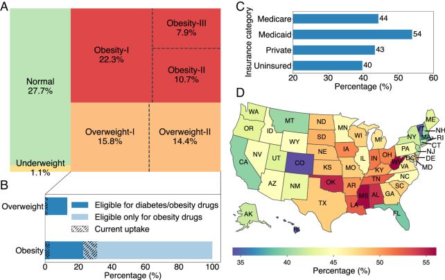
The US is in the throes of an obesity crisis. According to the CDC, some 73.6 percent of American adults are overweight, with a body mass index (BMI) at or over 25. That includes the 41.9 percent of Americans that are considered obese, with a BMI at or over 30.
Obesity is linked with an increased risk of a whole range of serious health problems, such as cardiovascular disease, heart failure, liver disease, depression, cancer, stroke, and diabetes, all of which can shorten a patient’s lifespan.
The distribution of the adult US population across BMI categories, (A) and national access rates to weight loss drugs (B, C, and D). (Pandey et al., PNAS, 2024)
Led by epidemiologist and data scientist Abhishek Pandey of Yale University, a team of scientists set out to quantify the impact of expanding access to weight-loss drugs on the US mortality rate from many of these obesity-related diseases.
They created a map of the BMI distribution across the US, and cross-referenced it with the percentage of Americans who are currently able to access weight-loss prescriptions. This allowed them to accurately quantify the mortality rate from obesity-related complications directly attributable to a lack of access to these prescriptions.
“Limited access stems from a combination of financial barriers, supply constraints, and restrictive insurance coverage,” the researchers write in their paper.
“Although insurance typically covers these medications for diabetes treatment, coverage for weight-loss is less consistent, often requiring patients to pay out-of-pocket or face restrictive insurance policies. Furthermore, 25.6 million Americans are uninsured and more than 80 million are inadequately insured. Currently those uninsured with diabetes or obesity have no access to these innovative weight-loss drugs, and access is challenging even for those with coverage.”
They determined that, if everyone who should be eligible for weight-loss prescriptions was able to obtain one, the obesity rate in the US would fall to 38 percent, and more than 50,000 lives would be saved annually.
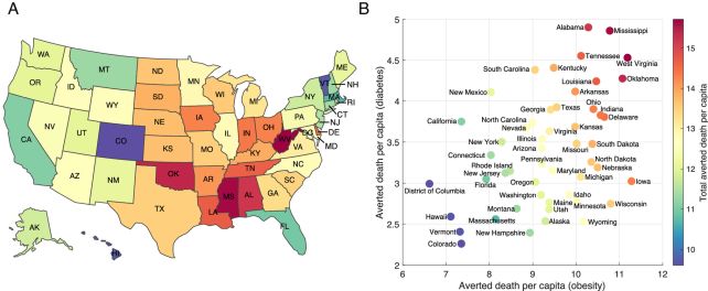
The state-level annual averted death rate per 100,000 people. (Pandey et al., PNAS, 2024)
This is the best-case scenario, in which neither cost nor supply are barriers to access. Even with these barriers in place, however, increased access would dramatically reduce the mortality rate from obesity comorbidities, including a reduction in deaths from type 2 diabetes by 11,769 people.
The US touts itself as one of the richest countries in the world, but the study highlights that, in spite of the country’s wealth, being poor can still kill you. The researchers believe that steps should be taken to address this devastating disparity.
“We need to ensure that drug prices are more aligned with manufacturing costs and increase production capacity to meet demand,” explains mathematician Burton Singer of Yale University. “At the same time, we must tackle the insurance and accessibility issues that prevent many people from getting the treatment they need.”
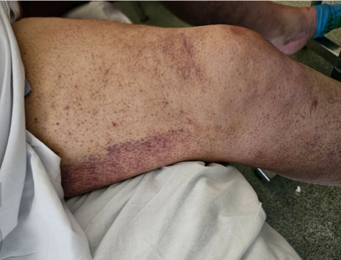
A disease associated with sailors in a bygone era is rearing once more from the depths of history in developed countries. In a recent stark example, a middle-aged Australian was diagnosed with scurvy.
The man sought hospital care for a painful rash on his legs that was accompanied by bruising and swelling. The doctors at Sir Charles Gairdner Hospital ruled out infections as well as inflammatory, immune, and blood disorders.
It wasn’t until physician Andrew Dermawan and colleagues questioned the patient further, days into his hospital stay, that the medical specialists discovered financial difficulties had impacted the man’s diet.
Scurvy results from a lack of vitamin C. Without sufficient amounts of this vital nutrient, wounds stop healing properly, and capillaries and gums start to bleed leading to loss of circulating blood cells.
The 50-year-old man had early signs of deficiency, with low white blood cell counts and blood in his urine despite not having any current urinary infection.
Such physiological changes can lead to weight loss, joint swelling, physical weakness, depression, and eventual risk of fatal bleeding.
It only takes about four weeks of less than 10 mg/day of vitamin C for symptoms of scurvy to emerge. Luckily treatment is simple, and the patient made a rapid recovery soon after he started taking 1000 mg of vitamin C along with other prescribed supplements.
Found in fresh fruit and vegetables, vitamin C can also be provided as supplements to reduce the risk of scurvy. (Akaradech Pramoonsin/Getty Images)
The man’s blood test revealed low levels of several vitamins and no traces of vitamin C at all. He had also ceased taking supplements prescribed after bariatric surgery because he couldn’t afford them.
“Our patient had multiple risk factors, namely, poor dietary habits, obesity, previous bariatric surgery, use of proton pump inhibitors and low-income status. His history of iron, vitamin D and folate deficiencies were also clues to his underlying nutritional deficiency,” Dermawan and team explain.
Other risk factors for scurvy include eating disorders, smoking, alcoholism, steroid use and kidney problems.
While this is only one example, there have been concerning signs this condition is returning to developed countries for some time now. Cases more than doubled in the UK between 2007 to 2017.
“Scurvy is a re-emerging diagnosis in the current era of a rising cost of living,” Dermawan and team warn in their case report.
With food increasing in price by around 3 percent in Australia and nearly 6 percent over the past year in places like the UK, an increasing number of people around the globe on low incomes are being forced to make tough decisions to get by.
“The increasing cost of living means that people are more reliant on lower-cost foods, which tend to be poor in nutritional value,” the team’s report continues.
These low nutrient foods tend to feel more filling and have a high calorie content, so it makes sense that people struggling with tight budgets would be more likely to rely on them. What may be less well known is that overcooking foods destroys nutrients like vitamin C, making access to raw fruits and vegetables important for maintaining the resources our body needs to function properly.
More common levels of vitamin C deficiencies have also been linked to troubles with memory and making decisions in older people. A 2022 study found vitamin C deficiency was associated with cognitive impairments.
“Previous research has shown that vitamin C plays a significant role in the functioning of the brain, with studies finding that vitamin C deficiency may be associated with cognitive impairment, depression and confusion,” Flinders University physician Yogesh Sharma said at the time.
“Given we know vitamin C deficiency is common among older hospitalized patients, medical professionals need to remain vigilant for this condition and confirm a patient’s vitamin C status in suspected cases.”
For most of our evolutionary history, human activity has been linked to daylight. Technology has liberated us from these ancient sleep-wake cycles, but there is evidence sunlight has left and continues to leave its mark.
Not only do we still tend to be awake in the daytime and sleep at night, we can thank light for many other aspects of our biology.
Light may have driven our ancestors to walk upright on two legs. Light helps explain the evolution of our skin colour, why some of us have curly hair, and even the size of our eyes.
As we’ll explore in future articles in this series, light helps shape our mood, our immune system, how our gut works, and much more. Light can make us sick, tell us why we’re sick, then treat us.
Million of years of evolutionary history means humans are still very much creatures of the light.
We stood up, then walked out of Africa
The first modern humans evolved in warm African climates. And reducing exposure to the harsh sunlight is one explanation for why humans began to walk upright on two legs. When we stand up and the Sun is directly overhead, far less sunshine hits our body.
Early Homo sapiens had extra Sun protection in the form of strongly pigmented skin. Sunlight breaks down folate ( vitamin B9), accelerates ageing and damages DNA. In our bright ancestral climates, dark skin protected against this. But this dark skin still admitted enough UV light to stimulate vital production of vitamin D.
However, when people colonised temperate zones, with weaker light, they repeatedly evolved lighter skin, via different genes in different populations. This happened rapidly, probably within the past 40,000 years.
With reduced UV radiation nearer the poles, less pigmentation was needed to protect sunlight from breaking down our folate. A lighter complexion also let in more of the scarce light so the body could make vitamin D. But there was one big drawback: less pigmentation meant less protection against Sun damage.
How our skin pigmentation adapted with migration patterns and changing light.
This evolutionary background contributes to Australia having among the highest rates of skin cancer in the world.
Our colonial history means more than 50 percent of Australians are of Anglo-Celtic descent, with light skin, transplanted into a high-UV environment. Little wonder we’re described as “a sunburnt country“.
Sunlight has also contributed to variation in human eyes. Humans from high latitudes have less protective pigment in their irises. They also have larger eye sockets (and presumably eyeballs), maybe to admit more precious light.
Again, these features make Australians of European descent especially vulnerable to our harsh light. So it’s no surprise Australia has unusually high rates of eye cancers.
We cannot shake our body clock
Our circadian rhythm – the wake-sleep cycle driven by our brains and hormones – is another piece of heavy evolutionary baggage triggered by light.
Humans are adapted to daylight. In bright light, humans cansee well and have refined colour vision. But we see poorly in dim light, and we lack senses such as sharp hearing or acute smell, to make up for it.
Our nearest relatives (chimps, gorillas and orangutans) are also active during daylight and sleep at night, reinforcing the view that the earliest humans had similar diurnal behaviours.
This lifestyle likely stretches further back into our evolutionary history, before the great apes, to the very dawn of primates.
The earliest mammals were generally nocturnal, using their small size and the cover of darkness to hide from dinosaurs. However, the meteorite impact that wiped out these fearsome reptiles allowed some mammalian survivors, notably primates, to evolve largelydiurnal lifestyles.
If we inherited our daylight activity pattern directly from these early primates, then this rhythm would have been part of our lineage’s evolutionary history for nearly 66 million years.
This explains why our 24-hour clock is very difficult to shake; it’s so deeply ingrained in our evolutionary history.
Successive improvements in lighting technology have increasingly liberated us from dependence on daylight: fire, candles, oil and gas lamps, and finally electric lighting. So we can theoretically work and play at any time.
However, our cognitive and physical performance deteriorates when our intrinsic daily cycles are disturbed, for instance through sleep deprivation, shift work or jet lag.
Futurists have already considered the circadian rhythms required for life on Mars. Luckily, a day on Mars is around 24.7 hours, so similar to our own. This slight difference should be the least of the worries for the first intrepid martian colonists.
Light is still changing us
In the past 200 years or so, artificial lighting has helped to (partly) decouple us from our ancestral circadian rhythms. But in recent decades, this has come at a cost to our eyesight.
Many genes associated with short-sightedness (myopia) have become more common in just 25 years, a striking example of rapid evolutionary change in the human gene pool.
And if you have some genetic predisposition to myopia, reduced exposure to natural light (and spending more time in artificial light) makes it more likely. These noticeable changes have occurred within many people’s lifetimes.
Light will no doubt continue to shape our biology over the coming millennia, but those longer-term effects might be difficult to predict.
Mike Lee, Professor in Evolutionary Biology (jointly appointed with South Australian Museum), Flinders University
Although our galaxy’s supermassive black hole is relatively placid, the center of the Milky Way wherein it resides is not a placid place. Its extreme location is rife with what can best be described as shenanigans on an epic scale.
Now it can add a powerful cosmic accelerator known as a PeVatron to its list of japes. An observatory high in the mountains of Mexico has recorded repeated emission of some of the highest-energy gamma rays ever recorded from a single point close to the galactic center.
The nature of this source, named HAWC J1746-2856, is unknown – but, over a period of seven years, the High-Altitude Water Cherenkov (HAWC) observatory recorded 98 gamma-ray events with energy levels exceeding 100 teraelectronvolts.
“These results are a glimpse at the center of the Milky Way to an order of magnitude higher energies than ever seen before,” says physicist Pat Harding of Los Alamos National Laboratory.
“The research for the first time confirms a PeVatron source of ultrahigh-energy gamma rays at a location in the Milky Way known as the Galactic Center Ridge, meaning the galactic center is home to some of the most extreme physical processes in the Universe.”
PeVatrons are what you get when you mix cosmic rays – mostly charged protons and atomic nuclei streaming through space almost at light speed – and giant, natural particle accelerators. Environments such as supernova remnants, stars being born, and the powerful magnetic fields around supermassive black holes can be PeVatrons.
If the particle accelerator is strong enough, it can accelerate the cosmic rays to extremely high energies, up to teraelectronvolt ranges – that’s a trillion electronvolts.
In spite of their power, such high-energy accelerators aren’t easy to find.
“A lot of those processes are so rare you wouldn’t expect them to be happening in our galaxy, or they occur on scales that don’t correlate with the size of our galaxy,” Harding explains. “For instance, a black hole eating another black hole would be an event only expected outside our galaxy.”
When the accelerated cosmic ray then decelerates suddenly, due to an interaction with something else in space like a magnetic field or a dust cloud, the energy it carries is released in the form of gamma radiation.
Gamma radiation cannot travel very far in Earth’s atmosphere, which means we can’t detect them directly from the ground.
However, when they enter our atmosphere, their interactions with other molecules distribute their intense energy, breaking them into a shower of harmless, lower-energy particles. These can be detected using underground Cherenkov detectors like HAWC. Physicists can then reconstruct the gamma ray that produced the shower, and even figure out where in the sky it came from.
HAWC is particularly sensitive to teraelectronvolt energies, and it has made several breakthrough detections, including the first detection of TeV gamma rays from the Sun.
A team led by physicist Sohyoun Yu Cárcaron of the University of Maryland found signs of PeVatrons in a wealth of HAWC data collected over 2,546 days. And, interestingly, 98 of those signals seem to have come from the same point source in the center of the Milky Way galaxy.
Named HAWC J1746-2856, the accelerator spits out the most powerful emission ever observed from the galactic center.
The team have yet to narrow down HAWC J1746-2856’s identity, with no known supernova remnants coinciding with the source’s location. There are two things in that vicinity that could be responsible for the emission – the supermassive black hole around which the galaxy revolves, Sagittarius A*; and a known, but unidentified gamma-ray emitter called HESS J1746-285, near a galactic feature known as the Radio Arc.
Although the researchers were unable to discern the nature of the source, their findings confirm the existence of a PeVatron in the galactic center.
The results tell us a few other things, too. They reveal the cosmic ray density is higher than the galactic average in the galactic center, for example, suggesting a source of freshly accelerated protons in the region.
But we may have to wait for observations from the next generation of Cherenkov detectors to help solve the strange mystery of HAWC J1746-2856.
If you want to live longer, you might want to push yourself just a little harder during your next gym visit: a new study reveals that putting extra strain on your body when you exercise matters more than squeezing in another session.
Researchers from the University of Basel in Switzerland and the University of Leicester in the UK have shown the intensity of your exercise can be more important than the time spent engaging in physical activity.
The team amassed three years of fitness tracker information covering a total of 7,518 adults in the US, with mortality data logged for an additional four years after that.
Higher intensity physical activity was found to be associated with comparitively lower risk of an early death from all causes, but the difference was most noticeable when it came to cardiovascular disease – think strokes, artery disease, and other heart problems.
“Higher intensity stimulates the cardiovascular system more,” says University of Basel sports scientist Fabian Schwendinger.
“This improves vascular function and cardiorespiratory fitness … the performance of the cardiovascular and respiratory systems.”
Boosting the speed of your regular jogs, or taking the stairs instead of the lift are just two ways daily activity can be given a healthy boost. To take one example from the data, an extra 150 minutes of brisk walking during the week could reduce mortality risk by as much as 28 percent, the study reports. That’s a significant benefit for not much extra effort.
The research chimes with previous studies that found greater intensity during exercising can have positive health effects, though the study also compared directly against the total duration of exercising.
“One of the great strengths of our study is that it included people with very different levels of fitness and health,” says Schwendinger.
“This means that everyone, regardless of whether they are very athletic or inactive, can benefit from the knowledge that intensity reduces mortality.”
The study authors also found that intense physical activity seems to be most beneficial when it’s done in one session, rather than spread out over the day.
To be clear, more exercise of any intensity is helpful. What’s more, there is such a thing as overdoing it. There will come a point when exercising harder won’t give you any extra years on the end of your life, and may actually start doing damage to your body instead.
“It’s not about people only living longer if they train extremely intensively, wear themselves out and are completely out of breath,” says Schwendinger.
Excessive alcohol intake can mess up the normal rhythm of the heart and bring on cardiac arrhythmias, a new study shows – adding to concerns about the negative health impacts of binge drinking.
The research by a team in Germany builds on previous studies looking at how alcohol can lead to faster and more irregular heartbeats.
We’ve known about holiday heart syndrome – heartbeat variations while drinking – for decades. Here, the team monitored partygoers in real time as they were boozing, tracking the consequences across specific phases.
Ahead of a planned night of heavy drinking, 193 volunteers were given mobile electrocardiogram (ECG) monitors to track their heart rates during the drinking period (hours 1–5) and the recovery period (hours 6–19).
“Clinically relevant arrhythmias were detected in over five percent of otherwise healthy participants, and primarily in the recovery phase,” says cardiologist Moritz Sinner from the Ludwig Maximilian University of Munich (LMU Munich).
The partying participants recorded peak blood alcohol values that averaged 1.4 grams per kilogram – a high enough blood alcohol content to impact multiple body systems.
As for the cardiac arrhythmias observed in 10 of the participants, they included atrial fibrillation (abnormal beating in the atrial chambers) and ventricular tachycardias (abnormal beating in the ventricles). Heart rates of over 100 beats per minute were recorded in some cases.
In one example, an otherwise healthy, 26-year-old male developed a case of atrial fibrillation about 13 hours after he stopped drinking, which lasted for 79 minutes. The man had no previous history of atrial fibrillation.
Four participants experienced some degree of heart block – interference with the ‘when to beat’ signals that normally travel from the atria to the ventricles. In the most serious case, a healthy 29-year-old woman experienced a third-degree heart block during the recovery phase that lasted 15.4 seconds.
We know that drinking affects the body’s autonomic nervous system, meaning increases in heart rates and stress levels, but it’s not yet clear what the consequences might be for overall health or long-term disease risk.
“Our data supports the understanding that an alcohol-induced modulation of the autonomic nervous system is mediating the arrhythmia incidence,” write the researchers in their published paper.
“Taken together, the holiday heart syndrome remains rare in otherwise healthy individuals, but should be recognized as a relevant health problem.”
Any change in the normal beat of the heart can potentially be dangerous, whether it’s caused by the death of a loved one or a bad reaction to medication. It’s important that future studies look in closer detail at why these changes might be happening, and what the consequences could be.
It’s another reason to always drink in moderation: too much alcohol has been linked to cardiovascular disease, shifts in our genes, extensive liver damage, an increased risk of cancer, and much more besides.
“Our study furnishes, from a cardiological perspective, another negative effect of acute excessive alcohol consumption on health,” says cardiologist Stefan Brunner, from LMU Munich.
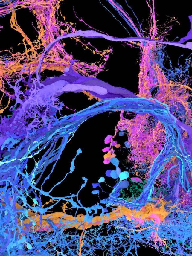
A new study reveals a missing area of brain activity in the minds of people with schizophrenia who hear voices.
The analysis of brain wave data suggests a combination of two neurological functions could trigger auditory verbal hallucinations.
Researchers from China found evidence of a breakdown in the ability to prepare the senses for specific words to be spoken. This on its own, however, isn’t enough; another area that filters our brain’s internal chatter is also enhanced in those with schizophrenia who experience unsettling auditory hallucinations.
Without the self-sound suppression along with the enhanced internal noise-associated signals, things can clearly get rather jumbled up in our minds.
“People who suffer from auditory hallucinations can ‘hear’ sounds without external stimuli,” the team explains. “Impaired functional connections between motor and auditory systems in the brain mediate the loss of ability to distinguish fancy from reality.”
Neuroscientist Fuyin Yang from Shanghai Jiao Tong University School of Medicine and colleagues examined the brains of 20 patients diagnosed with schizophrenia who experienced auditory hallucinations, and compared them to another 20 patients who were also diagnosed with schizophrenia and had not experienced hallucinations.
These patients were all taking antipsychotic medications and were in a stable condition throughout the experimental period. Earlier results from a group of people not diagnosed with schizophrenia were used as a control.
Comparing the brain activity data from electroencephalograms across the three groups of patients who were asked to hear and then speak a short, pregenerated syllable revealed the stark differences.
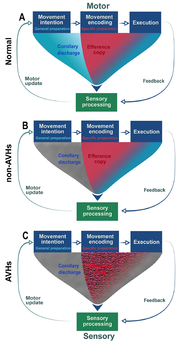
Both groups of schizophrenia patients showed comparatively reduced activity associated with our brain’s ability to predict the sound of our voice prior to our body uttering a single word. Known as a corollary discharge, this function typically gives our senses a chance to anticipate sounds as self-produced, and not treat them external.
But only the patients who reported hearing voices also had a hyperactive efference copy – the motor signal that instructs our bodies to speak, which the team describes as an internal auditory representation.
How the different groups of patients’ brains experience motor to sensory signals, with gray representing the absence of a signal. (Yang et al., PLOS Biology, 2024)
In the healthy controls and schizophrenia patients who don’t have auditory hallucinations, this signal is only enhanced around the syllable someone is prepared to say. But for those that do hear voices, the enhancement is more generalized, essentially increasing random internal brain chatter.
“Imprecise activation function of efference copy… results in the varied enhancement and sensitization of auditory cortex,” the researchers write in their paper.
So it appears that auditory hallucinations arise when the uninhibited corollary discharge misinterprets the neural activity caused by the failure of our brains to specify our internal signal to speak, Yang and team explain.
This leaves some people struggling to distinguish between external voices and their own thoughts, blurring the line between their internal and external realities.
With this new understanding of the mechanisms behind these auditory hallucinations, we can hopefully develop better treatments.
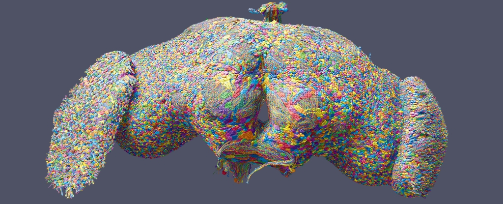
It’s an accomplishment that scientists have dreamed about for more than half a century. At last, a massive international team of researchers has mapped in exquisite detail every neuron in the brain of an adult animal with eyes and legs.
The nifty noggin, which anyone can now freely peruse, is no larger than a poppy seed, and yet it contains 139,255 neurons and 50 million connections.
This might seem like a small victory compared to a human brain, which contains 80 billion neurons and 100 trillion connections, but it’s a huge leap from where neuroscientists started in the 1960s.
If history is anything to go by, the research could be in the running for a future Nobel prize.
“This is a major achievement,” says neuroscientist Mala Murthy from Princeton University, who helped lead the team.
“There is no other full brain connectome for an adult animal of this complexity.”
Emory University biologist Anita Devineni, who was not involved in the current research, predicts this new map will “transform Drosophila neuroscience” in an accompanying commentary in Nature.
Scientists first started mapping the wiring of animal brains roughly half a century ago, and these initial attempts were focused on a very simple creature: the sightless and legless worm, Caenorhabditis elegans.
In 2002, the scientists behind this partial brain map of just 302 neurons shared a Nobel Prize.
In 2023, researchers completed a much larger brain map, including all 3,016 neurons in the larva of a fruit fly.
Once again, however, this animal could not walk or see.
An adult fruit fly is the next big step. Its brain networks, which underlie sight and the complicated movements of walking, are similar to those in humans, and they can now be explored in greater detail than ever before.
“Flies can do all kinds of complicated things like walk, fly, navigate, and the males sing to the females,” explains molecular biologist Gregory Jefferis from the University of Cambridge, who helped lead the research.
“Brain wiring diagrams are a first step towards understanding everything we’re interested in – how we control our movement, answer the telephone, or recognize a friend.”
The team that put this diagram together included researchers from 122 institutions, with major contributions from Princeton University, the University of Cambridge, and the University of Vermont.
The map required more than 7,000 slices of a female fruit fly brain and 21 million images, which could have filled 100 typical laptops with their data.
An artificial intelligence program, built at Princeton University, was essential in combing through the data.
The results are now compiled in no less than 9 related papers, with overlapping groups of authors.
One paper annotates the entire map. To correct for computer errors, a crowdsourcing project was built where researchers and volunteers globally could proofread the names and functions given to neurons. It would have taken one person 33 years working full time to make all 3 million or so manual edits.
Of all 8,453 known and named cell types in the diagram, covering 96 percent of all neurons, more than 4,500 are new to science.
“Just like you wouldn’t want to drive to a new place without Google Maps, you don’t want to explore the brain without a map,” explains neuroscientist Sven Dorkenwald, a 2023 graduate of Princeton.
“What we have done is build an atlas of the brain, and added annotations for all the businesses, the buildings, the street names. With this, researchers are now equipped to thoughtfully navigate the brain, as we try to understand it.”
In one accompanying paper, for instance, scientists used the annotated map to trigger neurons in a fruit fly’s computer brain, causing it to extend its proboscis for sugar, just like occurs in real flies when those neurons are activated.
Today, 6 different Nobel Prizes have been given to researchers studying the common fruit fly. A 7th could be on the way.
Early screening for neurodevelopmental disorders such as autism is important to ensure children have the support they need to gain the essential skills for daily life.
The American Academy of Pediatrics recommends that all children be screened for developmental delays, with additional screening for those who are preterm or have a low birth weight.
However, the US Preventive Services Task Force has called for more research into the effectiveness of current autism screening practices.
Primarily based on milestone checklists and symptoms, autism diagnoses also currently rely on observations of behavior that often manifests after crucial developmental stages have passed.
Researchers and clinicians are working to develop simple, reliable tools that could identify early signs or risk factors of a condition before symptoms are obvious.
While early screening can lead to the risk of overdiagnosis, understanding a child’s developmental needs can help guide families toward resources that address those needs sooner.
We areresearchers whostudy the role the microbiome plays in a variety of conditions, such as mental illness, autoimmunity, obesity, preterm birth and others.
In our recently published research on Swedish children, we found that microbes and the metabolites they produce in the guts of infants – both found in poop and cord blood – could help screen for a child’s risk of neurodevelopmental conditions such as autism.
And these differences can be detected as early as birth or within the first year of life. These markers were evident, on average, over a decade before the children were diagnosed.
Microbes as biomarkers
Biomarkers are biological indicators – such as genes, proteins or metabolites in blood, stool or other types of samples – that signal the presence of a condition at a certain point in time.
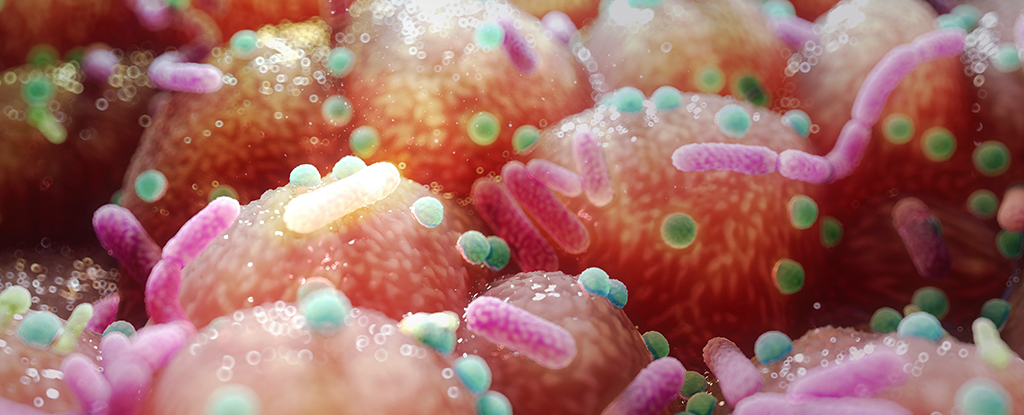
One potential biomarker for neurodevelopmental conditions such as autism are gut microbes. The connection between the gut and brain, or the gut-brain axis, is an area of considerable interest among scientists. Gut microbes play significant roles in health, including in immunity, neurotransmitter balance, digestive health and much more.
Gastrointestinal symptoms such as diarrhea, pain and constipation are common in children with autism and ADHD, with as many as 30% to 70% of autism patients also diagnosed with functional gastrointestinal disorders.
A small pilot study found that children with autism showed improvements in gastrointestinal and autism-related symptoms after having healthy microbes transferred into their guts, with some benefits lasting up to two years.
Your gut and your brain are intricately connected.
Most studies on the microbiome and neurodevelopmental conditions, however, are restricted to people who are already diagnosed with ADHD, autism or other conditions, and these studies often show mixed results.
These limitations raise an important question: Does the microbiome play a direct role in the development of autism and other neurodevelopmental conditions, or are changes in microbiome composition a consequence of the conditions themselves?
However, these studies have a notable limitation: They don’t examine microbial imbalances prior to diagnosis or symptom onset. Instead, these studies focus on children already diagnosed with autism, comparing them to their siblings and unrelated neurotypical children.
In most cases, dietary data and samples are collected several years after diagnosis, meaning the study cannot test for whether microbial imbalances cause autism.
Microbes matter
We wondered whether studying the bacteria residing in small children before they are diagnosed or show symptoms of autism or other conditions could give us a clue into their neurodevelopment.
So, we examined the cord blood and stool collected at approximately 1 year of age from participants of an ongoing study called All Babies in Southeast Sweden, which follows the health of approximately 17,000 children born between 1997 and 1999 and their parents.
We have followed these children since birth, nearly 1,200 of whom were later diagnosed with a neurodevelopmental disorder by age 23.
We found significant differences in bacterial composition and metabolite levels that developed before symptoms of neurodevelopmental conditions – such as gastrointestinal upset, crankiness and sleep problems – as well as formal medical diagnoses. These differences spanned many conditions, including autism, ADHD and speech disorders.
Next, we linked bacteria to neurotransmitters – chemical signals that help brain cells communicate – and vitamins such as riboflavin and vitamin B in the child’s stool.
Given previous research on children and adults already diagnosed with a neurodevelopmental disorder, we expected to find differences in the microbiome composition and health between those with and without neurodevelopmental conditions.
But we were surprised to discover just how early these differences emerge. We saw variability in the microbes and metabolites that affect immune and brain health, among others, in the stool collected from the diapers of children around 1 year of age and in umbilical cord blood collected at birth.
The imbalance in microbial composition – what microbiologists call dysbiosis – we observed suggests that incomplete recovery from repeated antibiotic use may greatly affect children during this vulnerable period. Similarly, we saw that repeated ear infections were linked to a twofold increased likelihood of developing autism.
Children who both repeatedly used antibiotics and had microbial imbalances were significantly more likely to develop autism.
More specifically, children with an absence of Coprococcus comes, a bacterium linked to mental health and quality of life, and increased prevalence of Citrobacter, a bacterium known for antimicrobial resistance, along with repeated antibiotic use were two to four times more likely to develop a neurodevelopmental disorder.
Parents should use antibiotics if they are prescribed and deemed necessary by their pediatrician. Rather, our study suggests that repeated antibiotic use during early childhood may signal underlying immune dysfunction or disrupted brain development, which can be influenced by the gut microbiome.
In any case, it is important to consider whether children could benefit from treatments to restore their gut microbes after taking antibiotics, an area we are actively studying.
Another microbial imbalance in children who later were diagnosed with neurodevelopmental disorders was a decrease in Akkermansia muciniphila, a bacterium that reinforces the lining of the gut and is linked to neurotransmitters important to neurological health.
Even after we accounted for factors that could influence gut microbe composition, such as how the baby was delivered and breastfeeding, the relationship between imbalanced bacteria and future diagnosis persisted.
And these imbalances preceded diagnosis of autism, ADHD or intellectual disability by 13 to 14 years on average, refuting the assumption that gut microbe imbalances arise from diet.
We found that lipids and bile acids were depleted in the cord blood of newborns with future autism. These compounds provide nutrients for beneficial bacteria, help maintain immune balance and influence neurotransmitter systems and signaling pathways in the brain.
Microbiome screening at well-child visits
Microbiome screening is not a common practice in well-child visits. But our findings suggest that detecting imbalances in beneficial and harmful bacteria, especially during critical periods of early childhood development, can provide essential insights for clinicians and families.
There is a long way to go before such screening becomes a standard part of pediatric care. Researchers still need validated methods to analyze and interpret microbiome data in the clinic.
It’s also unclear how bacterial differences change across time in children around the world – not just which bacteria are present or absent, but also how they may be shaping immune responses and metabolism.
But our findings reaffirm the growing body of evidence that the early gut microbiome plays a key role in shaping neurodevelopment.