Tag: Antibodies

  • Research highlights the need for continued surveillance of emerging SARS-CoV-2 variants and vaccines

    Research highlights the need for continued surveillance of emerging SARS-CoV-2 variants and vaccines

    [ad_1]

    Researchers at the Francis Crick Institute and the National Institute for Health and Care Research Biomedical Research Centre at UCLH have highlighted the importance of continued surveillance of emerging SARS-CoV-2 variants and vaccine performance as the virus continues to evolve.

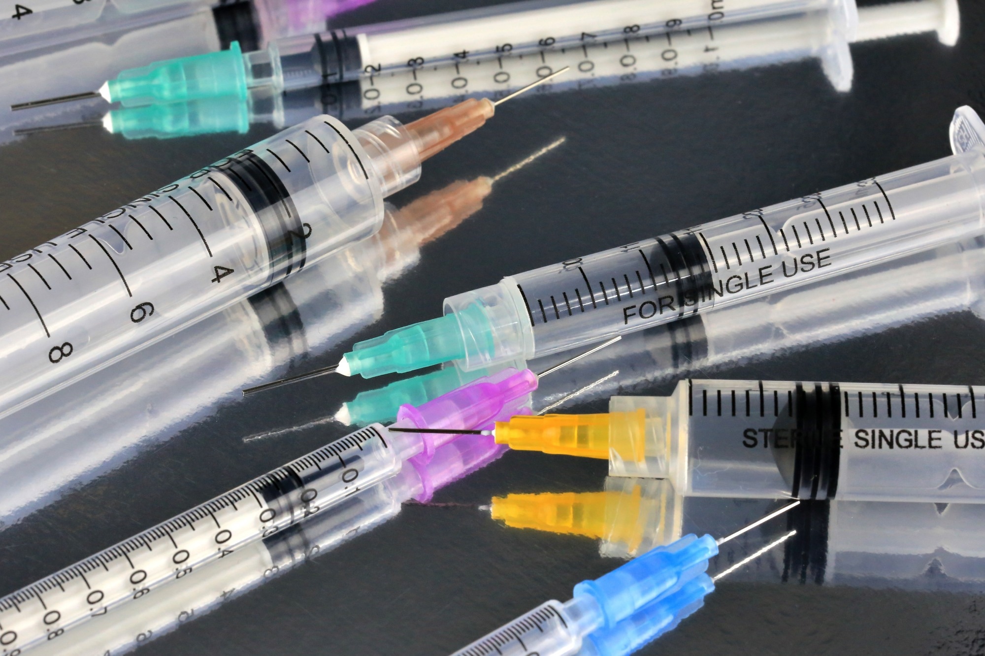
    Published today as a research letter in The Lancet, their study compared the newer monovalent COVID vaccine, which specifically targets the XBB variant of Omicron (as recommended by the World Health Organisation), with older bivalent vaccines containing a mix of an Omicron variant and the original strain of COVID-19, which the UK deployed in Autumn 2023 before turning to monovalent vaccines1

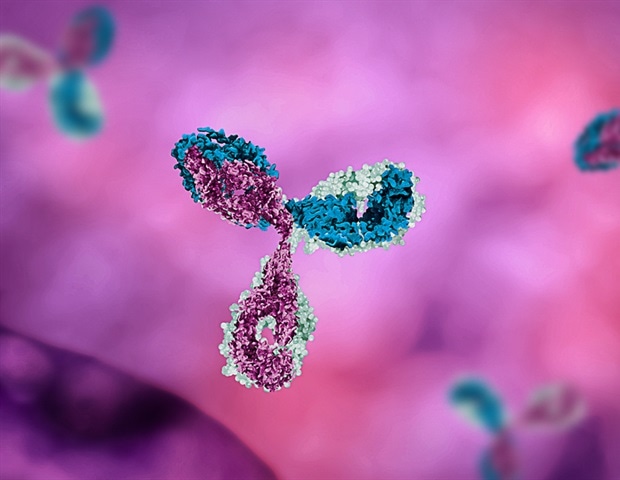
    The researchers found that both vaccines generated neutralizing antibodies against the most recent strain of Omicron, BA.2.86. However, the new monovalent vaccine generated higher levels of antibodies against a range of other Omicron variants.

    The team collected blood and nasal mucosal samples both before and after a fifth dose vaccination from 71 participants of the Legacy study, a research collaboration between the Crick and the NIHR University College London Hospitals Biomedical Research Centre. They compared the antibody levels before and after vaccination.

    All 36 participants who received the bivalent vaccine and 17 who received the monovalent vaccine had boosted levels of antibodies against all variants tested, including the newest strain BA.2.86, which caused a wave of infection this winter. But those with the newer monovalent vaccine had 3.5x higher levels of antibodies against the XBB and BQ.1.1 strains after their booster vaccination.

    Since the Omicron virus is highly transmissible and the virus replicates in the nose and throat, the researchers tested the levels of antibodies in the participants’ nasal cavity.

    They found that the monovalent vaccine increased their ability to produce mucosal antibodies against most of the tested variants, whereas the bivalent vaccine didn’t provide a significant boost.

    Neither vaccine increased neutralizing antibody levels in the nasal cavity against the newest variant, BA.2.86, suggesting that current vaccines may be less likely to stop transmission or prevent asymptomatic or mild illness, while still protecting against severe disease.

    This highlights the importance of careful vaccine updates and continuing to complement a vaccination program with the development of antibody drugs that work against all variants, as some more vulnerable people don’t respond well to vaccines.

    The UK’s strategy to deploy stocks of older vaccines paid off last year, as both vaccines provided equal protection against the newest strain. However, ongoing monitoring is needed, as the virus is continuing to evolve, so vaccine-induced antibodies might not work so well in the future. In the long run, vaccines that are effective against all new variants and can block COVID-19 being transmitted from person to person are needed.”


    Emma Wall, Senior Clinical Research Fellow at the Crick and Consultant in Infectious Diseases at UCLH

    David LV Bauer, Group Leader of the RNA Virus Replication Laboratory at the Crick, said: “The situation this winter could have been different if the newly emerged BA.2.86 and JN.1 variants were substantially distinct from older Omicron variants, but fortunately this wasn’t the case.

    “Most new variants arise quicker than most clinical trials can produce data. But laboratory analysis can provide a detailed picture very quickly. Continued surveillance will help us stay on top of viral evolution.”

    Source:

    Journal reference:

    Shawe-Taylor, M., et al. (2024) Divergent performance of vaccines in the UK autumn 2023 COVID-19 booster campaign. The Lancet. doi.org/10.1016/S0140-6736(24)00316-7.

    [ad_2]

    Source link

  • LAG-3 protein structure may be the key to unlocking new cancer treatments

    LAG-3 protein structure may be the key to unlocking new cancer treatments

    [ad_1]

    A molecular “snapshot” of a protein can be critical to understanding its function. Scientists at Stanford and NYU have published and investigated a new structure of the protein LAG-3 which could enable the development of new cancer treatments.

    Some cancerous tumors hijack proteins that act as “brakes” on our immune system and use them to form a sort of shield against immune recognition. Immunotherapy treatments have been created that turn off these “brakes” and allow our body to attack foreign-looking cancer cells. To further advance such treatments, researchers at Stanford University and New York University have published a new structure of one of these brake proteins, LAG-3. Their work contains key details of the molecule’s structure, as well as information about how the LAG-3 protein functions.

    Although over a dozen immunotherapies targeting LAG-3 are in development, and one is already FDA approved, knowledge of LAG-3’s structure and function has been incomplete.

    “Given the amount of time and resources being put into developing therapeutics that target LAG-3, it is astounding that we don’t yet have a full understanding of how this protein functions,” said Jennifer Cochran, the Addie and Al Macovski Professor in the School of Engineering and professor of bioengineering, and co-senior author on the study detailing LAG-3, published in Proceedings of the National Academy of Sciences.

    Getting a clear image of a protein might not seem like a big deal, but when it comes to proteins, form often begets function. If you know what a protein looks like at the atomic scale, you can begin to understand how it interacts with other molecules and design experiments to further deduce how it works. Studies like these are crucial to developing drugs that can optimally block their target’s function.

    A key structure

    Proteins like LAG-3, called immune checkpoints, exist to stop our immune system from attacking things they shouldn’t. In theory, our immune system should naturally recognize tumor cells as foreign. But a checkpoint protein shield can give cancer cover.

    Current immunotherapies aren’t chemical drugs, they’re lab-manufactured antibodies that attach to certain parts of these checkpoints, and essentially turn them off. Once the checkpoint is turned off, our immune system can recognize and target the cancer again.

    There are already approved antibody treatments that target two checkpoint proteins: CTLA-4 and PD-1. Both turn off our immune systems but in different ways. Because CTLA-4 and PD-1 were the first two checkpoint proteins found, they are quite well studied, and different approaches to inhibiting them for cancer therapy earned scientists the 2018 Nobel Prize in physiology or medicine.

    LAG-3 seems to work in an entirely different way. Scientists hope that those differences might make it a better or complementary target to treat certain types of cancer, said Jack Silberstein, the Stanford immunology PhD student who co-led the work.

    Because of that, Silberstein said, “there was all this excitement in the field. Groups rushed to make antibodies against LAG-3, without knowing entirely how LAG-3 or those antibodies functioned.”

    Silberstein and colleagues, including those in Stanford’s ChEM-H Macromolecular Structure Knowledge Center and the SLAC National Accelerator Laboratory, began working on LAG-3’s structure in 2019. A structure of LAG-3 was published by a different group in 2022 providing an initial glimpse of the protein, but it lacked crucial detail around sugar molecules that are key to LAG-3’s function, and detailed information on how the LAG-3 structure related to its biological activity.

    A painstaking process

    When Silberstein first started this project, “I quickly realized why there was no published structure. It’s a tremendously difficult protein to work with.”

    And the technique Silberstein used to get the structure, called X-ray crystallography, is extremely finicky. First, Silberstein had to grow a crystal made entirely out of LAG-3 protein. Then, in collaboration with Irimpan Mathews at the SLAC National Accelerator Laboratory, they fired X-ray beams at the crystal to create a 3D image of the molecule.

    LAG-3 is a spindly, flexible protein, so it’s difficult to get the molecules to stack in an orderly way. Silberstein estimates he made more than 10,000 crystals, of which 3,000 were fired with X-rays before the team finally solved the structure.

    It was a very intense, grind-it-out-for-three-years, bang-your-head-against-the-wall kind of thing.”


    Jack Silberstein, Stanford immunology PhD student

    But it paid off. The team’s structure confirmed that LAG-3 exists as a dimer, with two LAG-3 molecules coming together to form the functional checkpoint protein. The sugar residue that was elusive in previous structural efforts is a key element in the LAG-3 dimer interface and helps promote a different orientation of the LAG-3 protein.

    With the structure described, colleagues at New York University, including MD, PhD student Jasper Du and pathology Assistant Professor Jun Wang co-led critical experiments further elucidating LAG-3’s function. Other NYU colleagues, including Kun-Wei Chan and Xiang-Peng Kong, helped conduct electron microscopy studies to detail disruption of dimer formation by LAG-3 antibodies.

    Additional work by the team uncovered, for the first time, that an antibody that has been used for close to 20 years to demonstrate therapeutic efficacy in animal tumor models blocks the activity of LAG-3 by binding to the interface between two LAG-3 molecules, disrupting LAG-3 from forming its functional dimer. Intriguingly, LAG-3 antibodies in clinical development bind to other areas of the protein, away from this dimer interface.

    There will never be just one “cure,” because cancers are all different and involve a number of diverse biochemical pathways. Silberstein and Cochran envision a future where a tapestry of surgical, chemical, and immunological treatment approaches are employed, driven by basic science discoveries and medical innovations. Additional treatments targeting LAG-3 may very well be a part of that picture.

    [ad_2]

    Source link

  • Pregnant women with autoimmune conditions at a greater risk of developing adverse pregnancy outcomes, study suggests

    Pregnant women with autoimmune conditions at a greater risk of developing adverse pregnancy outcomes, study suggests

    [ad_1]

    In a recent review published in BMC Medicine, researchers analyzed systematic reviews conducted on the association between autoimmune diseases and pregnancy outcomes.

    Study: Autoimmune diseases and adverse pregnancy outcomes: an umbrella review. Image Credit: Africa Studio/Shutterstock.com
    Study: Autoimmune diseases and adverse pregnancy outcomes: an umbrella review. Image Credit: Africa Studio/Shutterstock.com

    Background

    Autoimmune diseases, particularly in women, have been associated with poor pregnancy outcomes as a result of environmental variables such as lifestyle changes, dietary changes, and exposure to certain infections and medicines. The unfavorable pregnancy outcomes associated with certain autoimmune disorders might improve, worsen, or remain constant during pregnancy.

    Autoimmune diseases can complicate pregnancy by enabling antibodies generated by the mother to infiltrate the fetal system, affecting the development of the fetal heart. Clinical management of autoimmune pregnancies necessitates multidisciplinary care and an appreciation of the risk of adverse pregnancy outcomes.

    About the review

    In the present review, researchers analyzed the impact of autoimmune disease prevalence on pregnancy outcomes, using systematic reviews to identify the strength and precision of these associations.

    The team searched the Cochrane Medline and Embase databases from inception through December 15, 2023, without language restrictions for systematic reviews evaluating the association between autoimmune disorders and pregnancy outcomes. They excluded systematic reviews, including those involving women conceiving through assisted reproduction therapy and those evaluating the relationship between drugs for autoimmune diseases and pregnancy outcomes. They also excluded literature reviews, scoping reviews, conference abstracts, and protocols.

    The researchers used the Joanna Briggs Institute (JBI) framework, following the Preferred Reporting Items for Overviews of Reviews (PRIOR) checklist. Two researchers independently performed data screening and extraction and appraised the identified records using the Assessment of Multiple Systematic Reviews Version 2 (AMSTAR 2) tool, consulting a third researcher to resolve discrepancies.

    The team evaluated systematic review quality using the Newcastle-Ottawa scale (NOS). They synthesized data quantitatively to estimate relative risks (RRs) and odds ratios (ORs) and performed random effect modeling for meta-analysis to obtain pooled effect estimates.

    Autoimmune disorders included celiac disease, inflammatory bowel disease (IBD), including ulcerative colitis and Crohn’s disease, psoriatic disorders (psoriasis and psoriatic arthritis), Sjögren’s syndrome, rheumatoid arthritis, systemic sclerosis, systemic lupus erythematosus (SLE), thyroid autoimmunity (Hashimoto’s thyroiditis and Grave’s disease), vitiligo, and type 1 diabetes mellitus (T1DM).

    Results

    Initially, the team identified 2,743 records, of which 2,351 underwent title-abstract screening and 92 underwent full-text screening after duplicate removal. As a result, they analyzed 32 records, including 709 primary studies, most of which were of moderate-high quality. They found a significant ectopic pregnancy risk among IBD patients (OR, 1.3), with similar risks for ulcerative colitis and Crohn’s disease.

    The team found increased miscarriage risk among females with systemic lupus erythematosus (OR, 4.9) and Sjögren’s syndrome (RR, 8.9), with the risk being higher (OR, 2.8) in the case of thyroid autoimmune conditions. Miscarriage risk was also significantly higher among women with celiac disease, rheumatoid arthritis, systemic sclerosis, and psoriasis, with OR values of 1.4, 1.3, 1.6, and 1.1, respectively. Female celiac disease patients had a significantly higher risk of recurrent gestational losses (OR, 5.8), exacerbated in thyroid autoimmunity presence (OR, 1.9).

    Gestational hypertension odds were higher among females with T1DM, psoriasis, and psoriatic arthritis, with OR values of 2.7, 1.3, and 1.5, respectively, enhanced by thyroid autoimmunity (OR, 1.3). The team found higher pre-eclampsia prevalence among females with type 1 diabetes mellitus (OR, 4.2), systemic lupus erythematosus (OR, 3.2), and systemic sclerosis or scleroderma (OR, 2.2). Women with IBD were at an increased risk of gestational diabetes (OR, 3.0). Cesarean section delivery was associated with T1DM (OR, 4.0) and SLE (OR, 2.1). Women with thyroid autoimmune disorders had higher odds of postpartum depression (OR, 2.0).

    Women with systemic sclerosis and celiac disease were at a higher risk of intrauterine growth restriction (IUGR), with OR values of 3.2 and 1.7, respectively. The OR values for small for gestational age (SGA) babies were 2.5 for SLE, 1.5 for rheumatoid arthritis, and 0.7 for T1DM patients. The OR values for stillbirth among women with SLE, T1DM, rheumatoid arthritis, celiac disease, and IBD were 17, 4.0, 2.0, 2.0, and 1.6, respectively.

    The team noted a higher risk for preterm birth among women with T1DM (OR, 4.4), systemic lupus erythematosus (OR, 2.8), systemic sclerosis (OR, 2.4), Sjögren’s syndrome (RR, 2.3), inflammatory bowel disease (OR, 1.8), rheumatoid arthritis (OR, 1.6), psoriatic arthritis (OR, 1.5), celiac disease (OR, 1.3), and psoriasis (OR, 1.2). They reported low-birth-weight babies among women with SLE (OR, 6.0) and systemic sclerosis (OR, 3.8). Neonatal mortality was associated with SLE (OR, 8.3), T1DM (OR, 2.3), and Sjögren’s syndrome (OR, 1.8).

    Conclusion

    Overall, the review findings showed that women with autoimmune disorders are at a high risk of unfavorable pregnancy outcomes. However, further research is required to develop more evidence-based, standardized recommendations and assist physicians and women in making educated decisions about treating these diseases before and throughout pregnancy.

    [ad_2]

    Source link

  • Is there a higher risk of depression among specific populations of patients with rheumatoid arthritis?

    Is there a higher risk of depression among specific populations of patients with rheumatoid arthritis?

    [ad_1]

    In a recent study published in JAMA Network Open, researchers assessed the risk of depression following the diagnosis of rheumatoid arthritis (RA).

    Study: Rheumatoid Arthritis and Risk of Depression in South Korea. Image Credit: Africa Studio/Shutterstock.com
    Study: Rheumatoid Arthritis and Risk of Depression in South Korea. Image Credit: Africa Studio/Shutterstock.com

    RA, a prevalent autoimmune disease, is characterized by systemic inflammation. The chronic nature of the disease necessitates lifelong treatment, often leading to comorbidities, including depression. Depression is highly prevalent among RA patients compared to the general population and has been associated with increased disease activity, pain exacerbation, elevated risk of myocardial infarction, less frequent remission, poor health-related quality of life, and higher health care utilization. Therefore, depression management and prevention are essential to improving the health and quality of life of RA patients.

    About the study

    In the present study, researchers examined associations of RA with subsequent depression risk in South Korea. They included subjects diagnosed with RA during 2010-17. Seropositive RA (SPRA) was defined using the International Classification of Diseases, Tenth Revision (ICD-10) codes and enrolment in the Rare and Intractable Diseases (RID) program.

    RID program enrollment for SPRA required a positive test result for anticyclic citrullinated peptide antibodies or rheumatoid factors and a physician report indicating the fulfillment of RA classification criteria. Seronegative RA (SNRA) was defined using ICD-10 codes and prescription of disease-modifying anti-rheumatic drugs (DMARDs) for ≥ 270 days.

    The team excluded subjects if they were aged under 20, had prior depression, missing data, or developed depression within a year post-index date. RA patients were matched to individuals without RA (controls) by sex, age, and index date. The study endpoint was a new diagnosis of depression. Participants were followed up from one year post-RA diagnosis until the diagnosis of depression, death, or December 31, 2019.

    The Kaplan-Meier method was used to estimate the cumulative incidence of depression. Differences between groups were evaluated using log-rank tests. Cox regression analyses estimated adjusted hazard ratios and 95% confidence intervals for the risk of depression. The association between depression risk and the type of DMARDs used was also evaluated.

    Analyses were adjusted for sex, age, smoking/alcohol status, income, body mass index, physical activity, diabetes, chronic kidney disease, hyperlipidemia, and hypertension. Besides, stratified analyses were conducted by sex, age, comorbidities, and health behaviors. Restricted mean survival time (RMST) differences were analyzed between groups by sex and age.

    Findings

    Overall, 230,922 participants aged 54.6, on average, were included for analysis. There were 38,487 RA patients and 192,435 controls; most participants were female (71%). Among RA patients, 11,645 were seronegative, and 26,842 were seropositive. RA patients were more likely to be non-drinkers and less likely to be obese. SPRA patients were more likely to be female, older, non-drinkers, and less likely to be obese than SNRA patients.

    The median follow-up duration was 4.1 years, during which 6,422 RA patients and 20,641 had newly developed depression. RA patients had a higher risk of depression than controls. Moreover, SPRA and SNRA groups had elevated depression risk compared to controls. Among RA patients with depression, 402 were prescribed biological or targeted synthetic DMARDs, and 6,020 were prescribed only conventional synthetic DMARDs.

    Notably, the incidence of depression among RA patients was consistently lower among recipients of targeted synthetic or biological DMARDs than non-recipients. Stratified analyses yielded findings consistent with the primary analysis. RMST differences were variable across age groups, with a higher difference in the ≥ 60-year age group.

    Conclusions

    In sum, the researchers observed a 1.66-fold increased depression risk among RA patients relative to those without RA. There was no significant difference in depression risk by the serologic status of RA, with both SNRA and SPRA groups exhibiting an increased risk. RA patients receiving targeted synthetic or biological DMARDs had lower risks than those who did not. Nevertheless, the study has a few limitations. Notably, disease activity was inaccessible, resulting in limited evaluation of RA severity.

    Moreover, information on depression levels at the index date was not available. Besides, because participation was limited to those undergoing health screening, participants might have been healthier or engaged more in healthy behaviors than the general population.

    Taken together, the findings suggest an increased depression risk among RA patients, irrespective of RA serologic status, age, sex, and behavioral factors, warranting consistent screening of RA patients and comprehensive healthcare to address their physical and mental well-being.

    [ad_2]

    Source link

  • Tryptophan digestion by gut microbiome spurs arthritis inflammation

    Tryptophan digestion by gut microbiome spurs arthritis inflammation

    [ad_1]

    A University of Colorado Department of Medicine faculty member says she and her colleagues have identified the means in which bacteria in the digestive system can break down tryptophan in the diet into an inflammatory chemical that primes the immune system towards arthritis.

    The research was co-authored by Kristine Kuhn, MD, PhD, Scoville Endowed Chair and head of the CU Division of Rheumatology. Several of her division colleagues collaborated on the paper, which was published in February in the Journal of Clinical Investigation.

    Tryptophan is an essential amino acid found in many protein-rich foods, including meats, fish, dairy products, and certain seeds and nuts. It has many uses in the body, including helping in the production of proteins, muscles, enzymes, and neurotransmitters – the nervous system’s chemical messengers. The body doesn’t make it; we get it from our diet.

    Many people think of tryptophan as the ingredient in turkey that supposedly makes us sleepy after a Thanksgiving feast. In fact, researchers say that although tryptophan plays a role in helping to regulate the sleep cycle, the amount that’s in turkey probably isn’t a significant cause of post-dinner drowsiness.

    Cause and effect

    Kuhn and her associates set out to learn how a substance that often is a force for good in the body is converted into a pathway to inflammatory diseases such as rheumatoid arthritis, which affects about 1% of the population. It can cause painful swelling of the hands and feet, and joint deformities if left untreated.

    It’s been known that the microbiome – the bacteria in our gut – can break down tryptophan into byproducts. Some of those byproducts are anti-inflammatory, but we’ve also associated some inflammatory causes of those products. We’re the first to highlight which products are contributing to inflammation, and how they are doing that.”


    Kristine Kuhn, MD, PhD, Scoville Endowed Chair and Head of the CU Division of Rheumatology

    She says the new research “builds upon some observations we had in patients with spondyloarthritis – not quite rheumatoid arthritis, but a closely related condition – where we found that changes in the microbiome were associated with increased production of these products called indoles, which are what bacteria make from tryptophan.” Similar changes were observed in arthritis studies involving mice, she says.

    “We put mice on antibiotics to wipe out their microbiome, and they didn’t get arthritis, and they didn’t have indole,” she says. “So we said, OK, what if they do have a microbiome and we put them on a diet with little tryptophan? The microbiome can’t break down tryptophan into indole, and the mice didn’t get arthritis. So two different ways, we showed that it’s tryptophan that’s broken down by the microbiome into indole.”

    Inflammatory flags

    So how does that work? “We found that when indole is present, the mice start to develop autoreactive T-cells that are more inflammatory. They have less of those regulatory T-cells that help maintain balance in the immune system, and they start to develop antibodies that are more pathogenic. We found that the antibodies had flags for being more inflammatory when indole was present.”

    The paper concludes that “blockade of indole generation may present a unique therapeutic pathway” for rheumatoid arthritis and spondyloarthritis. That’s all about finding the right path for the body’s tryptophan, Kuhn says.

    “If tryptophan hits our body’s cells, it tends to go get broken down into anti-inflammatory products versus when it hits the bacterial cells and goes more inflammatory. The ways we think about how this could lead to therapies are: How do you keep that balance tipped so that tryptophan goes towards that anti-inflammatory pathway? How can you manipulate intestinal bacteria to tip that balance? That’s where we’re interested in going in the future.”

    Does Kuhn’s research suggest we should be eating differently? “I get asked that a lot,” she says. “A diet that’s rich in plant-based fibers and lean meats – this whole Mediterranean diet – seems to push the microbiome into a healthier state, so that you are getting the anti-inflammatory properties of tryptophan, whereas the typical western diet seems to go more toward the inflammatory pathway.”

    As for other ways to protect against arthritis, Kuhn says that through research by her Division of Rheumatology colleagues, “we have started to understand the at-risk stage, where we can actually identify people who are likely to progress to rheumatoid arthritis within the next few years based on blood markers. There’s some data that suggests we could intervene during that period and prevent disease, but we’re not quite sure yet what are the right ways to intervene.”

    Source:

    Journal reference:

    Seymour, B. J., et al. (2024). Microbiota-dependent indole production stimulates the development of collagen-induced arthritis in mice. The Journal of Clinical Investigation. doi.org/10.1172/JCI167671.

    [ad_2]

    Source link

  • Man takes 217 COVID vaccines with no ill effects, shows immune boost

    Man takes 217 COVID vaccines with no ill effects, shows immune boost

    [ad_1]

    In a recent case report published in The Lancet Infectious Diseases, researchers described a case of a 62-year-old male who received 217 vaccinations against severe acute respiratory syndrome coronavirus 2 (SARS-CoV-2) in 29 months and examined his immunological responses. They found that hyper-vaccination did not cause adverse events or significantly affect the quality of adaptive immune responses while resulting in increased T-cells and spike-specific antibodies.

    Study: Adaptive immune responses are larger and functionally preserved in a hypervaccinated individual. Image Credit: Douglas Sacha / ShutterstockStudy: Adaptive immune responses are larger and functionally preserved in a hypervaccinated individual. Image Credit: Douglas Sacha / Shutterstock

    Background

    Booster vaccinations may potentially amplify immune responses, while persistent antigen exposure may induce immune tolerance. However, the advantages, constraints, and risks of recurrent vaccination in humans remain to be thoroughly investigated. In the present study, researchers investigated the immunological responses in an older man hyper-vaccinated against SARS-CoV-2.

    The case

    In this case study, a 62-year-old male from Magdeburg, Germany (referred to as HIM), engaged in deliberate hyper-vaccination against SARS-CoV-2, receiving 217 vaccinations over 29 months for personal reasons. This occurred outside a clinical study context and contrary to national recommendations. Despite an investigation by a public prosecutor for potential fraud, no criminal charges were filed. Notably, HIM’s immunological evaluation, initiated during the public prosecutor’s investigation, received active and voluntary cooperation from HIM and was ethically approved. Throughout the extensive hyper-vaccination, HIM reported no vaccine-related side effects, and routine clinical chemistry parameters displayed no abnormalities between November 2019 and October 2023. In the repeated negative SARS-CoV-2 tests, including antigen tests, polymerase chain reaction (PCR) test, and nucleocapsid serology, HIM showed no signs of past SARS-CoV-2 infection.

    Starting from the 214th vaccination, HIM’s anti-spike SARS-CoV-2 immunoglobulin G (IgG) levels were measured before and after vaccinations. The antibody peak occurred at the 214th vaccination, and there was a slight increase after the 217th vaccination. Additionally, HIM showed IgG4 subclass switching after the 215th vaccination, which is uncommon in regimens with adenoviral-based vaccines as the first dose.

    A total of 29 individuals who received three doses of a messenger ribonucleic acid (mRNA) vaccine formed the control group. As compared to controls, HIM exhibited mildly elevated levels of anti-spike IgM and IgA in the serum. However, in saliva samples, HIM showed detectable levels of anti-spike IgG, contrary to the control participants. HIM’s serum neutralization capacity was higher (5.4-fold for wildtype and 11.5-fold for Omicron B1.1.529 spike proteins) than the controls, indicating elevated quantities of spike-specific IgG. This observed difference was not attributed to antibody avidity as it remained comparable among the groups.

    HIM showed a slightly increased number of spike-specific B-cells, with the same phenotype as seen in single-cell RNA sequencing (scRNA-seq). No significant differences were observed in the rates of somatic hypermutation or clonal expansion. CD8+ T-cells specific to the spike epitope were about six-fold more frequent in HIM, with a preference for effector memory T-cells. Further, scRNA-seq of LTD-specific T-cells showed a more differentiated phenotype and increased clonal expansion compared to controls. Flow-cytometric analysis and metabolic profiling showed no significant abnormalities in 14 protein markers.

    LTD-specific CD8+ T-cells in HIM showed a proliferative capacity similar to control individuals, aligned with conserved numbers of T-cells with a phenotype like early differentiated stem cells. After epitope-specific stimulation, HIM displayed higher cytokine-positive cells, but the cytokine release per cell remained roughly equal. Cytokine analysis in the supernatant revealed the typical pattern of virus-specific CD8+ T-cells. Additionally, HIM’s CD8+ T-cells showed higher peptide sensitivity than the control group. Examination of spike-reactive CD4+ T-cells revealed a dearth of nucleocapsid-specific immunity, with similar cytokine-producing CD4+ T-cell amounts in HIM compared to the control group while retaining peptide sensitivity.

    Conclusion

    In conclusion, the present case report showed that hyper-vaccination against SARS-CoV-2 yielded no adverse events and elevated T-cell levels and spike-specific antibodies. Notably, the implicit quality of adaptive immune responses showed no significant effects. Although breakthrough SARS-CoV-2 infections were not observed in the individual, any causal link with the hyper-vaccination regimen remains unclear. The researchers emphasize that they do not advocate for hyper-vaccination as an approach to improve adaptive immunity.

    [ad_2]

    Source link

  • COVID booster in lactating mothers can provide antibodies for infants

    COVID booster in lactating mothers can provide antibodies for infants

    [ad_1]

    Lactating mothers who get the COVID-19 booster pass along the antibodies to their children via their breast milk – and potentially protect babies too young to receive the vaccine, a study from the University of Florida Institute of Food and Agricultural Sciences (UF/IFAS) and the UF College of Medicine found.

    The study is the third in a series that looks at antibody protection being transferred via breast milk from mothers who received their first two COVID-19 vaccinations and, now, the booster shot. The second publication reported the same antibody transfer via breast milk.

    “We think that breast milk may play an important role in protecting the infants during the first six months of life from COVID,” said Dr. Vivian Valcarce, a former UF College of Medicine researcher who worked on this study. She now is an assistant professor at the University of Alabama at Birmingham. “We continue to see babies being hospitalized from COVID-19 infections.”

    The study was published in February in Frontiers in Nutrition, and the study was funded by the Gerber Foundation and the Children’s Miracle Network.

    The study looked at how breast milk antibody protection changed when a mother received their first COVID-19 booster shot, said Joseph Larkin, UF/IFAS associate professor of microbiology and cell science and part of UF’s Emerging Pathogens Institute. Researchers looked at the antibody response and antibody functionality in breast milk and tested to see if antibodies were present after the babies drank breast milk with COVID-19 antibodies.

    Larkin said this study suggests that breastfeeding can provide COVID-19 antibodies for infants too young to receive a vaccination – and that the antibodies wane in people’s bodies over time, so getting a booster can provide prolonged protection to babies that drink breast milk.

    When babies are born, they have an immature immune system, so they rely heavily on mom’s immune system. Breastfeeding can serve as a gap in between while babies are building their own immune system.”


    Joseph Larkin, UF/IFAS associate professor of microbiology and cell science 

    Larkin said some antibodies are transferred to fetuses through the placenta, as well, but that initial protection also lessens over time.

    In this study, 14 lactating mothers and their babies were followed from before they received their COVID-19 booster until after they received their booster shots, Larkin said. Researchers tested the mothers’ blood to confirm their bodies made COVID-19 antibodies after a booster shot, tested breast milk to confirm the milk had antibodies in it and tested babies’ poop to confirm antibodies were present in the babies’ bodies.

    To see if the breast milk’s antibodies worked against COVID-19, breast milk was placed in a 96-well plate with a lab-safe COVID virus strain, and researchers found these antibodies from the mother disable the virus, said Lauren Stafford, a UF/IFAS graduate research assistant and Ph.D. candidate in microbiology and cell science.

    The study was a collaboration between UF/IFAS and the UF College of Medicine and included Dr. Josef Neu, professor of pediatrics within the division of neonatology at the UF College of Medicine.

    “This shows how important breast milk and breastfeeding is for infant health during a pandemic,” Valcarce said.

    Source:

    Journal reference:

    Valcarce, V., et al. (2024). COVID-19 booster enhances IgG mediated viral neutralization by human milk in vitro. Frontiers in Nutrition. doi.org/10.3389/fnut.2024.1289413.

    [ad_2]

    Source link

  • Study reveals safety of MS drugs during breastfeeding in child’s early years of life

    Study reveals safety of MS drugs during breastfeeding in child’s early years of life

    [ad_1]

    Certain medications for multiple sclerosis (MS) called monoclonal antibodies, taken while breastfeeding, may not affect the development of a child during the first three years of life, according to a preliminary study released today, March 4, 2024. The study will be presented at the American Academy of Neurology’s 76th Annual Meeting taking place April 13–18, 2024, in person in Denver and online. The study examined four monoclonal antibodies for MS: natalizumab, ocrelizumab, rituximab and ofatumumab.

    MS is a disease in which the body’s immune system attacks myelin, the fatty white substance that insulates and protects the nerves. Symptoms may include fatigue, numbness, tingling or difficulty walking.

    Most monoclonal antibody medications for multiple sclerosis are not currently approved for use while a mother is breastfeeding. Yet MS can develop during the childbearing years of life. Since the risk of MS relapses increases after giving birth, some mothers may need or want to restart these therapies, so it is important to determine whether these medications, through breast milk, have a negative impact on a child’s development.”


    Kerstin Hellwig, MD, study author of Ruhr University in Bochum, Germany

    For the study, researchers used the German MS and Pregnancy Registry to identify 183 infants born to mothers taking monoclonal antibodies while breastfeeding. Of this group, 180 had mothers with MS and the three had mothers with neuromyelitis optica spectrum disease (NMOSD). NMOSD is also a demyelinating disease, but it is rare and specifically affects the optic nerve, spinal cord or brain. 

    The infants were compared to another 183 infants, matched for exposure to MS medications shortly before or during pregnancy, born to mothers with the same diseases who did not take monoclonal antibodies while breastfeeding.

    Of those exposed to MS medications, 125 were exposed to natalizumab, 34 to ocrelizumab, 11 to rituximab and 10 to ofatumumab. Two infants were first exposed to natalizumab and then ocrelizumab. One infant was exposed to rituximab and then ocrelizumab.

    The first exposures to the medications through breastfeeding ranged from the day a child was born to the ninth month of life. Infants were breastfed for an average of five-and-a-half months while their mothers took these medications.

    For all infants, researchers then examined the number of hospital stays, antibiotic use, developmental delays such as problems with social and fine motor skills and delayed speech development, and the infants’ weight at follow-up visits during the first three years of life.

    After comparing infants exposed to the medications to infants not exposed, researchers found no differences in their health or development.

    “Our data show infants exposed to these medications through breastfeeding experienced no negative effects on health or development within the first three years of life,” Hellwig said.

    A limitation of the study was that only about a third of the infants were followed for the full three years. Therefore, Hellwig said, the results for the third year of life are less meaningful than for years one and two.

    [ad_2]

    Source link

  • Broad-spectrum neutralizing antibody mAb-39 targets conserved S2 epitope in SARS-CoV-2

    Broad-spectrum neutralizing antibody mAb-39 targets conserved S2 epitope in SARS-CoV-2

    [ad_1]

    The ongoing COVID-19 pandemic caused by SARS-CoV-2 has caused serious damage to public health and the global economy, and one strategy to combat COVID-19 has been the development of broadly neutralizing antibodies for prophylactic and therapeutic use. The most emergency-use authorized (EUA) therapeutic monoclonal antibodies, are more likely to lose their neutralizing activities as the viral epitopes (e.g. the receptor-binding domain, RBD) within spike protein of SARS-CoV-2 they target are more prone to mutate. By contrast, the S2 subunit of spike protein, has a much lower frequency of mutation than the RBD and exhibits a high degree of homology among human SARS-like CoVs. As such, developing antibodies targeting the more conserved S2 epitopes are able to exert an incredibly broad neutralization spectrum against SARS-like CoV viruses.

    This study is led by Dr. Suping Zhang (Shenzhen University), Dr. Zhenhong Lin (Chongqing University), and Dr Lanfeng Wang (Shanghai Institute of Immunity and Infection, Chinese Academy of Sciences). They isolated one neutralizing mAb, called mAb-39, against SARS-CoV-2 and its variants from convalescent adult. This mAb exhibited good potency in neutralization SARS-CoV2 original strain, previous and current circulating Omicron variants (e.g. Omicron BA.1, BA.2.86, BA.4, BA.5, EG.5.1) in a cell-based assay.

    This antibody binds to a highly conserved new epitope on the glycoprotein from SARS-CoV2.

    This is very exciting! The good activity and highly conserved epitope of this antibody suggested its potential for clinical translation”.


    Dr. Hang Su, Shenzhen University

    The team found the mAb-39 greatly improved the neutralizing activity of anti-RBD antibody, the same type of EUA therapeutic monoclonal antibodies, against the highly neutralization-resistant Omicron variants. The study also extended the knowledge to the neutralizing and protective epitopes of SARS-CoV-2.

    Source:

    Journal reference:

    Su, H., et al. (2024). A human monoclonal antibody neutralizes SARS-CoV-2 Omicron variants by targeting the upstream region of spike protein HR2 motif. hLife. doi.org/10.1016/j.hlife.2024.02.001.

    [ad_2]

    Source link

  • Newly identified antibodies target a hard-to-spot region of the influenza virus

    Newly identified antibodies target a hard-to-spot region of the influenza virus

    [ad_1]

    Researchers at the National Institutes of Health have identified antibodies targeting a hard-to-spot region of the influenza virus, shedding light on the relatively unexplored “dark side” of the neuraminidase (NA) protein head. The antibodies target a region of the NA protein that is common among many influenza viruses, including H3N2 subtype viruses, and could be a new target for countermeasures. The research, led by scientists at the National Institute of Allergy and Infectious Diseases’ Vaccine Research Center, part of NIH, was published today in Immunity.

    Influenza, or flu, sickens millions of people across the globe each year and can lead to severe illness and death. While vaccination against influenza reduces the burden of the disease, updated vaccines are needed each season to provide protection against the many strains and subtypes of the rapidly evolving virus. Vaccines that provide protection against a broad range of influenza viruses could prevent outbreaks of new and reemerging flu viruses without the need for yearly vaccine reformulation or vaccinations. 

    One way to improve influenza vaccines and other countermeasures is to identify new targets on the virus’s surface proteins in “conserved” regions-;portions that tend to be relatively unchanged between different strains of the virus. Influenza NA is a surface protein containing a globular head portion and a narrow stalk portion. The underside of the NA head contains a highly conserved region with targets for antibodies-;known as epitopes-;that make it vulnerable to antibody binding and inhibition of the virus, as well as not being impacted by mutations common in drug-resistant strains. This region is termed the “dark side” due to its partially hidden location and relatively unexplored characteristics.

    The researchers isolated human antibodies that target the NA dark side from the blood of two people who had recovered from influenza type A subtype H3N2, a major subtype of seasonal flu viruses. In lab tests, the antibodies inhibited propagation of viruses from subtype H2N2, the subtype that caused pandemic influenza in 1957-58, and H3N2 viruses from humans, swine, and birds. The antibodies also protected mice from lethal infection by a subtype H3N2 virus when given to the animals either one day before or two days after infection, showing that the antibody may treat and prevent influenza in this model. 

    The scientists analyzed the structure of two of the antibodies while bound to NA using advanced microscopy techniques known as cryogenic electron microscopy. Each antibody targeted different, nonoverlapping regions of the dark side, demonstrating that this region has multiple areas that may be useful to explore for countermeasure development. 

    These findings show that the NA dark side has unique, previously untapped epitopes that could be applied to the development of new vaccine and therapeutic strategies. They suggest that antibodies targeting the NA dark side could be useful in combination with antivirals or other types of antibodies for interventions against influenza, as they are effective against influenza viruses with drug-resistant mutations. The researchers also note that NA dark side targets could be included in the next generation of broadly protective vaccines against influenza.

    Source:

    Journal reference:

    Lederhofer, J., et al. (2024) Protective human monoclonal antibodies target conserved sites of vulnerability on the underside of influenza virus neuraminidase. Immunity. doi.org/10.1016/j.immuni.2024.02.003.

    [ad_2]

    Source link