Tag: Cell

  • New drug targets key mechanism in ALS, protects motor neurons

    New drug targets key mechanism in ALS, protects motor neurons

    [ad_1]

    A new pharmacological inhibitor can intervene in a central cell death mechanism that is responsible for the death of motor neurons and hence important for the progression of the motor neuron disease amyotrophic lateral sclerosis (ALS). A research team led by Prof. Dr Hilmar Bading, neurobiologist at Heidelberg University, examined a neuroprotective molecule that belongs to a novel drug class. It is able to inhibit the interactions of certain proteins and has been successfully tested in a mouse model of ALS and in brain organoids of ALS patients. “On the long road to an effective treatment for ALS patients, these findings from basic research may represent a significant step forward,” says Prof. Bading.

    ALS is a degenerative disease of the nervous system particularly affecting and harmful to motor neurons. As the disease progresses, the nerve cells controlling voluntary muscle movement die. That leads to a progressive wasting of the muscles responsible for moving and speaking, but also for eating and breathing. To date, says Prof. Bading, there is no effective drug treatment for ALS patients, who in most cases die within two to five years after the diagnosis.

    The FP802 molecule the Heidelberg scientists used in the study belongs to a new pharmacological class of drugs. These are “TwinF interface inhibitors”, which were discovered by Prof. Bading and his team at the Interdisciplinary Center for Neurosciences (IZN) of Heidelberg University. These inhibitors disrupt the physical interactions of two ion channel proteins, with the names NMDA receptor and TRPM4, which, due to a so-called protein pocket named “TwinF” by the Heidelberg scientists, form a protein-protein complex.

    NMDA receptors are found on the cell surface of nerve cells and are present both in the synapses, the contact points between the nerve cells, and outside these contact points. They are activated by a biochemical messenger substance, the neurotransmitter glutamate. The stimulation of synaptic NMDA receptors in the brain contributes to learning and memory processes, as well as to protecting nerve cells. Outside the synapses, however, the activation of these receptors leads to a damaging of nerve cells and to their death. The team around Hilmar Bading investigated the reasons for this in a prior study. They found out that TRPM4 confers toxic properties to the extrasynaptic NMDA receptors in the brain. Together these two proteins form a “death complex”, which also plays a role in ALS.

    The neuroprotective molecule FP802 binds to the TwinF protein pocket of TRPM4, blocks the contact areas of the interacting proteins, and thereby disrupts the fatal complex of NMDA receptors and TRPM4. The Heidelberg scientists have studied this new drug principle using an ALS mouse model as well as brain organoids of ALS patients. “With this completely new therapeutic concept in combating neurodegenerative diseases we were able to achieve remarkable outcomes,” says Prof. Bading. The scientist explains that it was possible to prevent cell death and hence the loss of spinal motor neurons of mice by giving them the neuroprotectant. This treatment improved their motor abilities, mitigated the progression of the disease and extended the lifespan of the animals.

    The discovery of this new pharmacological class of drugs opens up a promising path for fighting ALS. A long-term goal is to develop TwinF interface inhibitors for use in patients.”


    Hilmar Bading, Interdisciplinary Center for Neurosciences (IZN) of Heidelberg University

    In close cooperation with the startup FundaMental Pharma, a Biotech offshoot of the IZN Department of Neurobiology, the molecule FP802 is to be optimised for use in humans in the coming years and tested for efficacy in clinical trials. Dr Jing Yan, who was involved in the latest study, recently joined FundaMental Pharma in order to accelerate the further development of FP802.

    The research was funded by the German Research Foundation, the European Research Council and the Alexander von Humboldt Foundation. The results were published in the journal “Cell Reports Medicine”.

    Source:

    Journal reference:

    Yan, J., et al. (2024). TwinF interface inhibitor FP802 stops loss of motor neurons and mitigates disease progression in a mouse model of ALS. Cell Reports Medicine. doi.org/10.1016/j.xcrm.2024.101413.

    [ad_2]

    Source link

  • New immunotherapy borrows cancer’s tricks to unleash powerful T cells

    New immunotherapy borrows cancer’s tricks to unleash powerful T cells

    [ad_1]

    Immunotherapies using engineered T cells have ushered in a new era in cancer treatment, but they have their limits. They may cause side effects or stop working, and they do not work at all against 90% of cancers. 

    Now, scientists at UC San Francisco and Northwestern Medicine may have found a way around these limitations by borrowing a few tricks from cancer itself. 

    By studying mutations in malignant T cells that cause lymphoma, they zeroed in on one that imparted exceptional potentcy to engineered T cells. The team inserted a gene for this unique mutation into normal human T cells, which made them more than 100 times more potent at killing cancer cells. They kept the tumors at bay for many months, showing no signs of becoming toxic.

    While current immunotherapies work only against cancers of the blood and bone marrow, the new approach was able to kill solid tumors derived from skin, lung and stomach tissues in mice. The team has already begun working toward testing this new approach in people.

    The breakthrough was inspired by the martial arts principle of using an opponent’s strength against them, said Kole Roybal, PhD, a co-author of the study and associate professor in microbiology and immunology. 

    We’ve used the mutations that give cancer cells their staying power to engineer what we call a ‘Judo T-cell therapy’ that can survive and thrive in the harsh conditions that tumors create.” 


    Kole Roybal, PhD, co-author of the study and associate professor in microbiology and immunology

    The study appears Feb. 7 in Nature

    A solution hiding in plain sight

    Immunology has proved difficult against most cancers because a solid tumor creates an environment focused on sustaining itself, redirecting resources like oxygen and nutrients for its own benefit. Often, cancerous tumors hijack the body’s immune system, causing it to defend, rather than attack, the cancer. 

    Not only does this impair the ability of regular T cells to target cancer cells, it also undermines the effectiveness of engineered T cells that are used in immunotherapies, which quickly tire against the tumor’s defenses. For immunotherapy treatments to work under those conditions, “We need to give healthy T cells abilities that are beyond what they can naturally achieve,” said Roybal, who is also a member of the Gladstone Institute of Genomic Immunology. 

    Using such T cells from patients with lymphoma, the UCSF and Northwestern teams screened 71 mutations, eventually isolating one that proved both potent and non-toxic, subjecting it to a rigorous set of safety tests.

    “This approach performs better than anything we’ve seen before,” said Jaehyuk Choi, MD, PhD, an associate professor of medical dermatology, as well as biochemistry and molecular genetics, at Northwestern University Feinberg School of Medicine. 

    “Our discoveries empower T cells to kill multiple cancer types and have the potential to offer cures to people who have a poor prognosis,” he said, noting that because cell therapies live and grow inside the patient, they can provide long-term immunity against cancer.

    In collaboration with the Parker Institute for Cancer Immunotherapy and venture capital firm Venrock, Roybal and Choi have launched a new company, Moonlight Bio, to realize the potential of their “judo” approach. Their first project is developing a lung cancer therapy that they hope to begin testing in people within the next few years.

    “We see this as the starting point,” Roybal said. “There’s so much to learn from nature about how we can enhance these cells and tailor them to different types of diseases.”

    Source:

    Journal reference:

    Garcia, J., et al. (2024). Naturally occurring T cell mutations enhance engineered T cell therapies. Nature. doi.org/10.1038/s41586-024-07018-7.

    [ad_2]

    Source link

  • Monash-led study unveils potential long-term treatment for lupus

    Monash-led study unveils potential long-term treatment for lupus

    [ad_1]

    In a recent study published in Nature Communications, a group of researchers assessed Smith (Sm)-specific regulatory T cells (Tregs) efficacy in halting lupus nephritis (LN) by engineering and testing epitope-targeted Tregs for systemic lupus erythematosus (SLE) treatment.

    Study: Smith-specific regulatory T cells halt the progression of lupus nephritis. Image Credit: megaflopp/Shutterstock.com
    Study: Smith-specific regulatory T cells halt the progression of lupus nephritis. Image Credit: megaflopp/Shutterstock.com

    Background 

    Tregs play a crucial role in immune balance, with their dysfunction linked to autoimmune conditions like SLE. Treg therapies, especially those targeting specific antigens, have shown promise in controlling autoimmune responses. LN, a critical SLE manifestation, is often associated with the Sm autoantigen and specific human leukocyte antigen (HLA) haplotypes, suggesting a targeted therapeutic approach might be effective.

    Further research is needed to optimize Sm-specific Treg therapy for broader clinical application and to understand its long-term efficacy and safety in diverse SLE patient populations.

    About the study 

    The present study utilized a biophysical affinity binding assay. Researchers screened peptides derived from Sm proteins against HLA-DR15, identifying those with the highest affinity. This thorough approach enabled the calculation of binding affinities and half-life estimations, crucial for selecting epitopes with the potential to evoke a strong T-cell response.

    Ethical adherence was paramount, with all procedures conforming to the Declaration of Helsinki and receiving approval from relevant ethics committees. The study’s rigorous inclusion criteria ensured that only suitable participants, informed and consenting, contributed to the findings.

    Further investigations into the immunogenicity of top-ranking Sm epitopes utilized whole blood from an HLA-DR15 homozygous donor. This approach allowed for the differentiation of monocytes into dendritic cells and the subsequent activation of Cluster of Differentiation 4 (CD4)+ T cells, providing a robust platform for assessing T-cell responses to Sm epitopes.

    The process extended to detailed methodologies for expressing and purifying HLA-DR15, crucial for understanding the intricate interactions between Sm epitopes and the immune system. Protein crystallization and structural determination further illuminated the binding mechanisms at play, offering insights that could pave the way for novel therapeutic strategies.

    This comprehensive study not only identified potential targets for autoimmune therapy but also set a high standard for methodological rigor and ethical compliance, promising future research in this critical area of medicine.

    Study results 

    The researchers explored the potential of Tregs, particularly in SLE and its severe manifestation, LN. Tregs, known for their role in maintaining immune equilibrium, become a focus due to their decreased numbers or malfunction in autoimmune diseases. Targeted therapies using Tregs, especially those engineered to be antigen-specific, offer a promising approach to suppress the pathogenic autoactivity inherent in these conditions.

    The study embarked on identifying immunodominant Sm protein epitopes, given their association with LN and specific HLA haplotypes. Through a thorough screening process involving 145 overlapping peptides from Sm proteins, researchers identified a set of epitopes with strong binding affinity to HLA-DR15, with SmB/B’58-72 standing out for its binding stability and capacity to induce T-cell proliferation, marking it as a prime candidate for further exploration.

    Diving deeper, the team elucidated the crystal structure of the SmB/B’58-72 epitope in complex with HLA-DR15, revealing key amino acid residues pivotal for T-cell activation. This structural insight laid the groundwork for identifying high-affinity T-cell receptors (TCRs) specific to the SmB/B’58-72 epitope. Leveraging high-throughput sequencing and binding assays, they isolated TCRs with potent affinity, notably TCR1, which demonstrated significant clonal expansion and functional activity indicative of its therapeutic potential.

    The translational leap involved engineering Tregs with the identified Sm-specific TCRs using lentiviral vectors. These engineered Tregs not only retained their regulatory phenotype but also showcased enhanced specificity and suppressive capacity against Sm epitope-induced pro-inflammatory responses, both in vitro and in a humanized mouse model of LN. This specificity was further evidenced by their ability to form immune synapses upon encountering their target antigen, leading to effective T-cell activation and suppression of autoimmunity.

    Moreover, the study highlighted the therapeutic efficacy of these Sm-specific Tregs in suppressing SLE patient-derived autoreactivity, as demonstrated through in vitro cytokine profiling and in vivo models of LN. Mice treated with Sm-Tregs exhibited significantly less renal injury and proteinuria compared to those receiving polyclonal Tregs or no treatment, underscoring the potential of antigen-specific Tregs in restoring immune tolerance and halting disease progression.

    This research opens new avenues for targeted immunotherapy in autoimmune diseases, emphasizing the power of precision medicine in addressing complex disorders like SLE and LN. Harnessing the specificity and regulatory capabilities of Tregs presents a promising strategy for developing more effective and personalized treatments, marking a significant step forward in the battle against autoimmune diseases.

    [ad_2]

    Source link

  • New treatment target found for CDKL5 deficiency disorder

    New treatment target found for CDKL5 deficiency disorder

    [ad_1]

    Scientists at the Francis Crick Institute have found a new treatment target for CDKL5 deficiency disorder (CDD), one of the most common types of genetic epilepsy. 

    CDD causes seizures and impaired development in children, and medications are limited to managing symptoms rather than tackling the root cause of the disease. The disorder involves losing the function of a gene producing the CDKL5 enzyme, which phosphorylates proteins, meaning it adds an extra phosphate molecule to alter their function.

    Following recent research from the same lab showing that a calcium channel could be a target for therapy for CDD, the team has now identified a new way to potentially treat CDD by boosting another enzyme’s activity to compensate for the loss of CDKL5. 

    In research published today in Molecular Psychiatry, the scientists studied mice that don’t make the CDKL5 enzyme. These mice show similar symptoms to people with CDD like impaired learning or social interaction. 

    The researchers first identified that CDKL5 is active in nerve cells in mice but not in another type of brain cell called an astrocyte. In the nerve cells, they measured the level of phosphorylation of EB2, a molecule known to be targeted by CDKL5, to understand what happens when CDKL5 isn’t produced. 

    Interestingly, even in mice that don’t produce CDKL5, there was still some EB2 phosphorylation taking place, which suggested that another similar enzyme must also be able to phosphorylate it.

    By looking at enzymes similar to CDKL5, the researchers identified that one called CDKL2 also targets EB2 and is present in human neurons. In mice without both CDKL5 and CDKL2, the remaining EB2 phosphorylation almost fully dropped off.

    The researchers concluded that, although most activity comes from CDKL5, about 15% is from CDKL2, and the remaining <5% from another enzyme yet to be identified. 

    Their research suggests that increasing the level of CDKL2 in people who are deficient in CDKL5 could potentially treat some of the effects on the brain in early development. 

    CDD is a devastating condition that impacts young children from birth, and we don’t know a huge amount about why losing this one enzyme is so disastrous for the developing brain. Through this research, we’ve identified a potential way to compensate for the loss of CDKL5. If we can increase levels of CDKL2, we might one day be able to stop symptoms from developing or getting worse.”


    Sila Ultanir, Group Leader of the Kinases and Brain Development Laboratory, The Francis Crick Institute

    The researchers are now investigating if mice without CDKL5 can be treated by stimulating their brain cells to produce more CDKL2. The lab is also working with biotechnology companies to identify molecules that increase CDKL2 for potential new medicines for CDD. 

    Margaux Silvestre, former PhD student at the Crick and now postdoctoral researcher at the Max Planck Institute for Brain Research in Frankfurt, said: “Our discoveries offer fresh insights into the expression and regulation of CDKL5 in the brain. Moreover, the identification of CDKL2 as a potential compensatory enzyme provides hope for uncovering better treatments that could truly make a difference in the lives of the children with this devastating condition. This research owes its success to all the authors involved in the publication but also the unwavering support we received from the technical teams at the Crick – a big shoutout to them!”

    The research was funded by the Loulou Foundation, a private foundation dedicated to the development of therapeutics and eventual cures for CDD.

    Source:

    Journal reference:

    Silvestre, M., et al. (2024). Cell type-specific expression, regulation and compensation of CDKL5 activity in mouse brain. Molecular Psychiatry. doi.org/10.1038/s41380-024-02434-7.

    [ad_2]

    Source link

  • Immune cell networks key to success of personalized cancer treatment

    Immune cell networks key to success of personalized cancer treatment

    [ad_1]

    Through an analysis of tumor samples collected over time from patients with advanced melanoma, a Ludwig Cancer Research study has identified a set of preexisting conditions in tumors that predict whether such patients are likely to respond to a personalized immunotherapy known as adoptive T cell therapy (ACT) using tumor-infiltrating lymphocytes (TIL).

    Led by Ludwig Lausanne’s David Barras, Eleonora Ghisoni, Johanna Chiffelle, Denarda Dangaj Laniti and Branch Director George Coukos and reported in Science Immunology, the study also describes biomarkers that, with further vetting, could help clinicians select patients for TIL-ACT. In this therapy, TIL-;which kill cancerous cells-;are isolated from a patient, expanded in culture and then reinfused into the patient as a treatment.

    Given the aggressiveness of advanced melanoma, the potential value of TIL-ACT for patients who respond to it after failing immune checkpoint blockade immunotherapy and other available lines of therapy can’t be overstated. The question, of course, is who those people are and since only a fraction of patients currently benefit from the experimental therapy, it is vitally important to be able to quickly identify those who are unlikely to respond so that they can be quickly offered alternative treatments. Our study has taken a big step toward making that possible.”


    George Coukos

    The Lausanne Branch of the Ludwig Institute for Cancer Research is developing a number of strategies for personalized immunotherapies, ranging from cancer vaccines to personalized adoptive cell therapies (ACT) for a variety of cancers, including TIL-ACT.

    To explore how the tumors differed between patients who responded to treatment and others, the researchers collected tumor samples from patients before therapy started and then at various timepoints after they had undergone TIL-ACT treatment. They then examined differences between the global gene expression patterns of individual cancerous and noncancerous cells and conducted additional molecular analyses of cellular features and, most notably, interactions between cells in the context of their location within the tumors.

    “Through these analyses,” Barras explained, “we discovered the underlying tumor cell biology and characteristics of the tumor microenvironment that mediate responses to ACT.”

    The researchers show that tumors that responded best to TIL-ACT were those that were most riddled with mutations-;and therefore coruscated with neoantigens likely to be recognized by CD8+ (or killer) T cells. Further, as might be expected, the killer T cells in these tumors were in states with a potential for intense anti-tumor activation.

    “Our most significant finding in this context was that tumors with preexisting networks of immune cells were the ones most primed to respond to TIL-ACT, and patients whose tumors featured such networks were the ones who responded best to therapy,” said Dangaj. “That included a pair of patients enrolled in the trial whose tumors were completely cleared by the treatment.”

    Those networks consisted of killer T cells in close association with myeloid cells-;dendritic cells and macrophages-;that “present” antigens to killer T cells to guide them to their targets. These cells also hyperactivate them by binding a protein known as CD28 on the TILs to boost and sustain their functionality and secreting other T cell-stimulating factors. Moreover, these myeloid cells, like the killer T cells, were themselves in an activated state in responsive patients.

    The researchers found in examining tumor samples collected after treatment that successful TIL-ACT therapy further expanded and activated these immune cell networks. Macrophages additionally expressed a molecule name CXCL9 that likely bolsters stimulatory interactions with T cells.

    Notably, the findings reflect discoveries Coukos, Dangaj and colleagues have made in studying the responsiveness of ovarian tumors to an approved immunotherapy known as PD-1 checkpoint blockade.

    “Aside from the value of improved patient stratification, our discoveries on the cell and molecular biology of tumors that respond to TIL-ACT could help us devise treatment strategies to ‘precondition’ patients to respond to this therapy,” said Coukos. “That is a very exciting possibility, and one we are eager to pursue.”

    Source:

    Journal reference:

    Barras, D., et al. (2024). Response to tumor-infiltrating lymphocyte adoptive therapy is associated with preexisting CD8 + T-myeloid cell networks in melanoma. Science Immunology. doi.org/10.1126/sciimmunol.adg7995.

    [ad_2]

    Source link

  • Scientists decode how tiny mutations can derail development

    Scientists decode how tiny mutations can derail development

    [ad_1]

    Our genomes provide the instructions for proper growth and development. Millions of genomic switches, known as enhancers, control the location and timing of gene expression, which in turn ensures the correct proteins are made in the right cells at the right time throughout our lives. New research from University of California San Diego Assistant Professor Emma Farley’s lab shows how we can now predict which single base-pair changes to the DNA within our genomes will alter these instructions and disrupt development, causing extra digits and hearts.

    We now have genome sequences for over half a million people and counting. These genomes hold the key to how each of us comes to be and the promise of attaining precision medicine tailored to an individual’s own genetic makeup. Yet we cannot take full advantage of these datasets since we don’t understand a critical aspect of the genome: enhancers, which act as switches to control when and where our genes are expressed as proteins. Most genetic variants or mutations that cause disease lie within these enhancers. A central challenge has been to determine which sequence changes within enhancers matter and which do not. Thus far, pinpointing such causal enhancer variants has been akin to searching for a needle in a haystack.

    Publishing in the journal Nature, the Farley lab has addressed this challenge by achieving the ability to predict which changes to enhancers would cause changes in gene expression across thousands of enhancers and cell types. This ability to predict causal enhancer variants is rooted in a deep understanding of how enhancers function. The researchers showed that enhancers activate gene expression by binding proteins known as transcription factors very weakly. Adhering to this rule ensures enhancers activate gene expression, and thus protein production, at the right level, place and time. The Farley lab found that single-letter changes to our genome that strengthen the interaction of an enhancer with a transcription factor cause enhancers to switch on gene expression inappropriately and make proteins at the wrong level, time and/or place. Therefore, these single-letter changes to the enhancer DNA within our genome have dramatic effects on the genetic instructions, leading to extra fingers in mice and humans.

    The Farley lab identified three human families in which such mutations cause extra fingers and was able to predict which mutations would lead to even more fingers and more severe limb defects. Their ability to predict which enhancer variants will alter genomic instructions is not limited to limbs and generalizes to thousands of enhancers across cell types and species. In a complementary study published in Developmental Cell, the Farley lab showed that within marine animals known as sea squirts, single-letter changes that make heart enhancers stronger led to the development of a second beating heart.

    Pinpointing enhancer variants that alter the instructions for development encoded in a genome is key for seizing the full potential of genomic data for improving human health and obtaining the goals of precision medicine. Across thousands of enhancers, the Farley lab found that searching for DNA base-pair changes that make enhancers stronger enabled (up to) a seven-fold increase in their ability to find causal enhancer variants.

    Our study illustrates a key vulnerability in our genomes: single base-pair changes that make transcription factors bind to an enhancer even slightly stronger can cause developmental defects. Taking advantage of this knowledge will allow us to better predict which enhancer variants underlie disease in order to harness the full potential of our genomes for better human health.”


    Emma Farley, Faculty Member, Departments of Medicine (School of Medicine) and Molecular Biology (School of Biological Sciences), University of California San Diego 

    Farley is a recipient of the New Innovator Award and National Science Foundation CAREER Award, which funded this work. For the Nature paper, the first authors of this work are two UC San Diego graduate students, Fabian Lim (Biological Sciences) and Joe Solvason (Bioinformatics and Systems Biology), and postdoctoral scholar Genevieve Ryan. They were supported by Farley lab members: Sophia Le, Granton Jindal, Paige Steffen and Simran Jandu.

    The Developmental Cell paper was authored by postdoc Granton Jindal, graduate students Alexis Bantle (Biological Sciences) and Joe Solvason (Bioinformatics and Systems Biology), Jessica Grudzien, Agnieszka D’Antonio-Chronowska, Fabian Lim, Sophia Le, Benjamin Song, Michelle Ragsac, Adam Klie, Reid Larsen Kelly Frazer and Emma Farley.

    The research was funded by National Institutes of Health (DP2HG010013, T32HL007444, T32GM127235, T32GM133351, T32GM008666 and U01HL107442), National Science Foundation (2239957, CMMI1728497), American Heart Association (18POST34030077), UC San Diego Chancellor’s Research Excellence Scholars Program and California Institute for Regenerative Medicine (CIRM GC1R-06673-B).

    Source:

    Journal reference:

    Lim, F., et al. (2024). Affinity-optimizing enhancer variants disrupt development. Nature. doi.org/10.1038/s41586-023-06922-8.

    [ad_2]

    Source link

  • Resveratrol can combat Alzheimer’s via inflammatory suppression, study shows

    Resveratrol can combat Alzheimer’s via inflammatory suppression, study shows

    [ad_1]

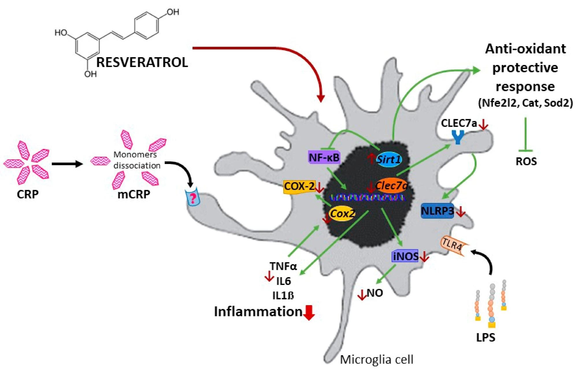
    In a recent study published in the journal Antioxidants, researchers investigated the mechanisms by which Resveratrol, a natural phenolic compound, can prevent and attenuate Alzheimer’s Disease (AD). They used the BV2 microglial cell lines established from C57BL/g transgenic murine models to elucidate the mechanistic benefits of Resveratrol against glia activation by proinflammatory monomeric C-reactive protein (mCRP). Their results highlight that Resveratrol inhibits lipopolysaccharides (LPS) and mCRP-induced-cyclooxygenase-2, thereby preventing the release of proinflammatory cytokines. It further upregulates the expression of the antioxidant enzymes, namely Cat and Sod2. Together, these results provide the mechanistic underpinning for the benefits of Resveratrol in combatting and controlling AD.

    Study: Resveratrol Activates Antioxidant Protective Mechanisms in Cellular Models of Alzheimer’s Disease Inflammation. Image Credit: Aimee Lee Studios / ShutterstockStudy: Resveratrol Activates Antioxidant Protective Mechanisms in Cellular Models of Alzheimer’s Disease Inflammation. Image Credit: Aimee Lee Studios / Shutterstock

    What is Resveratrol?

    Resveratrol, commonly found in red grapes and their derivatives (e.g., red wine), is a natural phenolic compound belonging to the stilbene family. Extensive preclinical research into the metabolite has revealed its potent anti-inflammatory, anti-neurodegenerative, antioxidant, and anti-aging properties. It is a common compound synthesized by more than 70 known plant species as a stress-response mechanism.

    Recent Resveratrol studies in animal models have shown that trans-resveratrol is capable of crossing the blood-brain barrier, suggesting that it might perform neuroprotective functions. This is augmented by the fact that older men and women who drink moderately are consistently observed to have lower dementia risk compared to lifetime abstainers. Unfortunately, the human body does not produce Resveratrol, and therapeutic dosages (150-250 mg/d) can only be acquired through oral supplementation.

    Scientists have attempted to elucidate this compound’s impacts on neurodegenerative and non-neural medical conditions to arrive at its mechanistic underpinning. However, Resveratrol’s mechanisms of action in humans remain a mystery, given the inconclusive findings of said studies. The compound is both hormetic and hydrophobic, limiting its absorption and bioavailability. Researchers have circumvented this by developing novel nanocarrier-based delivery systems showing significant promise in cancer- and neurotherapy. Murine models have further suggested that Resveratrol could substantially reduce oxidative stress and improve neurodegenerative outcomes via tumor necrosis factor α (TNFα) downregulation. However, these claims remain to be tested.

    Understanding the mechanisms by which Resveratrol exercises its neurological benefits might allow for the development of new interventions aimed at preventing or managing Alzheimer’s Disease (AD). It would further inform future clinical trials of the safe dosage range, given that the chemical can be cytotoxic in high concentrations.

    About the study

    In the present study, researchers attempt to evaluate the antioxidant protection mechanism of Resveratrol using BV2 microglia, which have been activated by monomeric C-reactive protein (mCRP). mCRP activation and overexpression are vital traits of most inflammation-activated diseases, and its prevention may delay or even reverse conditions like AD that progress in part due to inflammatory stress.

    Schematic representation of the protective mechanisms of resveratrol against the proinflammatory agent mCRP and LPS.Schematic representation of the protective mechanisms of resveratrol against the proinflammatory agent mCRP and LPS.

    The BV2 cell line used herein was established from C57BL/6 transgenic mice microglia, an established model for studying brain inflammation. mCRP was generated from pure CRP protein via urea/ ethylenediaminetetraacetic acid (EDTA) chelation, followed by dialysis. Escherichia coli 026:B26 was used as a lipopolysaccharide (LPS) strain. Resveratrol treatments on these primary cell cultures varied between 10-50 µM. mCRP assays utilized mCRP at 50 µg/mL. To avoid astrocyte damage, primary glial cultures were not subjected to nutrient (serum) starvation.

    Nitric oxide generation by glial cultures was determined using the colorimetric Griess reaction. The Enzyme-Linked Immunosorbent Assay (ELISA) was used to detect and measure tumor necrosis factor-alpha (TNF-α) and interleukin one-beta (IL1 ß) expressed in ng/mL and pg/mL, respectively. Western blotting assays were used to detect and identify other protein products produced by BV2 cells. BV2 cell RNA was then extracted and subjected to Real-Time Quantitative Polymerase Chain Reaction (qPCR) to determine relative gene expression.

    Finally, the immunofluorescence assay was used to measure the impact of Resveratrol on BV2 cell expression. Statistical analyses comprised two-way analysis of variance (ANOVA) and the Shapiro–Wilk test.

    Study findings

    Resveratrol was observed to significantly inhibit and reduce TNF-α production induced by mCRP and LPS, elucidating and validating its anti-inflammatory properties. The compound was further observed to suppress the activation of the nitric oxide pathway, preventing the generation of reactive oxygen species (ROS).

    Activation of the NLR family pyrin domain containing 3 (NLRP3) gene was also observed to be inhibited by Resveratrol. NLRP3 is the gene responsible for producing the cryopyrin protein, a crucial microglia cell sensor inflammasome activated during oxidative stress. Nuclear factor-κB (NF-κB) and Nos2 were observed to be downregulated on the addition of Resveratrol. Finally, Resveratrol was found to induce the expression of antioxidant genes, including Sirt1 and Nfe2I2.

    In summary, Resveratrol’s anti-AD effect was shown to arise due to a combination of oxidation suppression and antioxidant expression.

    Conclusions

    In the present study, researchers investigated the mechanisms by which Resveratrol, a plant metabolite found in over 70 species, can promote positive neurodegenerative outcomes, especially in AD. They used a combination of in vitro cell cultures, ELISAs, western blotting, and qPCR and revealed that Resveratrol both suppresses the generation of ROS and enhances the expression of antioxidant-protecting genes.

    “Resveratrol protected against the polarization of BV2 microglia into an activated phenotype induced by two critical proinflammatory agents, LPS and mCRP. The characterization of mCRP proinflammatory and pro-oxidant mechanisms in BV2 microglia showed the activation of the inflammatory/oxidative cascades of nitric oxide, NLRP3 inflammasome and COX-2 in this novel in vitro model. Resveratrol protective mechanisms against mCRP required the modulation of SIRT1, Nrf2, and NF-ĸB pathways that reduced downstream inflammatory mediators and, most notably, induced antioxidant enzymes. Resveratrol protective mechanisms against activation to proinflammatory phenotype by mCRP was confirmed in primary mixed glial cultures.”

    These findings highlight the potential for Resveratrol in future AD preclinical testing. Resveratrol and similar plant-derived metabolites may allow for the development of future clinical interventions against currently incurable diseases such as AD. However, extensive clinical trials are required to assess the effectiveness of Resveratrol on other oxidation-inducing genes and to arrive at a dosage that is safe for human use.

    Journal reference:

    • Bartra, C., Yuan, Y., Vuraić, K., Slevin, M., Pastorello, Y., Suñol, C., & Sanfeliu, C. (2024). Resveratrol Activates Antioxidant Protective Mechanisms in Cellular Models of Alzheimer’s Disease Inflammation. Antioxidants, 13(2), 177, DOI – 10.3390/antiox13020177, https://www.mdpi.com/2076-3921/13/2/177

    [ad_2]

    Source link

  • MSU scientists unveil a potential game-changer in the fight against glioblastoma

    MSU scientists unveil a potential game-changer in the fight against glioblastoma

    [ad_1]

    A team of Michigan State University scientists has unveiled a potential game-changer in the fight against glioblastoma, the most common and currently incurable form of brain cancer.

    A team of Michigan State University scientists has unveiled a potential game-changer in the fight against glioblastoma, the most common and currently incurable form of brain cancer.

    Their weapon of choice? A drug-like compound named Ogremorphin, or OGM. In laboratory experiments, OGM showed a remarkable ability to kill glioblastoma cells while leaving normal cells unharmed.

    Charles Hong, the chair of the Department of Medicine at MSU College of Human Medicine, who led the study, published in the journal Experimental Hematology and Oncology, declared it an “early but extremely promising path to a cure.”

    What makes OGM special lies in its precision. The researchers targeted an acid sensor called GPR68/OGR1 on the cancer cell membranes, disrupting a crucial signaling pathway that cancer cells rely on to survive and grow.

    Because glioblastoma cells acidify their tumor environment and then use the acid-sensing receptor to survive, the OGM compound essentially cuts off their lifeline. We haven’t found a single brain cancer cell line that it can’t kill.”


    Charles Hong, Chair of the Department of Medicine, MSU College of Human Medicine

    Hong led the study along with his College of Human Medicine colleagues Charles Williams and Leif Neitzel, as well as with researchers at the University of Maryland School of Medicine and the Johns Hopkins University School of Medicine.

    Hong believes this groundbreaking research isn’t confined to glioblastoma alone. Since other cancer types are also known to acidify their tumor environment to thrive and evade traditional therapies, this discovery could also lead to treatments targeting other types of cancer.

    The reality of brain cancer is that, even with the standard treatment that combines brain surgery, chemotherapy and radiation therapy, the median survival period is 15 to 18 months following diagnosis, with a five-year survival rate of around 10%. Such an outcome is due to cancer recurrence and treatment resistance.

    “We found an explanation for how an acidic tumor environment enables the cancer cells to survive and evade chemotherapy, and at the same time, we found a drug candidate that blocks this survival pathway to selectively kill them without touching normal cells,” Hong shared.

    “This is just a first step,” he added. “Developing a treatment for human glioblastoma patients will take years of research. We hope to have human trials within five years.”

    Source:

    Journal reference:

    Williams, C. H., et al. (2024). GPR68-ATF4 signaling is a novel prosurvival pathway in glioblastoma activated by acidic extracellular microenvironment. Experimental Hematology and Oncology. doi.org/10.1186/s40164-023-00468-1.

    [ad_2]

    Source link

  • Hydrogel with built-in antibiofilm and antioxidative functions promotes faster healing of infected chronic wounds

    Hydrogel with built-in antibiofilm and antioxidative functions promotes faster healing of infected chronic wounds

    [ad_1]

    Diabetic wounds often become chronically infected and are notoriously difficult to treat. Two primary reasons for this include the formation of a bacterial biofilm and high levels of oxidative stress. A novel hydrogel dressing was developed recently to combat both these undesirable characteristics and tested for its effects on diabetic-infected wound healing. The report appeared in Nature Communications.

    Study: Hydrogel dressings with intrinsic antibiofilm and antioxidative dual functionalities accelerate infected diabetic wound healing. Image Credit: New Africa/Shutterstock.com
    Study: Hydrogel dressings with intrinsic antibiofilm and antioxidative dual functionalities accelerate infected diabetic wound healing. Image Credit: New Africa/Shutterstock.com

    Background

    Wound healing is recognized to have four stages, namely, coagulation, inflammation, proliferation, and maturation. When this doesn’t happen, chronic wounds result. Most occur due to prolonged inflammation triggered by competing pro- and anti-inflammatory signals leading to loss of redox homeostasis.

    Chronic inflammation attracts leukocytes that secrete reactive oxygen species (ROS), a defense against microbial invasion. However, these ROS also antagonize wound healing by damaging living tissues and cells at various levels and promoting breakdown and further inflammation.

    In the worst cases, cells die within and around the wound site by apoptosis and other modes of programmed cell death because of excessively high ROS levels. Neighboring cells react to this and eventually die themselves, accounting for the severe necrosis, or tissue death, common to such wounds. This means that tissue debridement or even amputations, at times, becomes necessary to treat these wounds.

    Biofilm formation by microbes is another complication that gives rise to chronic wounds, preventing topical antioxidants from acting on the wound surface. Biofilms use up nutrients from the wound bed and secrete extracellular polymeric substances (EPS) that form a protective barrier against immune cells, antibiotics, and other antimicrobials. Moreover, they remain stable on the wound surface until medically removed.

    Biofilm microbes are, in fact, the primary species found in chronic wounds and are resistant to treatment in many cases. Most commonly, these are methicillin-resistant Staphylococcus aureus (MRSA) or carbapenem-resistant Pseudomonas aeruginosa (CRPA).

    Chronic wounds cost the economy over USD 50 billion in just the USA, in just one year. And this is only expected to increase as the population grows around the world. Diabetic wounds are among the most common types of chronic wounds and have, unfortunately, as high a risk of death as cancer, at about 31%.

    The effectiveness of ordinary wound dressings in chronic wounds is small. Dressings designed for chronic wounds have so far not been developed as stand-alone treatments. At present, specialized chronic wound dressings require the additional use of photothermal irradiation or release and leave significant amounts of antibiotics or metal ions in the wound.

    The current study was motivated by the need for improved chronic wound dressings that would be adequate by themselves, would not contaminate the wound, and would not produce unwanted discharge and moist wound matter.

    The researchers used a hydrogel, PPN, formed by crosslinked polyethylene glycol (PEG) hydrogel tethered with highly potent antibacterial cationic polymer, polyimidazolium (PIM), and the antioxidant N-acetylcysteine (NAC). The cationic hydrogel kills bacteria by absorbing them into its pore spaces and then contact killing by the pore walls.

    PPN was designed to have dual functionality, opposing both biofilm formation and oxidative stress in the wound bed. Both properties would act together in synergy to promote the healing of infected diabetic wounds.

    Very little of this hydrogel leaches into the wound, and it contains neither antibiotic nor metal compounds, ensuring the wound is uncontaminated by any of these once the dressing is taken off.

    What did the study show?

    PPN showed high antibacterial efficacy in vitro. The hydrogel formulations swelled up, absorbing 10-12 times their original weight of water within an hour. In two days, when tested in infected wounds on murine models, the hydrogels became dirty yellow, probably because of the absorption of fluid and dead bacteria in the wound. They remained structurally stable, however, indicating that they do not break down in the presence of infected wounds.

    The researchers tested these hydrogels on a human skin model that was grown in a 3D structure. This demonstrated improved keratinocyte differentiation in the presence of NAC. In addition, it speeded up re-epithelialization and, thus, wound closure. Notably, silver dressings have been shown to retard keratinocyte proliferation in chronic wounds. 

    Subsequently, they applied the dual-functionality hydrogel on infected wounds in diabetic rats, which closely resembled diabetic wounds in humans. The wounds were coated with a biofilm containing either MRSA or CRPA.

    The hydrogels showed excellent biocompatibility compared to silver dressings in current use. The infected wounds treated with the hydrogel showed rapid healing compared to those in control animals. Bacterial counts fell rapidly and steeply over the first three days and remained low over the next two weeks.

    In contrast, bacterial reduction was lower for both silver dressings and control dressings. The wounds were smaller and sloughing minimal in PPN-treated wounds compared to silver or control dressings or no treatment. In fact, untreated wounds showed biofilm formation and pus discharge with sloughing wounds, with evidence of reinfection.

    Wound healing factors were also found at higher levels in PPN-treated wounds than in untreated or control-treated wounds. More mature collagen was found in the PPN-treated wound, indicating better regeneration of skin structure. Both components of PPN were found to contribute to the improved results compared to only one.

    The hydrogel can be formulated in different ways for application to the healing of superficial or deep wounds. Its advantages include the absence of wound contamination and the fact that it does not require the use of photothermal irradiation or other healing modalities.

    What are the implications?

    PPN first removes bacteria from the wound site, allowing the number of inflammatory cells to drop. ROS levels are reduced by the NAC component, which allows them to diffuse into the hydrogel, providing an immune boost while relieving oxidative stress. Also, it encourages the release of wound-healing factors.

    Finally, the NAC stimulates keratinocyte differentiation and the restoration of a normal epithelial covering over the wound. All these promote wound healing.

    This PPN dressing is more potently antibacterial than silver dressing, with activity against MRSA and CRPA. It also does not cause further inflammation and accelerates wound healing. The feasibility of multiple formats for meeting different needs and its possible extension to other biomedical needs make this hydrogel a promising alternative for the treatment of chronic infected diabetic wounds.

    [ad_2]

    Source link

  • Epigenetic switch controls metastasis formation

    Epigenetic switch controls metastasis formation

    [ad_1]

    Scientists from the German Cancer Research Center (DKFZ) and Heidelberg University investigated in mice how spreading tumor cells behave at the site of metastasis: Some tumor cells immediately start to form metastases. Others leave the blood vessel and may then enter a long period of dormancy. What determines which path the cancer cells take is their epigenetic status. This was also confirmed in experiments with human tumor cells. The results of the study could pave the way for novel diagnostic and therapeutic applications.

    What makes cancer so dangerous? Cancer cells that leave the primary tumor to reach distant sites of the body where they may grow into daughter tumors, called metastases. While most primary tumors can be effectively treated, metastases are the real danger. Oncologists estimate that more than 90 percent of all cancer deaths in solid tumors are due to metastases.

    Researchers have been working for decades to understand and prevent the spread of tumor cells. However, the mechanisms that enable a cancer cell to survive in a distant organ and ultimately grow into a metastasis are still largely unknown.

    To spread throughout the body, cancer cells travel through blood and lymphatic system. Scientists at the DKFZ and at Heidelberg University have now developed a method to observe the behavior of migrating cancer cells in mice immediately upon arrival in the metastatic organ – in this case the lung.

    The team led by the two first authors Moritz Jakab and Ki Hong Lee discovered that some tumor cells, once they have arrived in the metastatic organ, leave the blood vessel and enter a resting state. Other cancer cells start to divide directly within the blood vessel and grow into metastases.

    This delicate fate decision of the metastasizing tumor cells is controlled by the endothelial cells that line the inside of all blood vessels. They release factors from the Wnt signaling pathway that promote the exit of tumor cells from the blood vessel and thereby initiate latency. When the researchers switched off the Wnt factors, latency no longer occurred.

    What distinguishes latent from growing metastasizing cancer cells?

    “At this point, we asked ourselves the question: Why do some cancer cells immediately form a metastasis, while others fall into a kind of sleep?” says Moritz Jakab. The dormant and metastasizing cancer cells did not differ genetically, nor in many other molecular aspects. But the researchers were able to detect a subtle difference: The methylation of the DNA differed between the two cell types. Tumor cells, whose DNA was less methylated, responded sensitively to the Wnt factors, which resulted in extravasation from the blood vessel and subsequent latency. On the other hand, the more methylated cancer cells did not respond to the Wnt factors, remained in the blood vessel and immediately started metastatic growth.

    To test this hypothesis, the team examined the DNA methylation status of various tumor cell lines. Indeed, they found that this directly correlated with their metastatic potential.

    These results are surprising and could have far-reaching consequences for tumor diagnosis and therapy. The results of the study could, for example, help to use certain methylation patterns as biomarkers to predict for patients how high the load of dormant cancer cells is and, thus, how likely the patient is to relapse after successful treatment of the primary tumor. But first we need to study whether natural human tumors behave in the same way as the employed cell lines or experimental tumors.”


    Hellmut Augustin, Senior Author

    Moritz Jakab, Ki Hong Lee, Alexey Uvarovkii, Svetlana Ovchinnikova, Shubharda L Kulkarni; Sevinc Jakab, Till Rostalski, Carleen Spegg, Simon Anders, Hellmut Augustin: Lung endothelium exploits suscepible tumour cell states to instruct metastatic latency.

    Source:

    Journal reference:

    Jakab, M., et al. (2024). Lung endothelium exploits susceptible tumor cell states to instruct metastatic latency. Nature Cancer. doi.org/10.1038/s43018-023-00716-7.

    [ad_2]

    Source link