Tag: Cell

  • CRISPR-Cas9 gene-editing tool repairs defective T cells to treat rare hereditary disease

    CRISPR-Cas9 gene-editing tool repairs defective T cells to treat rare hereditary disease

    [ad_1]

    Some hereditary genetic defects cause an exaggerated immune response that can be fatal. Using the CRISPR-Cas9 gene-editing tool, such defects can be corrected, thus normalizing the immune response, as researchers led by Klaus Rajewsky from the Max Delbrück Center now report in “Science Immunology.”

    Familial hemophagocytic lymphohistiocytosis (FHL) is a rare disease of the immune system that usually occurs in infants and young children under the age of 18 months. The condition is severe and has a high mortality rate. It is caused by various gene mutations that prevent cytotoxic T cells from functioning normally. These are a group of immune cells that kill virus-infected cells or otherwise altered cells. If a child with FHL contracts a virus – such as the Epstein-Barr virus (EBV), but also other viruses – the cytotoxic T cells cannot eliminate the infected cells. Instead, the immune response gets out of control. This leads to a cytokine storm and an excessive inflammatory reaction that affects the entire organism.

    “Doctors treat FHL with a combination of chemotherapy, immunosuppression and bone marrow transplantation, but many children still die of the disease,” says Professor Klaus Rajewsky, who heads the Immune Regulation and Cancer Lab at the Max Delbrück Center. He and his team have therefore developed a new therapeutic strategy. Using the CRISPR-Cas9 gene-editing tool, the researchers succeeded in repairing defective T cells from mice and from two critically ill infants. The repaired cytotoxic T cells then functioned normally, with the mice recovering from hemophagocytic lymphohistiocytosis. Rajewsky and his team have now published their findings in the journal “Science Immunology.”

    Gene repair strategy works in mice

    The starting point for the study were mice in which the team could mimic EBV infections. In these animals, the researchers altered a gene called perforin so that its function was completely lost or severely compromised – a common genetic defect in patients with FHL. When they then elicited a condition resembling an EBV infection, the affected B cells multiplied uncontrollably because the defective cytotoxic T cells were unable to eliminate them. As a result, the immune response went into overdrive and the mice developed hemophagocytic lymphohistiocytosis.

    The team next collected T memory stem cells – that is, long-lived T cells from which active cytotoxic T cells can mature – from the blood of the mice. The researchers used the CRISPR-Cas9 gene-editing tool to repair the defective perforin gene in the memory T cells and then injected the corrected cells back into the mice. The immune response in the animals quieted down and their symptoms disappeared.

    How long protection lasts is uncertain

    The first author of the paper, Dr Xun Li, used blood samples from two sick infants to test whether the strategy also works in humans. One had a defective perforin gene, the other a different defective gene.

    Our gene repair technique is more precise than previous methods, and the T cells are virtually unchanged after undergoing gene editing. It was also fascinating to see how effectively the memory T cells could be multiplied and repaired from even a small amount of blood.”


    Dr Xun Li, First Author

    Cell culture experiments showed that the infants’ repaired T memory cells were capable of a normal cytotoxic T cell response.

    This means the therapeutic mechanism works in principle. But before patients can benefit from this discovery, the team needs to first resolve open questions and test the treatment concept in clinical trials. “It is still uncertain how long the protective effect lasts,” says Dr Christine Kocks, a scientist in Rajewsky’s team. “Since the T memory stem cells remain in the body for a long time, we hope the therapy provides long-term or even permanent protection. It is also conceivable that patients could be treated with their repaired T cells over and over again.”

    The procedure is minimally invasive since only a small amount of blood is needed, and the mice did not require any preparatory treatment – unlike, for example, with a bone marrow transplant. “We very much hope that our mechanism of action is a breakthrough in treating FHL,” says Rajewsky, “either to gain more time for a successful bone marrow transplant or even as a treatment itself.”

    Source:

    Journal reference:

    Li, X., et al. (2024) Precise CRISPR-Cas9 gene repair in autologous memory T cells to treat familial hemophagocytic lymphohistiocytosis. Science Immunology. doi.org/10.1126/sciimmunol.adi0042.

    [ad_2]

    Source link

  • Yokogawa introduces CellVoyager High-Content Analysis System CQ3000

    Yokogawa introduces CellVoyager High-Content Analysis System CQ3000

    [ad_1]

    Yokogawa Electric Corporation (TOKYO: 6841) today announced the introduction of the CQ3000, a high-content analysis system for capturing high-definition 3D microscopic images of live cell cultures. Expanding the company’s CellVoyager™ family of products, the CQ3000 will be launched commercially later in 2024.

    The CQ3000 has been designed to capture 3D microscopic images of live cell cultures in high definition at high speed. When used together with Yokogawa’s CellPathfinder image analysis software, the CQ3000 can quantify and analyze intracellular organelles to assess cellular reactions and the effects of drug compounds. It enables highly efficient evaluation of cells in a wide range of applications, from basic research to drug discovery screening.

    “The CQ3000 is an enhancement to the CellVoyager series lineup that will accelerate the development of new drugs and streamline basic research in cutting-edge biology and medicine. Through the provision of products and solutions that contribute to drug discovery research and the advancement of individualized medicine and regenerative medicine, Yokogawa’s aims to ensure well-being and quality of life for all.”

    Hiroshi Nakao, Vice President and head of the Life Business Headquarters at Yokogawa Electric

    About CQ3000

    High-definition observation of living cells over long time periods

    The CQ3000 enables the stable and precise observation of live cell cultures over long time periods thanks to Yokogawa’s high-precision temperature control and dual spinning-disk with microlens that captures optical slice images (confocal images) with higher speed and better sensitivity, which minimizes damage to cells compared to other confocal systems on the market.

    High speed screening

    The CQ3000 can capture images of microplates at high speed thanks to enhancements to its stage control and auto-focus functions. When used in combination with optional dual camera and wide-field image capture feature, which uniformly captures the entire field of view, image capture time is reduced. This product enables the high-speed selection of promising chemical compounds from vast numbers of new drug candidates.

    High image quality

    By using Yokogawa’s proprietary water immersion lens mechanism, in addition to detailed observation at high magnification levels, it is also possible to capture bright images at low magnification levels. The use of an optional uniformizer achieves even illumination across the entire field of view, for the capture of vivid images.

    To learn more about the CQ3000 please visit: https://www.yokogawa.com/solutions/products-and-services/life-science/high-content-analysis/high-content-analysis-system-cq3000/

    [ad_2]

    Source link

  • New microscopy technology unveils detailed images of brain cancer tissue

    New microscopy technology unveils detailed images of brain cancer tissue

    [ad_1]

    Brigham and MIT researchers uncovered never-before-seen details in human brain tissue with new, inexpensive microscopy technology.

    Key takeaways:

    • Researchers have developed a new microscopy technology called decrowding expansion pathology (dExPath) to analyze brain tissue.
    • By pulling proteins apart with dExPath, researchers can stain proteins in tissue that could not be accessed before, highlighting nanometer sized structures or even cell populations that were previously hidden.
    • This “super-resolution imaging” technology has the potential to provide insights that could improve diagnostic strategies and patient outcomes.

    Researchers from Brigham and Women’s Hospital, a founding member of Mass General Brigham, and the Massachusetts Institute of Technology (MIT) have unveiled unprecedentedly detailed images of brain cancer tissue through the use of a new microscopy technology called decrowding expansion pathology (dExPath).Their findings, published in Science Translational Medicine, provide novel insights into brain cancer development, with potential implications for advancing the diagnosis and treatment of aggressive neurological diseases.

    “In the past, we have relied on expensive, super-resolution microscopes that only very well-funded labs could afford, required specialized training to use, and are often impractical for high-throughput analyses of brain tissues at the molecular level,” said Pablo Valdes, MD, PhD, a neurosurgery resident alumnus at the Brigham and lead author of the study. “This technology brings reliable, super-resolution imaging to the clinic, enabling scientists to study neurological diseases at a never-before-achieved nanoscale level on conventional clinical samples with conventional microscopes.”

    Researchers previously relied on costly, super-high-resolution microscopes to image nanoscale structures in cells and brain tissue, and, even with the most advanced technology, they often struggled to effectively capture these structures at the nanoscale level.

    Ed Boyden, PhD, the Y. Eva Tan Professor in Neurotechnology at MIT and co-senior author on this study, began addressing this problem by labeling tissues, and then chemically modifying them to enable uniform physical expansion of tissues. However, this expansion technology was far from perfect. Relying on enzymes known as proteases to break up tissue, scientists found that this chemical treatment with enzymes destroyed proteins before they could analyze them, leaving behind only a skeleton of the original structure, retaining only the labels.

    Working together, Boyden and E. Antonio Chiocca, MD, PhD, Neurosurgery Chair at Brigham and Women’s Hospital and co-senior author on this study, mentored Valdes during his training as a neurosurgeon-scientist, to develop novel chemistries with dExPath to address the limitations of the original expansion technology.

    Their new technology chemically modifies tissues by embedding them in a gel and ‘softening’ the tissues with a special chemical treatment that separates protein structures without destroying them and which allows tissues to expand. This provided exciting findings to the MIT and Brigham researchers, who routinely use commercially available antibodies to bind to and illuminate biomarkers in a sample. Antibodies, however, are large and many times cannot easily penetrate cell structures to reach their target. Now, by pulling proteins apart with dExPath, these same antibodies used for staining can penetrate spaces to bind proteins in tissue that could not be accessed before expansion, highlighting nanometer sized structures or even cell populations that were previously hidden.

    The human brain has several stop guards in place to protect itself from pathogens and environmental toxins. But these elements make studying brain activity challenging. It can be a bit like driving a car through mud and ditches. We cannot access certain cell structures in the brain because of barriers that stand in the way. That is just is one of the reasons that this new technology could be so practice changing. If we can take more detailed and accurate images of brain tissue, we can identify more biomarkers and be better equipped to diagnose and treat aggressive brain diseases.”


    E. Antonio Chiocca, MD, PhD, Chair of the Department of Neurosurgery, Brigham and Women’s Hospital

    To validate the effectiveness of dExPath, Boyden and Chiocca’s team applied the technology to healthy human brain tissue, high and low-grade brain cancer tissues, and brain tissues affected by neurodegenerative diseases including Alzheimer’s and Parkinson’s diseases. Investigators stained tissue for brain and disease specific biomarkers and captured images before and after expanding samples with dExPath.

    The results revealed uniform and consistent expansion of the tissue without distortion, enabling accurate analysis of protein structures. Additionally, dExPath effectively eliminated fluorescent signals in brain tissue called lipofuscin, which makes imaging of subcellular structures in brain tissues very difficult, further enhancing image quality. Further, dExPath provided stronger fluorescent signals for improved labeling as well as simultaneous labeling of up to 16 biomarkers in the same tissue specimen. Notably, dExPath imaging revealed that tumors previously classified as “low-grade” contained more aggressive features and cell populations, suggesting the tumor could become far more dangerous than anticipated.

    While promising, dExPath requires validation on larger sample sizes before it can contribute to the diagnosis of neurological conditions such as brain cancer. Valdes underscores that, although still in its early stages, his team aspires for this technology to eventually serve as a diagnostic tool, ultimately enhancing patient outcomes.

    “We hope that with this technology, we can better understand at the nanoscale levels the intricate workings of brain tumors and their interactions with the nervous system without depending on exorbitantly expensive lab equipment,” said Valdes who is now an assistant professor of neurosurgery and Jennie Sealy Distinguished Chair in Neuroscience at the University of Texas Medical Branch. “The accessibility of dExPath will bring enable super-resolution imaging to understand biological processing at the nanometer level in human tissue in neuro-oncology and in neurological disease such as Alzheimer’s and Parkinson’s, and one day, could even improve diagnostic strategies and patient outcomes.”

    Source:

    Journal reference:

    Valdes, P. A., et al. (2024) Improved immunostaining of nanostructures and cells in human brain specimens through expansion-mediated protein decrowding. Science Translational Medicine. doi.org/10.1126/scitranslmed.abo0049.

    [ad_2]

    Source link

  • Is there an association between vitamin D, immunocompetence, and aging?

    Is there an association between vitamin D, immunocompetence, and aging?

    [ad_1]

    In a recent review published in the journal Nutrients, researchers explored the role of an individual’s immunocompetence in the responsiveness to vitamin D.

    They discussed the modulation of immunocompetence via the epigenetic programming function of the vitamin D receptor (VDR) and its ligand and highlighted the impact of aging on immunocompetence.

    Study: Vitamin D and Aging: Central Role of Immunocompetence. Image Credit: Iryna Imago/Shutterstock.comStudy: Vitamin D and Aging: Central Role of Immunocompetence. Image Credit: Iryna Imago/Shutterstock.com

    Background

    Vitamin D plays a crucial role in bone health by regulating calcium homeostasis and preventing conditions like rickets and osteomalacia. However, its influence on immunity extends beyond this function.

    Vitamin D deficiency, linked to modern lifestyle factors like limited sun exposure, affects the endogenous production of active vitamin D metabolites.

    The inactive vitamin D3 is converted to active 1,25(OH)2D3 in the liver and kidneys, which acts as a hormone and affects various tissues. Notably, various cells, including those of the innate immune system, can produce 1,25(OH)2D3 locally, contributing to auto- and paracrine effects. This compound acts as a ligand of high affinity for VDR, regulating the expression of numerous genes.

    The vitamin D status, indicated by serum 25(OH)D3 levels, categorizes individuals into deficient, insufficient, or sufficient groups. Vitamin D responsiveness varies among people due to genetic and epigenetic factors influencing molecular responses.

    Low responders, constituting about 25% of the population, may have increased susceptibility to diseases related to compromised immunity. The VDR-based modulation of immunocompetence may contribute to aging and reduce the risk of age-related diseases.

    The present review offers insights into the immunomodulatory functions of vitamin D and its impact on various health aspects beyond bone metabolism.

    Vitamin D signaling

    VDR binds specifically to genomic DNA, recognizing the motif RGKTSA. In complex with retinoid X receptor (RXR), VDR preferentially binds to direct repeat sequences in the euchromatin. Various “pioneer factors” facilitate VDR in opening chromatin, which is crucial for efficient binding.

    Chromatin accessibility and VDR binding can be assessed using next-generation sequencing technologies, including ChIP-seq (chromatin immunoprecipitation sequencing) and ATAC-seq (assay for transposase-accessible chromatin using sequencing), especially in peripheral blood mononuclear cells.

    Genomic regions of vitamin D target genes demonstrate changes in chromatin accessibility and VDR binding after vitamin D3 supplementation.

    Enhancers and transcription start site regions, even at a considerable linear distance, can interact via DNA looping within the same topologically associating domain, influencing gene expression.

    VDR’s genomic actions involve protein-protein interactions with the Mediator complex and RNA polymerase II, influencing transcription. Vitamin D also exerts epigenomic effects, altering DNA methylation, histone modifications, and chromatin organization, dynamically shaping the cell’s epigenetic landscape.

    These genomic and epigenomic effects contribute to vitamin D’s modulatory role in hematopoiesis and immunocompetence, affecting human immune cells both in vitro and in vivo.

    Epigenetic programming of immune cells

    Throughout embryogenesis and adult cellular differentiation, stem and progenitor cells undergo epigenetic programming, determining the function of terminally differentiated cells. 1,25(OH)2D3 plays a crucial role in this process, influencing hematopoiesis and the differentiation of immune cells.

    Hematopoietic stem cells (HSCs) differentiate into various blood and immune cell types, and 1,25(OH)2D3 regulates embryonic HSC numbers.

    Various transcription factors influenced by vitamin D drive the differentiation of myeloid progenitor cells into granulocytes and monocytes. Vitamin D is also the differentiation of monocytes into dendritic cells and macrophages.

    Epigenetic programming by vitamin D contributes to innate immune cell adaptation, modulating responses to infections, inflammation, and diseases.

    Variability in vitamin D status and response index among individuals affects the epigenetic programming of monocytes and derived cells, emphasizing the potential of optimized vitamin D3 supplementation for supporting proper immune cell epigenetics and overall immunocompetence. However, further research is needed to validate this concept fully.

    Decline in immunocompetence during aging

    Aging involves accumulating molecular damage, resulting in cellular dysfunction and weakened organs. Immunocompetence, crucial for appropriate immune responses, declines with age, leading to increased susceptibility to infections and diseases.

    The thymus atrophies, diminishing the production of T-cells, and “inflammaging” ensues. However, interindividual differences exist, and some individuals may display relatively higher immunocompetence.

    Lower immunocompetence correlates with accelerated aging and heightened disease risks. Vitamin D sufficiency may protect against cancers by preserving immunocompetence.

    Adequate vitamin D levels could stabilize immune resilience, safeguard against diseases, and contribute to healthy aging by mitigating various hallmarks of aging, including inflammation and cellular stress.

    Conclusion

    In conclusion, the active form of vitamin D plays a crucial role in modulating the epigenome of immune cells, particularly in monocytes.

    The observed associations between vitamin D deficiency, increased disease risk, and accelerated aging may be attributed to diminished immunocompetence.

    Considering individual responsiveness, a precautionary daily vitamin D3 dose of 1 µg (40 IU)/kg body mass is suggested, exceeding general recommendations but staying within safe limits to strengthen immunocompetence. The researchers emphasize personalized vitamin D supplementation to safeguard against prevalent diseases and promote healthy aging.

    [ad_2]

    Source link

  • Unveiling the mystery behind rapid memory loss in cancer patients

    Unveiling the mystery behind rapid memory loss in cancer patients

    [ad_1]

    In a rare but serious complication of cancer, the body’s own immune system can start attacking the brain, causing rapid-onset memory loss and cognitive deficits. What triggers this sudden biological civil war was largely unknown.

    Now, researchers at University of Utah Health have found that some tumors can release a protein that looks like a virus, kickstarting an out-of-control immune reaction that may damage brain cells.

    Their findings published in Cell on Jan. 31, 2024.

    A rapid immune attack

    Jason Shepherd, Ph.D., associate professor of neurobiology at University of Utah Health and last author on the study, explains that the swift escalation of symptoms-;which can include memory and behavioral changes, loss of coordination, and even seizures-;is a hallmark of the disease, called anti-Ma2 paraneoplastic neurological syndrome. The disease is one of a group of cancer-related neurological syndromes that occur in less than one in 10,000 people with cancer. The precise symptoms of these diseases vary, but all involve rapid immune reactions against the nervous system. “The symptoms come in quickly and can be quite debilitating,” Shepherd says.

    This fascinating research illustrates how tumor cells can manipulate their environment. We hope that this innovative transdisciplinary research will positively impact both the lives of cancer patients and of those who experience neurological symptoms.”


    Neli Ulrich, Ph.D., executive director of the Comprehensive Cancer Center at Huntsman Cancer Institute at the University of Utah and a Jon M. and Karen Huntsman Presidential Professor in Cancer Research at the U

    Stacey L. Clardy, M.D., Ph.D., a neurologist at U of U Health and a coauthor on the study, adds, “Most patients begin to experience these unusual neurologic symptoms before they even know they have a cancer.”

    These rapid-onset symptoms are the result of the immune system suddenly starting to target specific proteins that are found in the brain. Scientists knew that this flare of immunity often targets a protein called PNMA2. But nobody knew why PNMA2 provokes such a strong immune response, which left researchers at a loss for ways to prevent it. “We do not understand what is happening at the cellular or molecular level to actually cause the syndrome,” Clardy says, “and understanding the mechanism of disease is crucial to developing better treatments.”

    A virus lookalike

    To figure out how PNMA2 kickstarts an immune reaction, Junjie Xu, a graduate researcher in neurobiology at U of U Health and the lead author on the study, examined the protein’s structure using advanced microscopy. When he saw the first clear image of the protein, he was “so, so excited,” Xu says. Multiple PNMA2 proteins had spontaneously self-organized into 12-sided complexes that bore a striking resemblance to the geometric protein shells of some viruses.

    One of the immune system’s healthy functions is to attack viruses, and PNMA2’s virus-like structure makes it particularly prone to being targeted as well, the researchers found. In fact, in experiments in mice, the immune system only attacked PNMA2 protein when it was assembled into virus-like complexes.

    Wrong place, wrong time

    The location of PNMA2 in the body is also a crucial piece of the puzzle, the scientists found. “This protein normally is only expressed in the brain, in neurons,” Xu says, “but some cancer cells can express it, which can trigger an immune response.”

    As long as PNMA2 stays in the brain, the immune system won’t react to it. But rarely, a tumor elsewhere in the body will start producing PNMA2 protein. And when the immune system detects PNMA2 protein outside the brain, it reacts like it would to any foreign invader. The immune system makes antibodies that bind to the unfamiliar substance, and those antibodies direct immune cells to attack.

    But, once activated, the immune system doesn’t just attack the PNMA2 produced by the cancer. It also targets the parts of the brain that produce PNMA2 normally, including regions involved in memory, learning, and movement. The brain normally has a degree of protection from the immune system, but cancer weakens that barrier, leaving the brain especially vulnerable to this immune onslaught.

    In future work, the researchers aim to figure out which aspect of the immune response leads to patients’ rapid cognitive decline-;the antibodies themselves, immune cells making their way into the brain, or some combination of the two.

    Understanding how the immune system causes neurological symptoms may help scientists design targeted treatments, Shepherd says. “If we show that PNMA2 antibodies are the culprit that really drives the neurological symptoms, you could devise a way to block those antibodies from getting into the brain or mop them up with something as a treatment… If you can alleviate some of those neurological symptoms, it really would be huge.”

    Source:

    Journal reference:

    Xu, J., et al. (2024) PNMA2 forms immunogenic non-enveloped virus-like capsids associated with paraneoplastic neurological syndrome. Cell. doi.org/10.1016/j.cell.2024.01.009.

    [ad_2]

    Source link

  • Scientists coax a deadly bacterium to destroy itself from the inside out

    Scientists coax a deadly bacterium to destroy itself from the inside out

    [ad_1]

    Northwestern University researchers have successfully coaxed a deadly pathogen to destroy itself from the inside out.

    In the new study, researchers modified DNA from a bacteriophage or “phage,” a type of virus that infects and replicates inside of bacteria. Then, the research team put the DNA inside Pseudomonas aeruginosa (P. aeruginosa), a deadly bacterium that is also highly resistant to antibiotics. Once inside the bacterium, the DNA bypassed the pathogen’s defense mechanisms to assemble into virions, which sliced through the bacterium’s cell to kill it.

    Building on a growing interest in “phage therapies,” the experimental work represents a critical step toward engineering designer viruses as new therapeutics to kill antibiotic-resistant bacteria. It also reveals vital information about the innerworkings of phages, a little-studied area of biology.

    The study will be published on Wednesday (Jan. 24) in the journal Microbiology Spectrum.

    “Antimicrobial resistance is sometimes referred to as the ‘silent pandemic,’” said Northwestern’s Erica Hartmann, who led the work. “The numbers of infections and deaths from infections are increasing worldwide. It’s a massive problem. Phage therapy has emerged as an untapped alternative to our reliance on using antimicrobials. But, in many ways, phages are microbiology’s ‘final frontier.’ We don’t know much about them. The more we can learn about how phage work, the more likely we can engineer more effective therapeutics. Our project is cutting-edge in that we are learning about phage biology in real time as we engineer them.”

    An indoor microbiologist, Hartmann is an associate professor of civil and environmental engineering at Northwestern’s McCormick School of Engineering and a member of the Center for Synthetic Biology.

    Desperate need for antibiotic alternatives

    Associated with an increase in antimicrobial use, the rise of antibacterial resistance is an urgent and growing threat to the global population. According to the Centers for Disease Control and Prevention (CDC), nearly 3 million antimicrobial-resistant infections occur each year in the United States alone, with more than 35,000 people dying as a result.

    The growing crisis has motivated researchers to look for alternatives to antibiotics, which are continually losing effectiveness. In recent years, researchers have started to explore phage therapies. But even though billions of phages exist, scientists know very little about them.

    For every bacterium that exists, there are dozens of phages. So, there is an astronomically large number of phages on Earth, but we only understand a handful of them. We haven’t necessarily had the motivation to really study them. Now, the motivation is there, and we are increasing the number of tools we have to dedicate to their study.”


    Erica Hartmann, Northwestern University

    Treatment without side effects

    To explore potential phage therapies, researchers either pinpoint or modify an existing virus to selectively target a bacterial infection without disrupting the rest of body. Ideally, scientists one day could tailor a phage therapeutic to infect a specific bacterium and design “a la carte” therapeutics with precise traits and characteristics to treat individual infections.

    “What’s powerful about phage is it can be very specific in the way that antibiotics are not,” Hartmann said. “If you take an antibiotic for a sinus infection, for example, it disrupts your entire gastrointestinal tract. A phage therapy can be designed to affect only the infection.”

    While other researchers have investigated phages therapies, almost all of those studied have focused on using phages to infect Escherichia coli. Hartmann, however, decided to focus on P. aeruginosa, one of the five most deadly human pathogens. Particularly dangerous for people with compromised immune systems, P. aeruginosa is a leading cause of hospital infections, often infecting patients with burn or surgery wounds as well as lungs in people with cystic fibrosis.

    “It is one of the highest priority, multi-drug resistant pathogens that many people are really concerned about,” Hartmann said. “It is extremely drug resistant, so there is an urgent need to develop alternative therapeutics for it.”

    Mimicking infection, bypassing defenses

    In the study, Hartmann and her team started with P. aeruginosa bacteria and purified DNA from several phages. Then, they used electroporation -; a technique that delivers short, high-voltage pulses of electricity -; to poke temporary holes in the bacteria’s outer cell. Through these holes, phage DNA entered the bacteria to mimic the process of infection.

    In some cases, the bacteria recognized the DNA as a foreign object and shredded the DNA to protect itself. But after using synthetic biology to optimize the process, Hartmann’s team was able to knock out the bacteria’s antiviral self-defense mechanisms. In these cases, the DNA successfully carried information into the cell, resulting in virions that killed the bacteria.

    “Where we were successful, you can see dark spots on the bacteria,” Hartmann said. “This is where the viruses burst out of the cells and killed all the bacteria.”

    After this success, Hartmann’s team introduced DNA from two more phages that are naturally unable to infect their strain of P. aeruginosa. Yet again, the process worked.

    Phage manufacturing in a cell

    Not only did the phage kill the bacteria, the bacteria also ejected billions more phages. These phages can then be used to kill other bacteria, like those causing an infection.

    Next, Hartmann plans to continue modifying phage DNA to optimize potential therapies. For now, her team is studying the phages expelled from the P. aeruginosa.

    “This is an important piece in making phage therapies,” she said. “We can study our phage in order to decide which ones to develop and eventually mass produce them as a therapeutic.”

    The study, “A synthetic biology approach to assemble and reboot clinically relevant Pseudomonas aeruginosa tailed phages,” was supported by the Walder Foundation, the National Science Foundation and the National Institutes of Health.

    Source:

    Journal reference:

    Ipoutcha, T., et al. (2024) A synthetic biology approach to assemble and reboot clinically relevant Pseudomonas aeruginosa tailed phages. Microbiology Spectrum. doi.org/10.1128/spectrum.02897-23.

    [ad_2]

    Source link

  • Researchers discover new class of compound that targets cancer stem cells

    Researchers discover new class of compound that targets cancer stem cells

    [ad_1]

    Many cancer therapies, in addition to producing numerous side effects, fail to achieve complete tumour remission, partly due to the presence of cancer stem cells, which are difficult to eradicate. These cells can self-renew and play a key role in tumor recurrence and metastasis processes, so there is significant interest in developing therapies that target this subset of tumour cells. 

    A collaboration between chemists from the Center for Research in Biological Chemistry and Molecular Materials (CiQUS), led by Prof. JL Mascareñas, and cell biologists from the CSIC (Instituto de Investigaciones Biomédicas Sols-Morreale, IIBM CSIC-UAM, Madrid), led by Dr. Bruno Sainz, has led to the discovery of a new class of compound that targets cancer stem cells and reduces their potential to generate tumors. For some years, Prof. Mascareñas’ laboratory has been conducting basic research on certain molecules based on metal complexes that can interact very selectively with DNA. These findings have allowed Dr. Sainz’s group to conduct extensive studies with mice implanted with patients’ tumors, demonstrating a powerful antitumor effect of these complexes. The scientists have demonstrated anticancer effects in pancreatic, colorectal, and osteosarcoma tumors, with low secondary toxicity, and studies on other types of cancer are currently underway. 

    Cancer stem cells rely on mitochondrial respiration to survive and evade the immune system’s defenses, which represents a metabolic Achilles’ heel. Mechanistic studies carried out this time suggest that the new compound, called Ru1, promotes a decrease in the expression of genes necessary for this respiration, the main energy source for these cells, causing them to lose their cancerous potential. Dr. Sainz’s group has also demonstrated that combined therapies with other antitumor agents are possible, resulting in additive effects. 

    The preliminary results of the scientific work, which also includes contributions from the USC’s ACUIGEN group, have just been published in a leading cancer research journal. All these studies have been made possible thanks to the support of different entities, including the Ignicia program (Xunta de Galicia), the Spanish Association Against Cancer, or the CaixaImpulse program (“la Caixa” Foundation), and the project is currently in an advanced stage for its transfer and preclinical valorization. 

    Source:

    Journal reference:

    Alcalá, S., et al. (2024). Targeting cancer stem cell OXPHOS with tailored ruthenium complexes as a new anti-cancer strategy. Journal of Experimental & Clinical Cancer Research. doi.org/10.1186/s13046-023-02931-7.

    [ad_2]

    Source link

  • Scientists uncover a way to “hack” neurons’ internal clocks to speed up brain cell development

    Scientists uncover a way to “hack” neurons’ internal clocks to speed up brain cell development

    [ad_1]

    The neurons that make up our brains and nervous systems mature slowly over many months. And while this may be beneficial from an evolutionary standpoint, the slow pace makes growing cells to study neurodegenerative and neurodevelopmental diseases -; like Parkinson’s disease, Alzheimer’s disease, and autism -; in the laboratory quite challenging.

    Currently, nerve cells derived from human pluripotent stem cells take months to reach an adultlike state in the lab -; a timeline that mirrors the slow pace of human brain development. (“Pluripotent stem cells” have the potential to develop into many other kinds of cells.)

    New research led by Memorial Sloan Kettering Cancer Center (MSK), however, has uncovered a way to “hack” the cells’ internal clocks to speed up the process. And the work is shedding new light on how cells’ developmental timetables are regulated.

    “This slow pace of nerve cell development has been linked to humans’ unique and complex cognitive abilities,” says Lorenz Studer, MD, Director of MSK’s Center for Stem Cell Biology and the senior author of two recent studies published in Nature and Nature Biotechnology. “Previous research has suggested the presence of a ‘clock’ within cells that sets the pace of our neurons’ development, but its biological nature had largely remained unknown -; until now.”

    New insights into nerve cell development

    Researchers, led by study first author Gabriele Ciceri, PhD, identified an epigenetic “barrier” in the stem cells that give rise to neural cells. (“Epigenetic changes” are ones that don’t alter the DNA code.) This barrier acts as a brake on the development process and determines the rate at which the cells mature. By inhibiting the barrier, the scientists were able to speed up the neurons’ development, they reported January 31 in Nature.

    While studying brain development in mice, I was struck by how neurons progress through a series of steps in a very precise schedule. But this schedule creates a big practical challenge when working with human neurons -; what takes hours and days in the mouse requires weeks and months in human cells.”


    Dr. Gabriele Ciceri, a senior research scientist in the Studer Lab at MSK’s Sloan Kettering Institute

    Furthermore, the team showed that this rate-setting epigenetic barrier is built into neural stem cells well before they differentiate into different types of neurons. They also found higher levels of the barrier in human neurons compared with mouse neurons, which may help explain differences in the pace of cell maturation in different species.

    Uncovering foundational biology

    That such discoveries were made at a cancer center isn’t as surprising as it might seem at first blush. The Studer Lab has long focused on harnessing advances in stem cell biology to develop new therapies for degenerative diseases and cancer -; both of which are strongly associated with aging.

    Moreover, MSK has long been a leader in “basic science” research -; that is, science that seeks to build fundamental understanding of human biology.

    About half of the National Institutes of Health (NIH) budget goes to funding basic science research. And the vast majority of drugs approved by the Food and Drug Administration in recent years involved publicly funded basic research, according to the NIH.

    “All of the major advances in cancer treatment in recent years -; immune checkpoint inhibitor therapy, CAR T cell therapy, cancer vaccines -; they’re all rooted in basic research,” says Joan Massagué, PhD, Director of the Sloan Kettering Institute and MSK’s Chief Scientific Officer. “Sometimes it can take years for the medical relevance of a particular discovery to become clear.”

    ‘A valuable research tool’

    A second study, led by Studer Lab graduate students Emiliano Hergenreder and Andrew Minotti and published January 2 in Nature Biotechnology, identified a combination of four chemicals that together can promote neuronal maturation. Dubbed GENtoniK, the chemical cocktail both represses epigenetic factors that inhibit cell maturation and stimulates factors that promote it.

    Along with helping to bring neurons to an adultlike state faster in the lab, the approach holds promise for other cell types, the researchers note.

    Not only was GENtoniK shown to speed the maturation of cortical neurons (involved in cognitive functions) and spinal motor neurons (involved in movement), but the chemicals were also able to accelerate the development of several other types of cells derived from stem cells, including melanocytes (pigment cells) and pancreatic beta cells (endocrine cells).

    “The generation of human neurons in a dish from stem cells provides a unique inroad into the study of brain health and disease,” the journal editors note in a research briefing that accompanied the study. “A major obstacle in the field arises from the fact that human neurons require many months to mature during development, making it difficult to recapitulate the process in vitro. The authors provide a valuable research tool by developing a simple drug cocktail that speeds up the maturation timeframe.”

    The findings could be particularly helpful in modeling disorders like autism that involve problems with synaptic connectivity, Dr. Studer says.

    Still, he notes, additional research is needed to develop models of neurodegenerative disorders that don’t occur until very late in life, such as Parkinson’s disease, which has long been a focus of Studer’s research.

    “Typically, a person is 60 to 70 years old when the disease begins. No baby gets Parkinson’s,” he says. “So, for those diseases, we need to be able to put the cells not just into an adult state but into an aged-like state. That’s something we’re continuing to work on.”

    Source:

    Journal references:

    [ad_2]

    Source link

  • Study explores the mediatory role of gut microbiota in metabolic syndrome and sleep disorders

    Study explores the mediatory role of gut microbiota in metabolic syndrome and sleep disorders

    [ad_1]

    In a systematic review published in Nutrientsresearchers described gut microbiota and metabolic alterations common to metabolic syndrome (MetS) and sleep disorders.

    Study: The Microbiota–Gut–Brain Axis in Metabolic Syndrome and Sleep Disorders: A Systematic Review. Image Credit: Kmpzzz/Shutterstock.comStudy: The Microbiota–Gut–Brain Axis in Metabolic Syndrome and Sleep Disorders: A Systematic Review. Image Credit: Kmpzzz/Shutterstock.com

    Background

    One of the key mediators of the association between sleep disorders and MetS is diet. Yet, scientific evidence on its impact on human metabolism and sleep is scarce.

    Moreover, studies have not unveiled the biological mechanisms governing the intricate crosstalk between neuroendocrine, immune, and metabolic pathways that connect sleep disorders to MetS.

    Furthermore, several factors, such as smoking, alcohol consumption, and poor dietary habits, may lead to gut dysbiosis, which, in turn, adversely affects the gut–brain axis. However, how precisely the gut microbiota affects sleep homeostasis and MetS remains unclear.

    Study methodology

    Researchers conducted two separate thorough literature searches in the Medline-Pubmed databases to review observational studies and randomized clinical trials (RCTs) published in the last ten years investigating the microbial composition in adults with MetS and sleep disorders. 

    The database search returned 117 articles, of which they selected 59 articles for extensive full-text search. The final sample set comprised 36 articles, 11 for sleep disorders and 25 for MetS.

    The gut-brain axis

    The communication between the gut and the brain is facilitated through multiple pathways. One such pathway involves the afferent vagus nerve, which innervates the gut and relays signals to the central nervous system (CNS).

    This nerve is responsive to various substances, such as microbial neurotransmitters, hormones, fatty acids, and cytokines.

    Among the diverse neuromodulators, acetylcholine (ACh), norepinephrine (NE), and γ-aminobutyric acid (GABA) are particularly notable.

    These are produced and metabolized by gut microbes, playing a crucial role in directly and indirectly stimulating the connection between the gut’s afferent neurons and the CNS.

    Specifically, studies have identified that Lactobacillus and Bifidobacterium spp. strains can synthesize GABA. This synthesis impacts neurological functions, including the modulation of sleep disorders and memory.

    Additionally, the gut microbe Clostridium sporogenes converts tryptophan (Trp) into 5-hydroxy-tryptophan, a precursor of serotonin.

    This conversion enhances the inhibitory neuroregulatory effect of L-tryptophan (Trp) by interacting with trace amine-associated receptors.

    Moreover, the gut microbiome is involved in the neuroprotective effects of melatonin against cognitive impairment caused by sleep deprivation (SD), as demonstrated in mouse studies.

    The gut microbiome also influences immune cell activity, both directly and indirectly, which in turn contributes to regulating the circadian clock.

    For example, Lactobacillus rhamnosum can stimulate regulatory T-cells both indirectly, through the modulation of immune signaling via microbial cell wall components like lipopolysaccharides (LPS), and directly, through pattern-recognition receptors (PRRs).

    Lastly, gut microbes are known to modulate the expression of genes that regulate circadian rhythms, such as Rev-ERBA.

    Host-microbial mechanisms influencing sleep disorders and MetS

    The studies included in this review demonstrated how the internal biological clock (or circadian rhythm) altered metabolic homeostasis, and any changes in nutritional and metabolic statuses affected the circadian rhythm; thus, this link was reciprocal. 

    Moreover, any perturbation to the delicate circadian pattern leads to internal desynchrony and organ failure, as commonly observed in sleep disorders, such as sleep apnea, narcolepsy, insomnia, and circadian rhythm sleep disorders, categorized based on their clinical manifestations.

    Several controlled trials addressed the need to establish a cause-and-effect association between sleep duration and gastrointestinal (GI) disorders.

    They found that gut microbial neurometabolites and amino acids, such as Trp and alpha-lactalbumin (A-LAC), affected the sleepgut–brain axis.

    Thus, many studies have shown that intake of Trp-rich foods, such as milk, is linked to improved sleep quality.

    In an RCT, Schaafsma et al. showed that three weeks of supplementation of a dairy-based product in subjects with sleep disorders effectively ameliorated their Pittsburgh Sleep Quality Index (PSQI) score and reduced their cholesterol levels.

    Intriguingly, fecal samples collected at the end of the study showed an abundance of Bifidobacteraceae. This gut microbe produces an active form of GABA; thus, it is a crucial player in the stress/anxiety/sleep axis.

    MetS is an ensemble of dyslipidemia, hypertension, central obesity, disrupted insulin sensitivity, and low-grade systemic inflammation and is a well-recognized marker of microbial dysbiosis in MetS.

    In addition, MetS patients exhibit a deficiency in short-chain fatty acid (SCFA) producing gut microbes. 

    Some studies included in this review showed that metabolic impairments observed in MetS were due to a decline in bacterial deconjugation activity of primary bile acids.

    Other studies showed that microbial-derived metabolites called branched-chain aromatic amino acids (BCAAs), e.g., leucine, were involved in obesity-associated insulin resistance via an mTOR-dependent mechanism.

    More and more studies have also pointed out the importance of feeding time and rhythmicity in shaping gut microbiota communities that can achieve this.

    Thus, only long-term dietary interventions may permanently alter the gut microbial composition to ameliorate MetS.

    Moreover, multiple animal studies and studies with human subjects demonstrated that higher ingested dietary fiber intake leads to a higher prevalence of bacterial SCFA producers in the gut, which are beneficial for glucose homeostasis and ameliorating metabolic parameters in MetS.

    Interestingly, this effect correlates with the enrichment of Bifidobacterium observed in the case of sleep improvements. 

    Conclusions

    Overall, this review highlights the importance of diets rich in fiber to modulate the beneficial bacteria in the gut microbiota composition of subjects with MetS and sleep disorders.

    In sleep disorders, a potential common microbial signature is the lower abundances of butyrate (a SCFA) producers, especially Faecalibacterium prausnitzii, coupled with a reduction in some members of the Lachnospiraceae family, like Roseburia, and an enrichment in the Bacteroidetes phylum. 

    This pattern is similar to the observed decrease in SCFA producers in MetS. Since MetS cohorts examined in this review were larger, more controlled, and better taxonomically defined, their microbial pattern is more consistent for further investigation. 

    [ad_2]

    Source link

  • Immune protein Ku70 key in fighting bowel cancer

    Immune protein Ku70 key in fighting bowel cancer

    [ad_1]

    In a recent study published in the journal ScienceAdvances, researchers conducted a plethora of experiments on human cell lines and transgenic murine models aimed at investigating the relationship between the Ku70 DNA repair protein and intestinal cancer. Their results highlight the potent tumor-suppressive function of the protein. Mutations in the gene encoding the protein or downregulations in expression substantially increased the risk of subsequent spontaneous intestinal cancer. Colitis and colitis-associated colorectal cancer risks were similarly heightened.

    This study furthers progress in understanding the Ku70-mediated Ras-ERK signaling pathway and Ku70 activation’s molecular mechanisms. These, in turn, could form the basis for the future development of DNA-based therapeutics.

    Study: Ku70 senses cytosolic DNA and assembles a tumor-suppressive signalosomeStudy: Ku70 senses cytosolic DNA and assembles a tumor-suppressive signalosome

    What is Ku70?

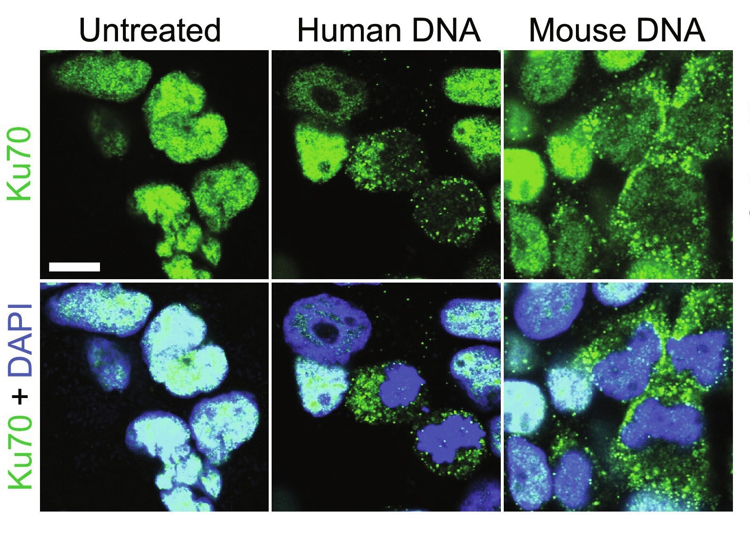
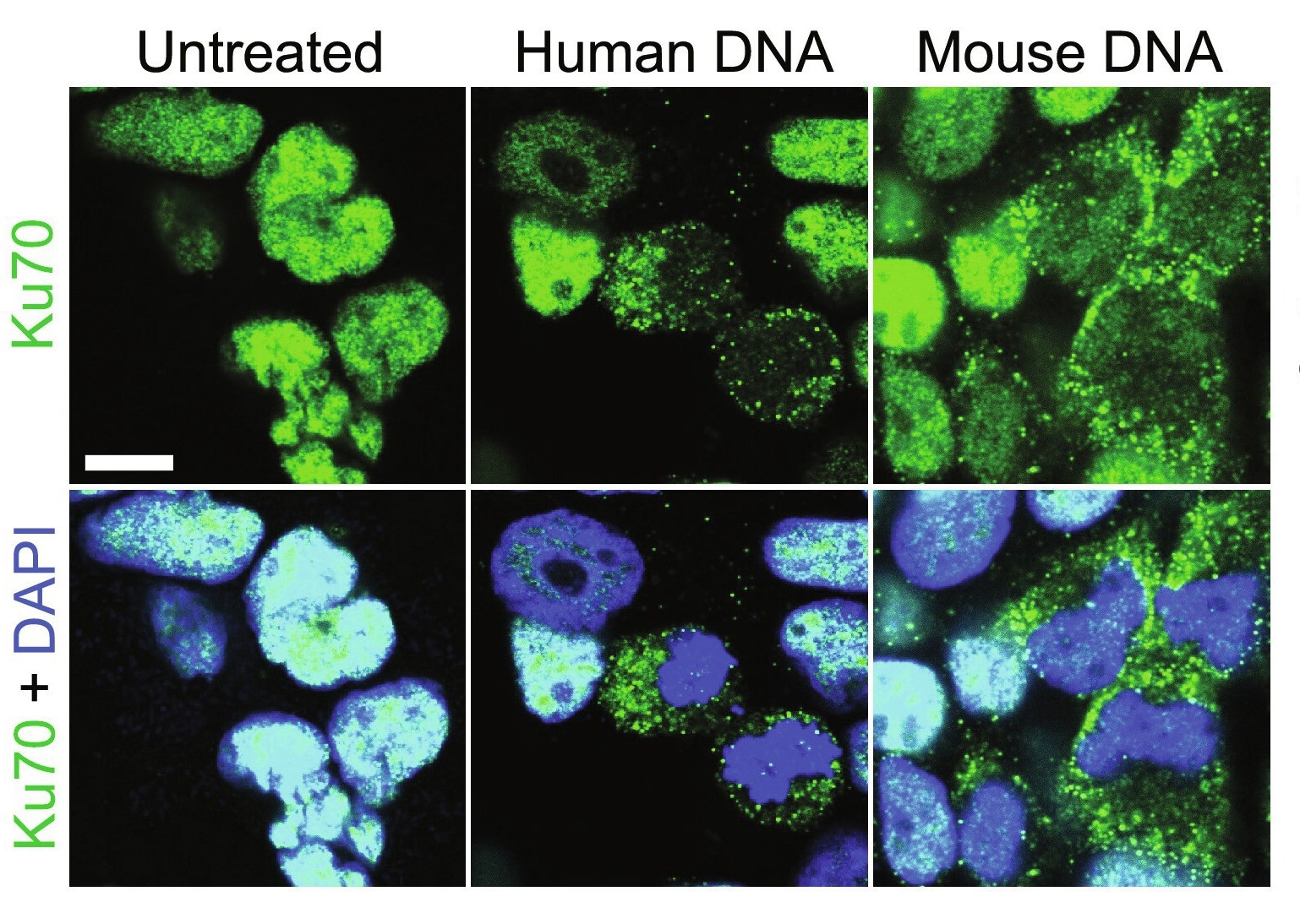
    Ku70 is a DNA repair subunit protein that helps repair DNA via the non-homologous end-joining (NHEJ) pathway. In humans, the protein is encoded by the XRCC6 gene, which studies have revealed is evolutionarily conserved. Ku70 can be found both in the cell nucleus and cytoplasm and, until recently, was thought to be restricted to its primary (DNA repair) role. Over the last decade, however, a growing body of literature suggests secondary protein functions, including antimicrobial and anti-tumor.

    Cytoplasmic Ku70 has been shown to produce type III interferons (IFNs) in response to bacterial or viral DNA in both human and murine model systems. It has additionally been shown to bind to Rickettsia conorii, a human bacterial pathogen, thereby facilitating its neutralization by nonphagocytic mammalian cells.

    Recently, research has suggested that the protein may also have a tumor-suppressive role. In an unrelated experiment, genetically altered mice lacking the Ku70 gene were found remarkably susceptible to hepatocellular carcinoma and spontaneous T-cell lymphoma development. Another study revealed genetic deletion of Ku70 to enhance colorectal cancer risk. Unfortunately, the Ku70 gene is implicated in growth, with its deletion resulting in stunted murine growth. Since smaller body size and poor growth would have confounded interpretation of these results, the association between Ku70 and cancer remains speculative and hitherto unconfirmed.

    About the study

    The present study aims to elucidate any association between Ku70 protein expression and intestinal cancer risk. Once the association is identified, the mechanism underlining Ku70’s protective anti-tumor function is explored. The experimental sample group consisted of wild-type (WT), Ku70+/−(heterozygous for Ku70), Ku70−/−(homozygous recessive), and C57BL/6NcrlAnu transgenic mice. All four mice types were equally divided into case (AOM-DSS) and control (untreated) cohorts.

    The study began with the experimental induction of colitis and colitis-associated colorectal tumorigenesis in the case-cohort via the intraperitoneal injection of 10 mg of azoxymethane (AOM). This was followed five days later by administering 1.5% Dextran Sodium Sulfate (DSS) for six days. Fourteen days after AOM administration, mice were euthanized, and their intestines and colon tissues were harvested for methodological validation and downstream analysis.

    Induced cancers were identified and characterized using histology, immunohistochemistry, and microscopy techniques. Ku70 and related proteins (e.g., cytokines) were identified and quantified using immunoblotting and enzyme-linked immunosorbent assays (ELISAs), respectively. Quantitative Real Time-Polymerase Chain Reaction (qRT-PCR) was used to isolate, amplify, and identify RNA of interest within the colon tissue. RNA was further purified using lithium chloride (LiCl) precipitation. Genomic DNA obtained from mouse feces was used to identify and characterize gut microbiome assemblages using 16S ribosomal RNA (rRNA) gene sequencing.

    Separately, cell line-based lines of evidence were obtained by constructing a plasmid containing the Ku70 gene, which was then transformed into NEB 5-alpha competent Escherichia coli. Finally, the recombinant E. coli was used to transfect the study subjects, namely HEK293T human cell lines and colon cells cultured from harvested colon tissue.

    Cell line analyses incorporated genomics, immunoblotting, and immunofluorescence techniques used for the murine models and included coimmunoprecipitation, proliferation, and organoid culture analyses. Active RAS was screened as a confounding variable using the Active Ras Detection Kit. RAS is a gene family whose mutations are estimated to account for 95% of pancreatic and 45% of colorectal cancers.

    Study findings

    This study’s highlight is validating that the cytosolic DNA sensor Ku70 has the secondary role of tumor suppression. Reductions in Ku70 expression or mutation in its gene were rapidly followed by tumorigenesis in both murine models and cell cultures. This study further unravels the mechanism of action of Ku70, which depicts an unexpectedly high mutation co-occurrence with genes encoding ARAF, RAF1, HRAS, NRAS, and BRAF, RAS family genes previously implicated in intestinal cancers.

    “Our study suggests that the Ku70-ERK signaling pathway is tumor suppressive, which is in contrast to the observation that Ras/Raf mutations, which are common in colorectal cancer, drive aberrant activation of downstream ERK-MAPK signaling.”

    Study findings further suggest that Ku70 may function in a cell-specific manner – epithelial and stromal cells from patients with Crohn’s disease or colorectal cancer display decreased Ku70 gene expression even in homozygous dominant conditions. Parallelly, Ku70 was found to form a cytosolic signalosome consisting of Ras, Raf, and Ku70, which docks at the endosome’s membrane and mediates the MEK-ERK-Cdc25A-CDK1 signaling axis activation, thereby resulting in an antitumorigenic effect.

    “…activation of the Ras-ERK pathway protects mice against colitis (83) and inhibits mammalian cell proliferation. Further studies are required to elucidate which cell types undergo Ras-ERK signaling for the progression of colorectal cancer and which cell types undergo Ku70 signaling for the attenuating of colorectal cancer.”

    “We speculate that the activation of the Ku70-mediated Ras-ERK signaling might be initiated by the cytoplasmic DNA arising from the gut microbiome introduced into the host cells following a rupture of the intestinal barrier. However, it is also possible that damaged nucleus and/or mitochondria may be a source of cytoplasmic DNA that triggers Ku70-mediated Ras-ERK signaling.”

    Journal reference:

    • Pandey, A., Shen, C., Feng, S., Tuipulotu, D. E., Ngo, C., Liu, C., Kurera, M., Mathur, A., Venkataraman, S., Zhang, J., Talaulikar, D., Song, R., Wong, L., Teoh, N., Kaakoush, N. O., & Man, S. M. (2024). Ku70 senses cytosolic DNA and assembles a tumor-suppressive signalosome. Science Advances, DOI – 10.1126/sciadv.adh3409, https://www.science.org/doi/10.1126/sciadv.adh3409

    [ad_2]

    Source link