Research suggests that widely used smartwatches could be used to monitor Parkinson’s disease stages in patients.
Wearable technologies could be leveraged to measure the progression of Parkinson’s disease and other neurodegenerative disorders, expediting new treatments for people with the condition.
This is critical for patient outcomes, as there are currently no existing drugs to slow the disease’s progression.
The study, performed by the University of Rochester Medical Center, found that a common Apple Watch paired with an iPhone can detect changes in Parkinson’s disease symptoms in individuals in the early stages of the disease.
Jamie Adams, MD, lead author of the study, explained: “Digital measures hold the promise to provide objective, sensitive, real-world measures of disease progression in Parkinson’s disease.
“This study shows that data generated by smartwatches and smartphones can remotely monitor and detect changes in multiple domains of the disease. These digital assessments could help evaluate the efficacy of future therapies.”
What is Parkinson’s disease?
Parkinson’s disease is a progressive neurological disorder primarily affecting movement control. It is characterised by the degeneration of dopamine-producing neurons in a region of the brain critical for regulating movement.
The decrease in dopamine levels leads to symptoms that typically worsen over time. Parkinson’s disease has no cure, but treatments can help manage its symptoms.
There are five Parkinson’s disease stages, as described by the Hoehn and Yahr scale:
Stage 1: This is the earliest stage where symptoms are mild and usually affect only one side of the body. Common signs include tremors, slight changes in posture, facial expressions, and walking.
Stage 2: Symptoms begin to worsen and affect both sides of the body. Tremors, rigidity, and movement difficulties become more noticeable. Daily tasks may take longer, and patients may experience changes in facial expressions and speech. However, balance is still maintained, and patients can live independently.
Stage 3: This stage marks the progression to mid-stage Parkinson’s. Loss of balance and slower movements are significant features. Patients may experience frequent falls due to impaired reflexes. While individuals can still perform daily activities, they require more assistance and may struggle with tasks like dressing and eating.
Stage 4: Symptoms are severe and disabling. While patients may still be able to walk or stand unassisted, they require substantial help with daily activities. Movement becomes increasingly limited, and patients may need assistance with personal care.
Stage 5: This is the most advanced stage. Patients may be unable to stand or walk and typically require a wheelchair or are bedridden. Full-time care is necessary, as patients are unable to perform daily activities independently. Cognitive issues, such as dementia, may also develop at this stage.
The speed of progression through the five Parkinson’s disease stages varies among individuals, and the rate of progression can be influenced by factors such as age, overall health, and the effectiveness of treatments.
Traditional tools to measure the progression of Parkinson’s disease are subjective and collect information during clinical visits.
This means the tools do not accurately reflect the day-to-day symptoms of the patients, potentially slowing the development of new therapies.
Alternatively, smartwatches can passively monitor an array of Parkinson’s disease symptoms, such as gait and tremors.
Additionally, information can be collected through finger-tapping tasks and voice recordings to measure speech-related symptoms.
Testing the potential of smartwatches
In the WATCH-PD study, researchers tracked early-stage Parkinson’s patients for 12 months.
Data from devices revealed significant declines in gait, increased tremors, and minor speech changes. The smartwatch detected reduced arm swing and recorded daily step counts, aligning with other long-term studies of the disease.
The study aimed to replicate a multi-centre clinical trial involving early, untreated Parkinson’s patients, with contributions from the pharmaceutical industry, regulators, researchers, and patients.
Supported by the Michael J. Fox Foundation, the WATCH-PD study has been extended for an additional 18 months.
Adams added: “This study brings us closer to having meaningful digital measures for future use in Parkinson’s clinical trials, which may speed up therapeutic development and get treatments to our patients faster.”
A $25m contribution is set to significantly advance cancer research at Queen’s University.
With support from Murray and Cara Sinclair, the university will build research capacity in an area of existing strength, establish advanced facilities, create training opportunities, and work to improve cancer care and treatments locally, nationally, and around the world.
In recognition of their contribution, the Queen’s Cancer Research Institute (QCRI) will be renamed the Cara & Murray Sinclair Cancer Research Institute (SCRI).
Patrick Deane, Principal and Vice-Chancellor at Queen’s, commented: “The funding will enhance the Cara & Murray Sinclair Cancer Research Institute’s ability to discover new potential treatments, test new drugs, and evaluate the impact that these treatments have on patients.
“It will also help develop highly skilled trainees who will be the next leaders in cancer research.”
Uniting experts to advance cancer research
The SCRI is the only Canadian centre uniting experts from three key disciplines—cancer biology and genetics, clinical trials, and cancer care and epidemiology—to share knowledge, advance treatments, and evaluate patient impact.
The SCRI’s clinical trials division is already an international leader in cancer research and drug development, and the institution is home to world-class researchers.
These include Dr Paul Kubes, the Canada Excellence Research Chair in Immunophysiology and Immunotherapy, and several other Canada Research Chairs.
New state-of-the-art facilities
The funding will support two new science facilities equipped to help researchers advance new discoveries and therapies for patients.
A state-of-the-art imaging facility will give researchers a real-time view of the immune system interacting with cancer cells. It will advance our understanding of how cancer cells defend against the immune system and resist treatments, allowing researchers to advance new drugs and therapies.
A second specialised biomanufacturing facility will be developed to enable personalised cellular immunotherapy treatments that harness the power of the body’s own immune system to target and destroy cancer cells.
These therapies are among the most promising new approaches to cancer treatment, and this facility aims to make them faster available to Canadian patients for clinical trials.
Training the experts of the future
A new training programme will be established to enable students and early-career researchers to gain hands-on experience and mentorship from senior cancer researchers in a multidisciplinary environment, so they can emerge as highly skilled leaders in the field.
“From basic science research and testing new drugs in trials to assessing the value of treatments, this gift supports the institute’s approach to taking cancer research from labs all the way to patients, and back,” says Andrew Craig, SCRI Director.
“This gift has the potential to dramatically improve the way that we treat cancer.”
Researchers funded by Cancer Research UK have pioneered a fluorescent dye that assists surgeons in prostate cancer surgery.
The glowing dye, developed by University of Oxford experts, sticks to prostate cancer cells to help surgeons remove them in real time.
During the first stage of the ProMOTE study, the dye enabled surgeons to remove all cancerous tissues, reducing the probability of the cancer recurring and preserving healthy tissues.
This helps to avoid life-changing side effects following prostate cancer surgery and picks up cancerous tissues not detected by the naked eye or other clinical methods.
With around 52,300 cases of prostate cancer each year in the UK alone, giving surgeons an extra pair of eyes could have significant impacts on improving cancer treatment outcomes.
Nuffield Professor of Surgery at the University of Oxford, and lead author of the study, Professor Freddie Hamdy, explained: “We are giving the surgeon a second pair of eyes to see where the cancer cells are and if they have spread.
“It’s the first time we’ve managed to see such fine details of prostate cancer in real time during surgery.
“With this technique, we can strip all the cancer away, including the cells that have spread from the tumour, which could give it the chance to come back later.
“It also allows us to preserve as much of the healthy structures around the prostate as we can to reduce unnecessary life-changing side-effects like incontinence and erectile dysfunction.
“Prostate surgery is life-changing. We want patients to leave the operating theatre knowing that we have done everything possible to eradicate their cancer and give them the best quality of life afterwards. I believe this technique makes that possibility a reality.”
This helps ensure complete removal of cancerous tissues while preserving healthy prostate areas, reducing the risk of cancer recurrence and minimising postoperative side effects.
The dye and marker molecule target Prostate-Specific Membrane Antigen (PSMA), a protein on the surface of prostate cancer cells.
The marker, derived from a ‘minibody’, binds exclusively to PSMA. This technology was developed by Oxford scientists in collaboration with ImaginAb Inc., based in Inglewood, California.
Improving prostate cancer surgery
To test the novel method, 23 men with prostate cancer received an injection of fluorescent dye before undergoing robot-assisted radical prostatectomy.
Surgeons utilised an imaging system that shines a special light on the prostate and surrounding areas, causing the cancer cells to glow.
For several patients, the dye revealed clusters of cancer cells that had spread from the tumour and were invisible to the naked eye.
Future potential
Although still in early clinical development, this marker dye could become a routine tool for surgeons, enabling them to visualise and remove every part of the cancer during prostate cancer surgery.
The imaging system could be integrated into robot-assisted surgical tools. Additionally, the marker dye could potentially be adapted for other cancers by modifying the protein used to attach to cancer cells.
Further clinical trials are underway with larger patient groups to determine if this technique can remove more prostate cancer while preserving more healthy pelvic tissue compared to current surgical methods.
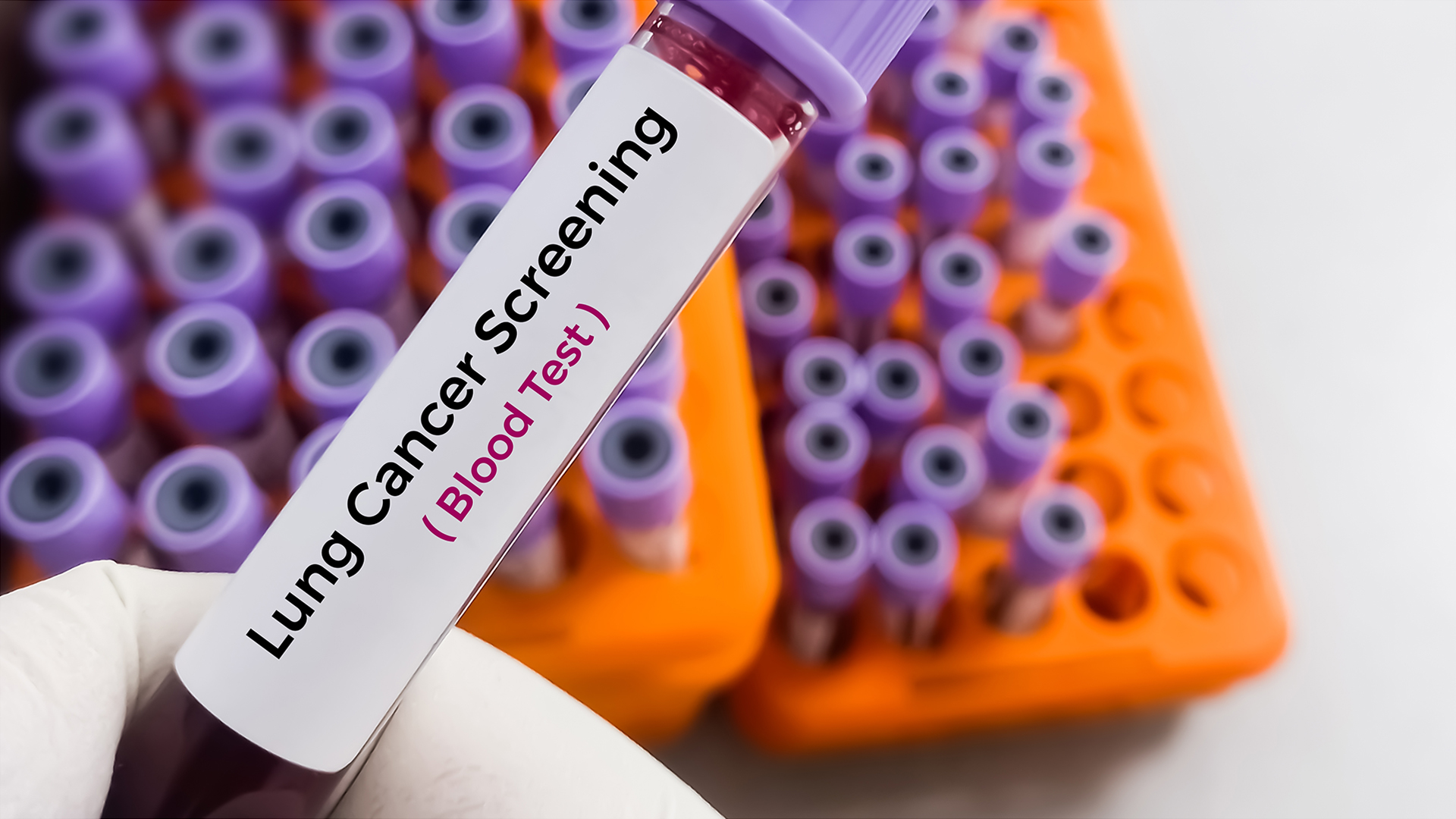
Scientists from the Johns Hopkins Kimmel Cancer Center have developed an AI blood test that accurately identifies lung cancer.
The team has performed a prospective study to demonstrate that AI technology could effectively identify people who are more likely to have lung cancer based on DNA fragment patterns in blood.
This new blood test could improve screening and reduce death rates by identifying high-risk patients who would benefit from follow-up CT scans, according to the team’s computer modelling.
The study’s corresponding author, Victor Velculescu, MD, PhD, explained: “We have a simple blood test that could be done in a doctor’s office that would tell patients whether they have potential signs of lung cancer and should get a follow-up CT scan.”
The US Preventive Services Task Force recommends screening for 15 million people aged 50-80 with a smoking history, but only 6%-10% comply each year.
Velculescu notes that reluctance is due to the time taken to perform and the radiation exposure involved in the screenings.
AI blood test development
To address these challenges, Velculescu and his team developed an AI blood test over the past five years.
This test identifies lung cancer by detecting patterns in DNA fragments in the blood. In healthy cells, DNA is neatly packaged, like a rolled-up ball of yarn, but in cancer cells, it is disorganised.
When cells die, their DNA fragments enter the bloodstream. The test distinguishes between the chaotic DNA fragments from cancer cells and the orderly fragments from healthy cells.
Excellent accuracy
To train the AI blood test, the team performed a study involving around 1,000 patients with and without lung cancer who met traditional screening criteria.
The researchers trained AI software to identify DNA fragment patterns in the blood of 576 people, with and without lung cancer and confirmed its accuracy in another group of 382.
The novel blood test has a 99.8% negative predictive value, meaning it would miss only 2 in 1,000 cases.
Their simulations suggest that increasing lung cancer screening rates to 50% within five years could quadruple detected cases and boost early detection by 10%, potentially preventing 14,000 cancer deaths over that period.
Researchers have developed an AI-powered speech analysis tool that accurately detects early signs of dementia.
Developed by experts from UT Southwestern Medical Center, the early screening tool effectively identified mild cognitive impairment and dementia in a Spanish-speaking cohort.
Munro Cullum, PhD, the corresponding author of the research, emphasised the potential of the technology: “Analysing a sample of speech obtained during some brief, routine neuropsychological tests shows promise in our ability to quickly screen for signs of cognitive impairment, particularly in population-based research studies.
“Machine learning-based tools such as this may play an increasingly important role in the future of cognitive screening for dementia.”
What is dementia?
Dementia is a broad term encompassing various brain disorders characterised by a decline in cognitive function severe enough to interfere with daily life.
It affects memory, thinking, orientation, comprehension, calculation, learning capacity, language, and judgment.
Alzheimer’s disease is the most common cause of dementia, accounting for 60-70% of cases, followed by vascular dementia, dementia with Lewy bodies, and frontotemporal dementia.
The global impact of dementia is substantial. According to the World Health Organization (WHO), over 55 million people live with dementia worldwide, and this number is expected to rise to 78 million by 2030 and 139 million by 2050 due to ageing populations.
The societal and economic burden is profound, with an estimated cost of $1.3 trillion annually, projected to double by 2030.
Research highlights the need for better diagnostic tools, effective treatments, and supportive care systems to manage the growing dementia crisis.
Now, researchers are leveraging the untapped potential of AI to improve dementia diagnostics.
Training the AI tool
To develop the AI speech analysis technology, the team collected data from 195 Spanish speakers who were part of a clinical trial in Spain.
All participants were initially evaluated and classified as having normal cognition, mild cognitive impairment (MCI), or dementia. Due to incomplete data or poor audio quality, 21 participants were excluded.
The final cohort included 174 participants with a mean age of 74, slightly more females (56%) than males. They were split into a training group of 122 (70%) and a test group of 52 (30%).
Researchers trained independent machine learning models using data from the training group participants, who completed four language tasks.
Neuropsychological performance and audio recordings were collected via the AcceXible platform, a proprietary web-based tool for disease detection through speech analysis.
Detecting early signs of dementia
The final model of the speech analysis algorithm was then used for the test group, and it was able to differentiate cognitively normal participants from those with dementia or MCI with an overall accuracy of 88.4% and 87.5%, respectively.
The final model outperformed one of the current standard-of-care screening measures known as the Mini-Mental State Examination (MMSE).
The novel technique may not only enhance the detection of breast cancer metastasis, but as it’s non-invasive, it could also improve cancer care significantly by reducing the need for needle or surgical biopsies.
Study leader Basak Dogan explained: “Most breast cancer deaths are due to metastatic disease, and the first site is usually an axillary lymph node.
“Determining nodal status is critical in guiding treatment decisions, but traditional imaging techniques alone do not have enough sensitivity to rule out axillary metastasis.
“That often requires patients to undergo invasive procedures that involve radioisotope and dye injection followed by surgery to remove and test whether the axillary nodes harbour cancer cells.”
Developing the AI model
To train the AI tool, the researchers employed dynamic contrast-enhanced breast MRI exams from 350 newly diagnosed breast cancer patients at UT Southwestern and the Moody Center for Breast Health.
All patients had known nodal status, and the images combined with various clinical measures were used to trail the AI model to identify axillary metastasis.
Accurately detecting metastatic breast cancer
The research showed that the AI technique was considerably better at identifying patients with axillary metastasis than MRI or ultrasound.
Moreover, the model would help avoid 51% of benign or unnecessary surgical sentinel node biopsies in clinical practice while correctly detecting 95% of patients with axillary metastasis.
Dogan added: “That’s an important advancement because surgical biopsies have side effects and risks, despite having a low probability of a positive result confirming the presence of cancer cells.
“Improving our ability to rule out axillary metastasis during a routine MRI – using this model – can reduce that risk while enhancing clinical outcomes.”
The team are now working to improve the image analysis process and include more varied data to validate the findings.
Temple University, together with its ecosystem of partners, is moving research results along the path to commercialisation, impacting diverse communities.
The rise of Temple University’s research enterprise has generated a significant increase in discoveries for diverse communities. These range from potentially curative therapies for HIV and other viruses, genetically targeted treatments for cardiovascular disease, digital health solutions for autism and chronic obstructive pulmonary disease, and a virtual reality system to combat the impact falls have on the elderly population.
Advancing nascent technology out of research institutions presents an exciting opportunity for the world, but it is a journey. One that requires a community of champions that need to align to realise the potential impact of each discovery.
Community-focused innovation development Innovation needs a community to thrive. This includes scientific and professional experts to help navigate the complex process of bringing an idea to market, investors and other funders that enable new businesses to carry the university’s innovation forward, and entrepreneurs who often work for equity until the company is sufficiently capitalised to pay its employees. Temple University, core to its mission, relies on this ecosystem of partners to move its research results onto the path towards commercialisation to deliver them to the community and beyond, and in doing so, brings growth to Philadelphia’s economy through these new businesses and products that make those solutions available to the public.
This partnership-driven approach applies to each phase of Temple’s commercialisation strategy. Within the university, ensuring Temple researchers have a clear pathway to develop their ideas is paramount. Temple re-engineered its commercialisation process and staffing structure to provide that enhanced onramp to innovators. Temple’s surrounding industry and entrepreneurship community were integrated as the necessary activators for the commercialisation process. The university established partnerships that enable early-stage proof-of-concept support with the benefit of a market signal through engagement with industry experts, enabling technology de-risking by achieving the critical milestones that can lead to further investment. An early-stage investment programme was developed with a leading early-stage capital provider in Pennsylvania, enabling Temple to participate in investments supporting new companies spun out of Temple research.
In March 2024, the university launched its Innovation Nest to reinforce the value of its community of partners and stakeholders. The Innovation Nest is the university’s first dedicated space to support the advancement of discoveries and incubation of companies generated by Temple’s research enterprise. With nearly 9,000 square feet of space, the facility houses Temple’s commercialisation team that provides concierge service to its research community, an incubator with flexible options for Temple spinout and affiliated startup companies, and an event venue with associated programmes designed to engage, educate, and expand the surrounding innovation ecosystem.
With its community of partners and the launch of the Innovation Nest, Temple stands poised to make an indelible mark on the global stage. From concept to reality, Temple’s innovations have the power to address pressing needs and spark transformative change — one breakthrough at a time. Amid this flurry of innovation, a disruptive platform technology focused on treating venous thromboembolic disease developed at the Lewis Katz School of Medicine stands as a shining example — a testament to Temple’s enduring legacy of innovation and impact.
Opening blocked arteries deep within lungs
Blood clots take a devastating toll on health, claiming over 100,000 lives annually in the United States alone and millions worldwide. It remains the number one cause of preventable death in hospitals. Yet, despite their deadly impact, these clots remain under-treated.
Accounting for a staggering 15% of all in-hospital deaths, 30 to 50% of survivors of these clots are left grappling with long-term disabilities, chronic thromboembolic pulmonary disease, and post-thrombotic syndrome. The toll is not merely in lives lost but in the impact on the quality of life for those who endure.
The challenge lies in the limitations of existing therapies, haunted by the spectre of high bleed rates combined with major adverse events that compromise their safety and temper their efficacy. For too long, clinicians have been hampered by the constraints of available treatments, leaving patients vulnerable to the ravages of this silent killer.
Enter Thrombolex. In a landmark collaboration with Temple University, Thrombolex unveiled its revolutionary BASHIR™ Endovascular catheters in 2020, heralding a new era in the treatment of thromboembolic diseases. This groundbreaking family of endovascular catheters has demonstrated unprecedented clinical efficacy, offering a beacon of hope to patients and practitioners alike.
In a multicentre clinical trial (RESCUE) involving 109 patients with pulmonary embolism, the BASHIR™ endovascular catheters showed unparalleled efficacy and safety compared to prior studies of other contemporary devices used for treating pulmonary embolism. With outcomes poised to improve patient survival, safety, and morbidity while significantly reducing the total cost of care within the healthcare system, Thrombolex’s innovation promises to rewrite the narrative for patients worldwide.
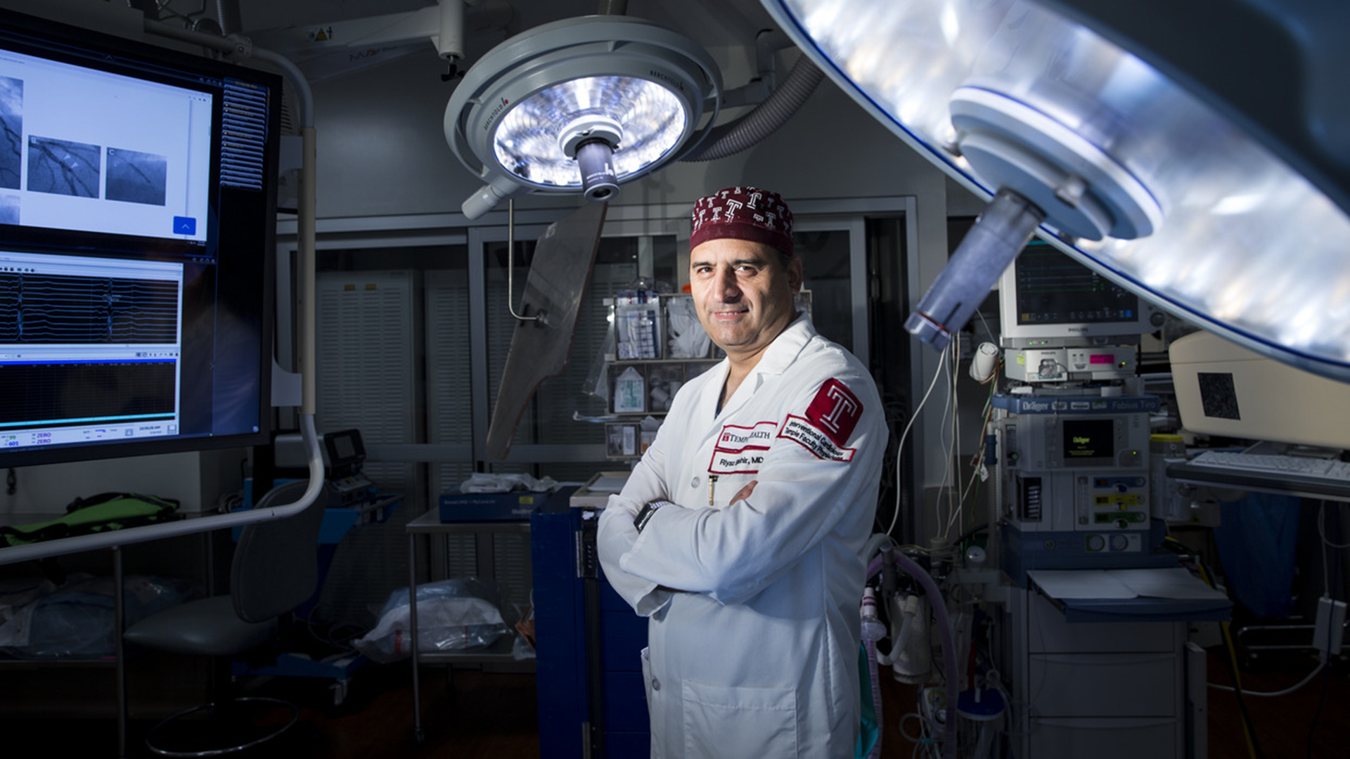
Riyaz Bashir, MD, FACC, RVT, said: safety profile and ease of use of this novel technology is a major milestone in the treatment of acute pulmonary embolism globally. Dr Bashir is a professor of medicine, the director of an interventional cardiology fellowship, and the director of the vascular and endovascular medicine division of cardiovascular diseases at Temple University Hospital.
Developed to address the pressing unmet clinical needs in treating thromboembolic diseases, including pulmonary embolism (PE) and Deep Vein Thrombosis (DVT), the BASHIR™ endovascular catheters represent a critical leap forward in medical science. With approximately $100bn in healthcare treatment costs globally, the urgency of this technology’s introduction cannot be overstated.
The innovative design of the BASHIR™ catheters is the fruit of a collaboration between Thrombolex and Dr Bashir. Differentiated by its ease of use, superior efficacy, and excellent safety profile, this platform technology promises to democratise access to life-saving treatment, transcending geographic and infrastructural barriers. Dr Bashir said: “The unique design of this device allows treatment with a very low dose of clot-dissolving medicine, which has the potential to markedly expand the role of this therapy in improving outcomes of these patients.”
With eight FDA-cleared devices currently in its product line, Thrombolex’s patented design boasts an expandable infusion basket that promptly restores blood flow and uniformly delivers low-dose medication at the site of the clot. Thrombolex has recently developed a breakthrough therapy protocol to treat patients in a single session, eliminating the need for an ICU stay, reducing the dosage of medication and further benefiting hospitals that are already resource-constrained. The catheters’ ease of use and significantly lower costs offer a lifeline to healthcare institutions grappling with the burden of treating patients with venous thromboembolic disorders.
Temple University’s international reach
As Thrombolex looks to expand its reach beyond the shores of the United States, eyes turn to Europe, a key market ripe for innovation. With over 50 institutions actively utilising BASHIR™ catheters in the US and a market penetration rate far below potential, the promise of Thrombolex’s groundbreaking platform technology is poised to shine bright on the global stage.
In the relentless pursuit of progress, Thrombolex stands at the vanguard of medical innovation, offering hope to millions and paving the way for a future where blood clots are no longer a silent killer but a conquered foe. Michael Cerminaro, co-founder, president, and CEO of Thrombolex, said: “We are very proud of our partnership with Temple University and the co-inventor of our novel platform technology, Dr Riyaz Bashir.”
He stated: “Our new RAPID-PE clinical study for the treatment of acute pulmonary embolism in a single session is a testament to our dedication to clinical research and an opportunity for us to have a material positive impact on the field of medicine.”
While Temple University nurtures homegrown innovators like Thrombolex, Philadelphia itself beckons to European enterprises seeking expansion into the Americas. Temple’s European Network of Research and Innovations Centers and Hubs (‘Enrich’) Virtual Landing programme provides a seamless gateway for European tech entrepreneurs to establish North American operations. This initiative offers tailored support, facilitating integration into industry-specific ecosystems and providing essential business development assistance, including product road mapping. An example is a company called Splendo Health, which is engaging with cardiologists and Temple Hospital to validate their concept for the US healthcare market.
Moreover, the Sbarro Health Research Organization (SHRO), nestled within the Innovation Nest, spearheads international programming and networking initiatives. This impactful collaboration elevates the global impact of both institutions in research and innovation. The Innovation Nest stands as a testament to the power of partnership and was made possible through the unwavering financial support of both the Commonwealth of Pennsylvania and SHRO.
With each groundbreaking partnership and every pioneering endeavour, Temple University continues to carve out a path toward a future defined by progress and possibility. In an ever-evolving landscape of higher education, this institution stands poised to lead the way, shaping not only its own legacy but also the trajectory of innovation on a global scale. With international campuses in Japan and Rome as well as spinouts eyeing international expansion, Temple’s influence is set to transcend beyond Philadelphia and redefine the realms of possibility for diverse communities.
Please note, this article will also appear in the 18th edition of our quarterly publication.
In a pivotal advancement in the understanding of microplastics and nanoplastics, a meta-study has revealed alarming evidence of their adverse effects on human health.
Spearheaded by Dal Yöntem of Koç University and Müfide Aydoğan Ahbab of the University of Health Sciences Türkiye, the study scrutinised 130 research papers, bringing to light the urgent need for further investigation into the pressing environmental and public health concern of plastic pollution.
The investigation revealed that microplastics and nanoplastics have profound impacts on human health, increasing the risk of various diseases and conditions.
The researchers explained: “If more people were aware of the potential threat of microplastic and nanoplastic pollution, and the associated increased risk of life-threatening conditions like cancer and the fatal form of dementia, Alzheimer’s, they would almost certainly call for and take more action.”
The growing prevalence of plastic pollution
Microplastics and nanoplastics exist in two primary forms: primary particles, commonly found in everyday products such as toothpaste and sunscreen, and secondary particles, resulting from the degradation of plastic waste.
While primary particles tend to have smoother surfaces, secondary particles exhibit a diverse range of shapes, posing a heightened risk to human cells.
Moreover, the pervasive presence of microplastics and nanoplastics in oceans, soil, air, and the human food chain underscores the urgent need for comprehensive mitigation strategies.
The presence of microplastics and nanoplastics is widely recognised as a significant hazard to mitochondrial wellbeing and activity, potentially elevating the risk of various health conditions such as Alzheimer’s disease and strokes.
Despite the global concern surrounding plastic pollution, the precise implications for human health remain largely unexplored. The limited detection methods available have constrained the understanding of human exposure.
Investigating the impact of exposure is crucial to discerning thresholds of exposure and toxicity, as well as devising strategies to mitigate its detrimental effects on the body.
Research indicates that microplastics and nanoplastics can instigate mitochondrial dysfunction primarily by inducing oxidative stress, leading to diminished adenosine triphosphate (ATP) production—the essential energy source for all cells.
This disruption in cellular processes can culminate in cell demise. Furthermore, microplastics and nanoplastics have been observed to interact with mitochondria directly, causing structural harm and functional impairment.
These interactions trigger various cellular responses, ranging from inflammation to apoptosis, exacerbating the overall toxicity of microplastics and nanoplastics.
Additionally, exposure may interfere with the dynamic processes responsible for maintaining mitochondrial health, potentially diminishing mitochondrial mass and further compromising cellular health and function.
Urgent call for enhanced research and regulatory action
The meta-study’s findings underscore regulators’ need to reassess the environmental and health risks associated with plastic pollution.
Despite the gravity of the issue, significant gaps persist in understanding the toxicity of microplastics and nanoplastics.
Current research predominantly focuses on primary microplastics and nanoplastics, particularly those composed of polystyrene (PS) plastic, leaving secondary particles and alternative plastic polymers largely unexplored.
Furthermore, the impact of common additives and absorbed environmental pollutants remains inadequately understood, highlighting the pressing need for further investigation.
Yöntem and Aydoğan Ahbab explained: “Everybody needs to look at their own use of plastic products. Governments and organisations need to develop policies that reduce plastic waste and promote sustainable alternatives.
“This collective effort will require cross-disciplinary collaboration encompassing environmental science, toxicology, public health, policymaking, and more. This is no longer just about the impact on our planet; we urgently need to find out more about microplastics and nanoplastics to safeguard our own and future generations’ health.”
As evidence mounts regarding the detrimental effects of microplastics and nanoplastics on human health, concerted efforts are needed to address this global environmental and public health crisis.
The findings demonstrate how AI techniques can assist physicians in providing early risk HHC risk assessment for metabolic dysfunction-associated steatotic liver disease (MASLD) patients, helping to provide more personalised care.
Study co-author Aniket Alurwar, clinical informatics specialist at the UC Davis Center for Precision Medicine and Data Sciences, commented: “MASLD can lead to HCC, but the disease is quite sneaky, and it’s often unclear which patients face that risk.
“It doesn’t make sense to biopsy every patient with MASLD, but if we can segment for risk, we can track those people more closely and perhaps catch HCC early.”
Addressing the threat of MASLD
MASLD, formerly known as nonalcoholic fatty liver disease (NAFLD), presents a significant health challenge, particularly as it is intricately linked with metabolic disorders such as Type 2 diabetes.
Nine open-source algorithms were tested, and five were selected for additional evaluation and model development.
These chosen algorithms were trained using deidentified health data from 1,561 UC Davis Health MASLD patients, among whom 227 later developed HCC.
Subsequently, these top five algorithms underwent validation using data from 686 UC San Francisco patients sourced from deidentified medical records.
Among these patients, 176 were diagnosed with HCC. Ultimately, the Gradient Boosted Trees algorithm emerged as the most accurate prediction model, demonstrating superior statistical accuracy, sensitivity, and specificity.
Identifying new risk factors
While advanced liver fibrosis remains a prominent risk indicator for HCC, typified by elevated Fibrosis-4 Index (FIB-4) scores, the team’s analysis unearthed additional predictors, including high cholesterol, hypertension, bilirubin levels, and alkaline phosphatase (ALP) activity.
The team discovered various pathways leading to HCC, with high FIB-4 levels being the most evident. Interestingly, some patients with low FIB-4 levels but elevated cholesterol, bilirubin, and hypertension also developed HCC.
However, according to current guidelines, such patients would not typically receive preventive care measures.
This multifactorial approach significantly enhanced the predictive accuracy of the Machine Learning model to 92.23%.
With an impressive accuracy rate, the pilot model stands as a testament to the potential of AI in healthcare.
Notably, the model identified ‘low-risk’ MASLD patients who may still face heightened HCC susceptibility, challenging conventional screening protocols.
A roadmap to future AI advancements
Looking ahead, the UC Davis team remains committed to refining their model. By integrating clinical notes and exploring advanced AI techniques like natural language processing, they aim to further enhance predictive accuracy.
Ultimately, their vision extends to seamlessly integrating these advancements into electronic health records, empowering clinicians with real-time risk assessments.
Denis Lacombe, CEO of the EORTC, explains the importance of patient-centric clinical research in cancer treatment and the commitment of the organisation to improving quality of life.
Cancer treatment has long been a challenge in the field of medicine, but it is one the world continuously rises to with innovative and persistent research dedicated to solutions.
At the European Organisation for Research and Treatment of Cancer (EORTC), the mission is to improve cancer patients’ survival and quality of life. The EORTC is a non-governmental organisation, utilising 60 years of experience to run an extensive, international academic programme spanning over 30 countries.
The Innovation Platform Editor Maddie Hall sat down with Denis Lacombe, CEO of the EORTC, to discuss the organisation’s dedication to patient-centric clinical research and the pragmatic clinical trials that are transforming the future of cancer treatment.
Denis Lacombe
Objectives of the organisation
The primary objective of the EORTC is to leave no one behind. As such, the organisation addresses clinical situations that have been comparatively neglected, such as rare cancers or elderly patients. We are also unique in our attempt to tackle a large variety of different cancers in different organs.
Our scientific strategy centres around treatment, choosing to maximise our expertise in this area while other organisations focus on cancer prevention and early detection. Through multidisciplinary clinical trials, we take a comprehensive approach to cancer treatment, addressing drugs, radiation, surgery, and the research fundamental to each, establishing EORTC as Europe’s largest cancer clinical trial organisation.
Ongoing and recent projects
At any given time, EORTC runs over a hundred clinical cancer trials, meaning that we are consistently watching a clinical trial mature and come to fruition. Whether this is by improving our knowledge of a specific cancer or directly informing doctors and patients about changing treatment, the organisation is continually advancing the standard of patient-centric care.
There are about ten clinical trials and other programmes supported by the European Union (EU) at any time. EORTC does not receive structural support from the EU but applies on a competitive basis.
One ongoing programme that is proving hugely successful is IMMUcan, which is geared to access the biological material of approximately 3,000 cancer patients with five different types of cancer. IMUCAN aims to use this data, alongside innovative technology, to understand the microenvironment of cancer cells.
Three pragmatic trials have recently been approved under the Horizon programme: A large clinical trial addressing brain tumours, retroperitoneal sarcoma, and the optimisation of hormone therapy for metastatic prostate cancer. These initiatives aim to address patient-centric clinical questions. Increasingly, trials are considering quality of life, patient-reported outcomes, and even patient preferences as an objective.
Treatment optimisation and pragmatic trials
The optimisation of treatments is a significant part of our current research strategy. When a drug comes to the market, it is not apparent which patients would benefit from the treatment and which would not. Similarly, the optimal dose, duration, or how to integrate it best in existing treatment approaches may not yet be documented or fully informed. Consequently, much research is needed on how treatments can be optimised to avoid unnecessary toxicity without benefit. The substantial costs of cancer treatments are relevant in this respect, as effort should be taken to ensure that collective resources are not spent on treating a patient unduly with a drug used, for instance, at a dose with more toxicity than necessary. EORTC is taking a de-escalation approach, addressing overtreatment, and decreasing toxicity to improve patient quality of life. We are increasingly attempting this in the context of pragmatic clinical trials. For instance, we are discussing with stakeholders and regulators the potential to adopt a more economical and efficient approach to data collection, allowing us to collect and analyse only the side effects of relevance.
Treatment optimisation is patient-centric; since we are not assessing drugs’ efficacy but aiming at obtaining a more favourable ratio between benefits and risks, the eligibility criteria for pragmatic clinical trials are much broader and closer to the real-world experience of treating the patient population. These trials do not intend to register a treatment or establish a new therapeutic system. Instead, they aim to inform doctors, patients, and healthcare systems how best to utilise therapeutic interventions. This allows us to ask pragmatic questions like: • Can we reduce the dose? • What is the optimal duration of treatment? • Can this be part of a combination treatment?
More specifically, rare and ultra-rare cancers are also often unaddressed, and trial designs have adapted to such clinical situations. A lot of methodological research is required to assess the practicalities.
The question of feasibility is also a high priority at EORTC since these pragmatic trial programmes run differently from the classic regulatory trials. This is a factor that we have consistently discussed with international partners, such as the National Cancer Institute (NCI) in the US, as well as the European Medicine Agency (EMA). The result has been the creation of the Cancer Medicines Forum, a partnership between EMA and EORTC designed to consider the optimisation of new anticancer agents beyond their licensing. It helps discuss with all stakeholders, including the commercial sector, as EORTC considers treatment optimisation a spectrum that should be addressed comprehensively and where the commercial sector can target some of the optimisation questions concerning a treatment from its inception.
Embracing new technologies to improve patient-centric care
An essential aspect of EORTC is our multidisciplinary approach, meaning we have services dedicated to technological research. A recent focus has centred around imaging technology, with our imaging platform enabling us to conduct specific research on the backbone of our pragmatic clinical trials and garner the information we need to specialise in cancer treatment.
The advantage of pragmatic trials is that, though we aim for as realistic an environment as possible, it remains controlled. Consequently, we retain the ability to control the data and the quality. This allows us to learn and understand new technologies, how they work, and eventually, how they can be modified for tailored treatments.
Embracing new technologies requires collaboration with the technology industry. We are currently collaborating with a technology company on a minimal residual disease programme. When a disease has not yet metastasised, there are still circulating cells and DNA; this programme aims to identify these as soon as possible to orient treatment. Our role is to validate the new approaches developed from clinical research to test their utility and, ultimately, their clinical relevance.
Global partnerships
The organisation is, in itself, already an international collaboration with extensive reach. Historically, EORTC has coordinated large global trials in partnership with organisations with a similar independent agenda. We have a long history of collaboration with organisations such as NCI), with whom we are currently joining forces to conduct the aforementioned trial on retroperitoneal sarcoma.
Thanks to global partnerships, conclusions can be drawn much faster, enabling quick access to a large pool of patients. They grant us insight into research from all over the world, with multiple investigators searching for solutions at once, making programmes scientifically richer and more relevant.
Similarly, EORTC has had several very successful collaborations with clinical trial groups in Canada, Australia, and Japan. Each has been an incredibly enriching experience; programmes are typically embraced by the majority, indicating the importance and relevance of the research. These global partnerships create a healthy learning environment, sharing culture, language, and cancer treatment standards.
For instance, we have an extensive fellowship programme, welcoming fellows from across the globe to spend up to three years in our Brussels office. Fellows join us, transfer knowledge, and learn about clinical research in our clinical environment. These fellows facilitate many of the programme’s successes, possessing a detailed understanding of our capacities and limitations, as well as improving future collaboration.
We join forces internationally, not only to coordinate specific clinical trials but also to conduct methodological research programmes, where we address questions such as: How could we do that better? For 20 years, EORTC, the NCI, and the clinical trial group in Canada have partnered on the RECIST programme. This aims to assess the efficiency of anticancer agents and how we can measure tumour shrinkage. Global partnerships such as this represent to the majority of the oncology community that we can conduct excellent methodological research, but clinical research that is trustworthy for future applications by others.
Alongside the clinical and methodological partnerships, we work with other organisations on cancer policy. Collaboration is a significant branch of EORTC, stimulated in every area of cancer treatment with which we engage. There is so much more that can be achieved when organisations join forces and compile resources; this is how we ultimately affect change.
EORTC in 2024: Prioritising patient-centric care
Our primary objective is to continue delivering this multidisciplinary clinical trial agenda for patients and doctors. In challenging times, with regulation changes and an unstable economic environment, we must continue prioritising patient-centric care. The effects of the Coronavirus pandemic persist, so EORTC aims to maintain this independent agenda and ensure our sustainability through fundraising and continued application for EU grants.
On 5 April 2024, we are holding a workshop about treatment optimisation at the EMA. We hope events such as these, combined with our global partnership, will increase the visibility of this concept and enable us to work with stakeholders on new cancer policies.
In particular, we would like to address how to better care for rare and ultra-rare cancer patients, recognising the challenges this patient population faces in accessing new drugs and treatments rarely registered for them. This will be an essential priority in 2024 and beyond in our attempt to solve this unmet need.
Europe is ageing, and cancer remains primarily a disease affecting the second half of life. Thus, an essential aspect of our current and future scientific strategy will concern elderly patients. Drugs are developed to treat the average adult patient population, meaning we lack the data on how to treat the elder stages of life. In some years, we will take their place; we must understand how to treat our parents, grandparents, and eventually ourselves efficiently.
EORTC is striving for the future of cancer treatment. As an organisation, we place significant emphasis on the next generation of clinical investigators, with the expertise and the facilities to provide an environment that can teach the skills not taught at medical school. Through courses, conferences, and workshops, we aim to establish the future generation of scientists and enable them to begin their careers with a pre-existing level of understanding.
Please note, this article will also appear in the seventeenth edition of our quarterly publication.