Tag: Immunology

  • New immunotherapy borrows cancer’s tricks to unleash powerful T cells

    New immunotherapy borrows cancer’s tricks to unleash powerful T cells

    [ad_1]

    Immunotherapies using engineered T cells have ushered in a new era in cancer treatment, but they have their limits. They may cause side effects or stop working, and they do not work at all against 90% of cancers. 

    Now, scientists at UC San Francisco and Northwestern Medicine may have found a way around these limitations by borrowing a few tricks from cancer itself. 

    By studying mutations in malignant T cells that cause lymphoma, they zeroed in on one that imparted exceptional potentcy to engineered T cells. The team inserted a gene for this unique mutation into normal human T cells, which made them more than 100 times more potent at killing cancer cells. They kept the tumors at bay for many months, showing no signs of becoming toxic.

    While current immunotherapies work only against cancers of the blood and bone marrow, the new approach was able to kill solid tumors derived from skin, lung and stomach tissues in mice. The team has already begun working toward testing this new approach in people.

    The breakthrough was inspired by the martial arts principle of using an opponent’s strength against them, said Kole Roybal, PhD, a co-author of the study and associate professor in microbiology and immunology. 

    We’ve used the mutations that give cancer cells their staying power to engineer what we call a ‘Judo T-cell therapy’ that can survive and thrive in the harsh conditions that tumors create.” 


    Kole Roybal, PhD, co-author of the study and associate professor in microbiology and immunology

    The study appears Feb. 7 in Nature

    A solution hiding in plain sight

    Immunology has proved difficult against most cancers because a solid tumor creates an environment focused on sustaining itself, redirecting resources like oxygen and nutrients for its own benefit. Often, cancerous tumors hijack the body’s immune system, causing it to defend, rather than attack, the cancer. 

    Not only does this impair the ability of regular T cells to target cancer cells, it also undermines the effectiveness of engineered T cells that are used in immunotherapies, which quickly tire against the tumor’s defenses. For immunotherapy treatments to work under those conditions, “We need to give healthy T cells abilities that are beyond what they can naturally achieve,” said Roybal, who is also a member of the Gladstone Institute of Genomic Immunology. 

    Using such T cells from patients with lymphoma, the UCSF and Northwestern teams screened 71 mutations, eventually isolating one that proved both potent and non-toxic, subjecting it to a rigorous set of safety tests.

    “This approach performs better than anything we’ve seen before,” said Jaehyuk Choi, MD, PhD, an associate professor of medical dermatology, as well as biochemistry and molecular genetics, at Northwestern University Feinberg School of Medicine. 

    “Our discoveries empower T cells to kill multiple cancer types and have the potential to offer cures to people who have a poor prognosis,” he said, noting that because cell therapies live and grow inside the patient, they can provide long-term immunity against cancer.

    In collaboration with the Parker Institute for Cancer Immunotherapy and venture capital firm Venrock, Roybal and Choi have launched a new company, Moonlight Bio, to realize the potential of their “judo” approach. Their first project is developing a lung cancer therapy that they hope to begin testing in people within the next few years.

    “We see this as the starting point,” Roybal said. “There’s so much to learn from nature about how we can enhance these cells and tailor them to different types of diseases.”

    Source:

    Journal reference:

    Garcia, J., et al. (2024). Naturally occurring T cell mutations enhance engineered T cell therapies. Nature. doi.org/10.1038/s41586-024-07018-7.

    [ad_2]

    Source link

  • Wayne State researchers secure $1.4 million DoD grant for prostate cancer study

    Wayne State researchers secure $1.4 million DoD grant for prostate cancer study

    [ad_1]

    A team of researchers from Wayne State University was awarded a $1.4 million, three-year grant from the U.S. Department of Defense for the study, “Cytochrome c acetylation drives prostate cancer aggressiveness and Warburg effect.”

    The study, led by Maik Hüttemann, Ph.D., professor of molecular medicine and genetics, and biochemistry, microbiology and immunology at Wayne State University’s School of Medicine, aims to establish the role of the protein cytochrome c, which the team proposes is central in two hallmarks of cancer: switching from aerobic to glycolytic metabolism – also known as the Warburg effect – and evasion of apoptosis.

    According to the National Cancer Institute of the National Institutes of Health, in 2023 it was estimated that more than 288,000 men would be diagnosed with prostate cancer and 34,700 would die in the United States, making it the second most common cancer in men. In the past decade, diagnoses of prostate cancer increased from 3.9% to 8.2%, with African American men having the highest incidence and mortality rates of the disease compared to white, Hispanic and Asian men. Cytochrome c was previously suggested to be a molecular determinant of prostate cancer health disparities, and this study will further explore this hypothesis.

    The research team proposes that cytochrome c transitions from a non-acetylated form in a normal prostate to a K53-acetylated cytochrome c in cancer.

    What we are proposing is that this transition causes switching from aerobic metabolism to Warburg metabolism because the modification renders cytochrome c less effective in transferring electrons in the electron transport chain, and at the same time making it incapable of triggering apoptosis. Warburg and evasion of apoptosis are two key features of cancer cells. This funding from the Department of Defense will allow us to develop an antibody as a prognostic and diagnostic tool and to mechanistically study the pathways leading to acetylation of cytochrome c, with the ultimate goal of identifying novel therapeutic targets that could result in developing a drug to overcome treatment resistance as a stand-alone or combination therapy.”


    Maik Hüttemann, Ph.D., professor of molecular medicine and genetics, and biochemistry, microbiology and immunology, Wayne State University’s School of Medicine

    “This important funding from the U.S. Department of Defense supports high-impact research needed to advance our understanding of how to detect and treat prostate cancer,” said Ezemenari M. Obasi, Ph.D., vice president for research at Wayne State University. “The work that Dr. Hüttemann and his collaborators are doing will improve health equity and reduce disparities in prostate cancer and may ultimately enhance the quality and length of life for those impacted by prostate cancer.”

    Collaborators on this project include Izabela Podgorski, Ph.D., professor of pharmacology, Wayne State University School of Medicine; Elisabeth Heath, M.D., associate director, Department of Oncology, Wayne State University School of Medicine; Seongho Kim, Ph.D., professor of oncology, Wayne State University School of Medicine; and Dongping Shi, M.D., chief and medical director, Detroit Medical Center Sinai-Grace Hospital.

    The grant number for this U.S. Department of Defense grant is HT94252410073.

    [ad_2]

    Source link

  • Immune cell networks key to success of personalized cancer treatment

    Immune cell networks key to success of personalized cancer treatment

    [ad_1]

    Through an analysis of tumor samples collected over time from patients with advanced melanoma, a Ludwig Cancer Research study has identified a set of preexisting conditions in tumors that predict whether such patients are likely to respond to a personalized immunotherapy known as adoptive T cell therapy (ACT) using tumor-infiltrating lymphocytes (TIL).

    Led by Ludwig Lausanne’s David Barras, Eleonora Ghisoni, Johanna Chiffelle, Denarda Dangaj Laniti and Branch Director George Coukos and reported in Science Immunology, the study also describes biomarkers that, with further vetting, could help clinicians select patients for TIL-ACT. In this therapy, TIL-;which kill cancerous cells-;are isolated from a patient, expanded in culture and then reinfused into the patient as a treatment.

    Given the aggressiveness of advanced melanoma, the potential value of TIL-ACT for patients who respond to it after failing immune checkpoint blockade immunotherapy and other available lines of therapy can’t be overstated. The question, of course, is who those people are and since only a fraction of patients currently benefit from the experimental therapy, it is vitally important to be able to quickly identify those who are unlikely to respond so that they can be quickly offered alternative treatments. Our study has taken a big step toward making that possible.”


    George Coukos

    The Lausanne Branch of the Ludwig Institute for Cancer Research is developing a number of strategies for personalized immunotherapies, ranging from cancer vaccines to personalized adoptive cell therapies (ACT) for a variety of cancers, including TIL-ACT.

    To explore how the tumors differed between patients who responded to treatment and others, the researchers collected tumor samples from patients before therapy started and then at various timepoints after they had undergone TIL-ACT treatment. They then examined differences between the global gene expression patterns of individual cancerous and noncancerous cells and conducted additional molecular analyses of cellular features and, most notably, interactions between cells in the context of their location within the tumors.

    “Through these analyses,” Barras explained, “we discovered the underlying tumor cell biology and characteristics of the tumor microenvironment that mediate responses to ACT.”

    The researchers show that tumors that responded best to TIL-ACT were those that were most riddled with mutations-;and therefore coruscated with neoantigens likely to be recognized by CD8+ (or killer) T cells. Further, as might be expected, the killer T cells in these tumors were in states with a potential for intense anti-tumor activation.

    “Our most significant finding in this context was that tumors with preexisting networks of immune cells were the ones most primed to respond to TIL-ACT, and patients whose tumors featured such networks were the ones who responded best to therapy,” said Dangaj. “That included a pair of patients enrolled in the trial whose tumors were completely cleared by the treatment.”

    Those networks consisted of killer T cells in close association with myeloid cells-;dendritic cells and macrophages-;that “present” antigens to killer T cells to guide them to their targets. These cells also hyperactivate them by binding a protein known as CD28 on the TILs to boost and sustain their functionality and secreting other T cell-stimulating factors. Moreover, these myeloid cells, like the killer T cells, were themselves in an activated state in responsive patients.

    The researchers found in examining tumor samples collected after treatment that successful TIL-ACT therapy further expanded and activated these immune cell networks. Macrophages additionally expressed a molecule name CXCL9 that likely bolsters stimulatory interactions with T cells.

    Notably, the findings reflect discoveries Coukos, Dangaj and colleagues have made in studying the responsiveness of ovarian tumors to an approved immunotherapy known as PD-1 checkpoint blockade.

    “Aside from the value of improved patient stratification, our discoveries on the cell and molecular biology of tumors that respond to TIL-ACT could help us devise treatment strategies to ‘precondition’ patients to respond to this therapy,” said Coukos. “That is a very exciting possibility, and one we are eager to pursue.”

    Source:

    Journal reference:

    Barras, D., et al. (2024). Response to tumor-infiltrating lymphocyte adoptive therapy is associated with preexisting CD8 + T-myeloid cell networks in melanoma. Science Immunology. doi.org/10.1126/sciimmunol.adg7995.

    [ad_2]

    Source link

  • Turbocharged CAR-T cells melt tumours in mice — using a trick from cancer cells

    Turbocharged CAR-T cells melt tumours in mice — using a trick from cancer cells

    [ad_1]

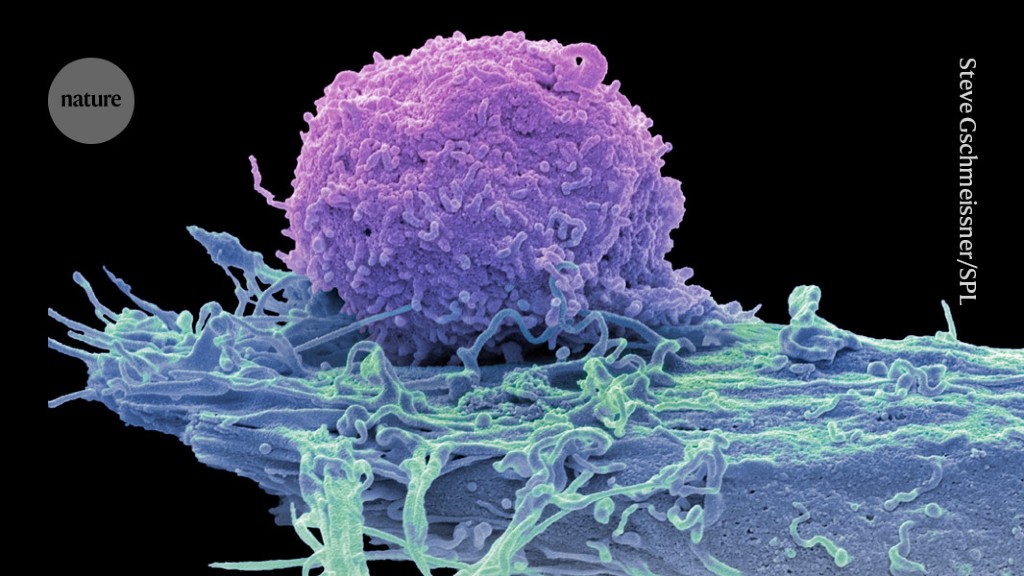
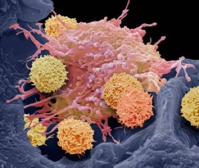
    Coloured scanning electron micrograph (SEM) of a T-cell ( purple) and a brain cancer cell ( oligodendroglioma).

    A cancer cell (blue; artificially coloured) is targeted by an engineered immune cell (purple), which can be enhanced by mutations originally discovered in cancer cells.Credit: Steve Gschmeissner/Science Photo Library

    Cancer cells are the ultimate survivors, riddled with mutations that let them thrive when healthy cells would die. These same mutations can boost the ability of game-changing cell therapies to quash cancer, a study in mice shows1.

    Among these therapies are chimeric antigen receptor (CAR) T cells, which are already used to treat several types of blood cancer. The new study shows that engineered CAR T cells carrying a mutation that was first found in cancerous T cells can vanquish tumours that don’t respond to current CAR-T therapies.

    “It’s a beautiful piece of work and opens the door for better CAR-T therapies in the future,” says Madeleine Duvic, a dermatologist and cancer researcher at the MD Anderson Cancer Center in Houston, Texas, who was not involved in the work.

    “Natural T-cell function isn’t good enough. We need to explore the extremes of T-cell function,” says Kole Roybal, an immunologist at the University of California, San Francisco, and co-author of the new paper. What better place to start than with the mutations that turn healthy T cells into hardier, cancerous ones?

    The new approach was published today in Nature.

    Cancer versus cancer

    In the past few decades, scientists have developed bespoke cell therapies by harnessing the cancer-killing power of immune cells such as T cells. The most advanced of these treatments, CAR-T-cell therapies, rely on T cells collected from people with cancer. The cells are edited to express CAR proteins, which enable the T cells to seek and destroy cancer cells. The T cells are then re-infused into the person they came from.

    These living drugs have taken the research community by storm, and the US Food and Drug Administration has approved several CAR-T cell therapies for blood cancers such as lymphomas and multiple myeloma. But scientists are still struggling to work out whether these cells can be used to kill ‘solid’ cancers, such as breast and lung tumours.

    Pulling from cancer’s playbook, Roybal and his colleagues incorporated 71 mutations, found in cancerous T cells, into CAR T cells. When they looked at how these perturbations affected T-cell function, one mutation stood out. The CAR T cells carrying an aberrant protein dubbed CARD11–PIK3R3 infiltrated well into tumours and had long-lasting cancer-killing activity.

    “It’s a very special molecule, it seems to be able to beat all the tests we put to it,” says study co-author Jaehyuk Choi, a dermatologist at Northwestern University in Evanston, Illinois.

    Potent cells

    The team treated mice carrying blood and solid cancers with several T-cell therapies boosted with CARD11–PIK3R3, and watched the animals’ tumours melt away. Researchers typically use around one million cells to treat these mice, says Choi, but even 20,000 of the cancer-mutation-boosted T cells were enough to wipe out tumours.

    “That’s an impressively small number of cells,” says Nick Restifo, a cell-therapy researcher and chief scientist of the rejuvenation start-up company Marble Therapeutics in Boston, Massachusetts.

    There is a risk that the supercharged cells will transform into cancers. But the animal data do not fuel any safety concerns, says Restifo, and the CARD11–PIK3R3 mutation seems to amp up edited T cells only when cancer cells are nearby, helping to mitigate worries about rogue immune cells.

    Choi and Roybal have co-founded Moonlight Bio in Seattle, Washington, to move these cells towards use in people with cancer. They hope to have edited cells in clinical trials in two to three years. But the bigger opportunity is the chance to find other cancer mutations that will make T-cell therapies tick.

    “A lot of people are going to think ‘Oh, this is such a good idea. Why didn’t I do this?’,” says Restifo.

    [ad_2]

    Source link

  • a blood-based immune system is discovered in the gut

    a blood-based immune system is discovered in the gut

    [ad_1]

    Access options

    Rent or buy this article

    Prices vary by article type

    from$1.95

    to$39.95

    Prices may be subject to local taxes which are calculated during checkout

    doi: https://doi.org/10.1038/d41586-024-00245-y

    Subjects

    Latest on:


    [ad_2]
    Source link

  • CRISPR-Cas9 gene-editing tool repairs defective T cells to treat rare hereditary disease

    CRISPR-Cas9 gene-editing tool repairs defective T cells to treat rare hereditary disease

    [ad_1]

    Some hereditary genetic defects cause an exaggerated immune response that can be fatal. Using the CRISPR-Cas9 gene-editing tool, such defects can be corrected, thus normalizing the immune response, as researchers led by Klaus Rajewsky from the Max Delbrück Center now report in “Science Immunology.”

    Familial hemophagocytic lymphohistiocytosis (FHL) is a rare disease of the immune system that usually occurs in infants and young children under the age of 18 months. The condition is severe and has a high mortality rate. It is caused by various gene mutations that prevent cytotoxic T cells from functioning normally. These are a group of immune cells that kill virus-infected cells or otherwise altered cells. If a child with FHL contracts a virus – such as the Epstein-Barr virus (EBV), but also other viruses – the cytotoxic T cells cannot eliminate the infected cells. Instead, the immune response gets out of control. This leads to a cytokine storm and an excessive inflammatory reaction that affects the entire organism.

    “Doctors treat FHL with a combination of chemotherapy, immunosuppression and bone marrow transplantation, but many children still die of the disease,” says Professor Klaus Rajewsky, who heads the Immune Regulation and Cancer Lab at the Max Delbrück Center. He and his team have therefore developed a new therapeutic strategy. Using the CRISPR-Cas9 gene-editing tool, the researchers succeeded in repairing defective T cells from mice and from two critically ill infants. The repaired cytotoxic T cells then functioned normally, with the mice recovering from hemophagocytic lymphohistiocytosis. Rajewsky and his team have now published their findings in the journal “Science Immunology.”

    Gene repair strategy works in mice

    The starting point for the study were mice in which the team could mimic EBV infections. In these animals, the researchers altered a gene called perforin so that its function was completely lost or severely compromised – a common genetic defect in patients with FHL. When they then elicited a condition resembling an EBV infection, the affected B cells multiplied uncontrollably because the defective cytotoxic T cells were unable to eliminate them. As a result, the immune response went into overdrive and the mice developed hemophagocytic lymphohistiocytosis.

    The team next collected T memory stem cells – that is, long-lived T cells from which active cytotoxic T cells can mature – from the blood of the mice. The researchers used the CRISPR-Cas9 gene-editing tool to repair the defective perforin gene in the memory T cells and then injected the corrected cells back into the mice. The immune response in the animals quieted down and their symptoms disappeared.

    How long protection lasts is uncertain

    The first author of the paper, Dr Xun Li, used blood samples from two sick infants to test whether the strategy also works in humans. One had a defective perforin gene, the other a different defective gene.

    Our gene repair technique is more precise than previous methods, and the T cells are virtually unchanged after undergoing gene editing. It was also fascinating to see how effectively the memory T cells could be multiplied and repaired from even a small amount of blood.”


    Dr Xun Li, First Author

    Cell culture experiments showed that the infants’ repaired T memory cells were capable of a normal cytotoxic T cell response.

    This means the therapeutic mechanism works in principle. But before patients can benefit from this discovery, the team needs to first resolve open questions and test the treatment concept in clinical trials. “It is still uncertain how long the protective effect lasts,” says Dr Christine Kocks, a scientist in Rajewsky’s team. “Since the T memory stem cells remain in the body for a long time, we hope the therapy provides long-term or even permanent protection. It is also conceivable that patients could be treated with their repaired T cells over and over again.”

    The procedure is minimally invasive since only a small amount of blood is needed, and the mice did not require any preparatory treatment – unlike, for example, with a bone marrow transplant. “We very much hope that our mechanism of action is a breakthrough in treating FHL,” says Rajewsky, “either to gain more time for a successful bone marrow transplant or even as a treatment itself.”

    Source:

    Journal reference:

    Li, X., et al. (2024) Precise CRISPR-Cas9 gene repair in autologous memory T cells to treat familial hemophagocytic lymphohistiocytosis. Science Immunology. doi.org/10.1126/sciimmunol.adi0042.

    [ad_2]

    Source link

  • Why autoimmune disease is more common in women: X chromosome holds clues

    Why autoimmune disease is more common in women: X chromosome holds clues

    [ad_1]

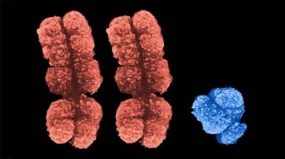
    A coloured scanning electron micrograph of an X chromosome in shades of blue on a black background.

    Protein–RNA complexes that shroud some copies of the X chromosome (artificially coloured) contribute to the female bias in prevalence of autoimmune disease.Credit: Lennart Nilsson, TT/Science Photo Library

    Why are women so much more susceptible to autoimmune diseases than men? A new explanation for the discrepancy has emerged: a molecular coating typically found on half of a woman’s X chromosomes — but not in males’ cells — might be provoking unwanted immune responses1.

    The coating, a mix of RNA and proteins, is central to a developmental process called X-chromosome inactivation. Researchers had previously implicated sex hormones and flawed gene regulation on the X chromosome as drivers of the autoimmune disparity. But the discovery that proteins central to X-chromosome inactivation can themselves set off immunological alarm bells adds yet another layer of complexity — and could point to new diagnostic and therapeutic opportunities.

    “This really adds a new mechanistic twist,” says Laura Carrel, a geneticist at the Pennsylvania State College of Medicine in Hershey.

    The study was published today in Cell1.

    Medical mystery

    Women account for around 80% of all cases of autoimmune disease, a category that includes conditions such as lupus and rheumatoid arthritis. What explains this sex bias has long been a mystery, however.

    “It’s a question that’s been irking immunologists and rheumatologists for the past 60 or 70 years,” says Robert Lahita, a rheumatologist at the Hackensack Meridian School of Medicine in Nutley, New Jersey.

    A prime suspect is the X chromosome: in most mammals, including humans, a male’s cells typically include only one copy, whereas a female’s cells typically carry two.

    (This article uses ‘women’ and ‘female’ to describe people with two X chromosomes and no Y chromosome, reflecting the language of the study, while acknowledging that gender identity and chromosomal make-up do not always align.)

    X-chromosome inactivation muffles the activity of one X chromosome in most XX cells, making their ‘dose’ of X-linked genes equal to that of the XY cells typical in males. The process is highly physical: long strands of RNA known as XIST (pronounced ‘exist’) coil around the chromosome, attracting dozens of proteins to form complexes that effectively muzzle the genes inside.

    Not all genes stay mum, however, and those that escape X inactivation are thought to underpin some autoimmune conditions. Additionally, the XIST molecule itself can initiate inflammatory immune responses, researchers reported in 20232. But that is not the whole story.

    XISTential questions

    Almost a decade ago, Howard Chang, a dermatologist and molecular geneticist at Stanford University School of Medicine in California and a co-author of the current study, noticed that many of the proteins that interact with XIST were targets of misguided immune molecules called autoantibodies.

    These rogue actors can attack tissues and organs, leading to the chronic inflammation and damage characteristic of autoimmune diseases. Because XIST is normally expressed only in XX cells, it seemed logical to think that the autoantibodies that attack XIST-associated proteins might be a bigger problem for women than for men.

    To test this idea, Chang and his colleagues turned to male mice, which don’t usually express XIST. The team bioengineered the mice to produce a form of XIST that did not silence gene expression but did form the characteristic RNA–protein complexes.

    The team induced a lupus-like disease in the mice and found that animals that expressed XIST had higher autoantibody levels than those that didn’t. Their immune cells were also on higher alert, a sign of predisposition to autoimmune attacks, and they showed more extensive tissue damage.

    Immune-system overdrive

    Notably, the same autoantibodies were also identified in blood samples from people with lupus, scleroderma and dermatomyositis — evidence that XIST and its associated proteins are “something that our immune systems have trouble ignoring”, says Allison Billi, a dermatologist at the University of Michigan Medical School in Ann Arbor.

    Montserrat Anguera, a geneticist at the University of Pennsylvania in Philadelphia, points to the human data as validation that the XIST-related mechanisms observed in mice have direct relevance to human autoimmune conditions, with implications for disease management. For example, diagnostics targeting these autoantibodies could assist clinicians in detecting and monitoring various autoimmune disorders.

    “This is a cool start,” she says. “If we could use this information to expedite the diagnosis ,it would be amazing.”

    [ad_2]

    Source link