Nature, Published online: 07 May 2024; doi:10.1038/d41586-024-01220-3
Efforts to develop an electronic newspaper providing information at the touch of a button took a step forward 50 years ago, and airborne bacteria in the London Underground come under scrutiny, in the weekly dip into Nature’s archive.
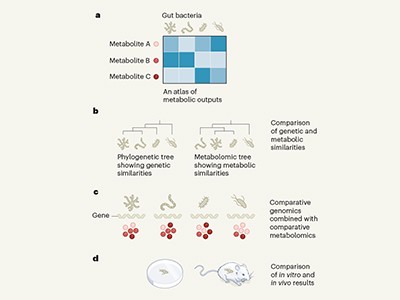
• Gut bacteria can affect biological processes at body sites far from the gut.
• The extent to which gut bacteria can affect reproductive tissues is not fully understood.
• Argaw-Denboba et al.1 report that altering the community of gut bacteria in male mice had negative consequences for the health and lifespan of offspring.
• Abnormalities in sperm RNA and the placenta were some of the changes associated with changes to male gut microbes.
• More work will be needed to uncover all the mechanisms underlying this phenomenon.
LIISA VEERUS & MARTIN J. BLASER: The power of paternal bacteria
The microbial communities living in and on animal hosts have become a notable focus of scientific research in recent decades. Studies have explored the many interactions that these microbiomes have with their hosts, and the consequent implications for health and disease. Argaw-Denboba and colleagues now present work that contributes to the emerging field of cross-generational microbiome effects. They investigated how the gut microbiome of male mice might affect the health of the animals’ offspring through changes in the paternal germline tissue, which contains the cells that form sperm. The authors’ observations pointing to a gut–germline axis could, if confirmed, shift the focus of microbiome research from the current mother–newborn model2 towards a new mother–father–newborn interactive system.
After changing the community of gut microbes in prospective fathers by administering either gut-specific (primarily non-absorbable) antibiotics or laxatives, the authors showed that the sperm from a father with a perturbed gut microbiome triggered changes in the placenta (which forms from cells of the embryo) that developed in its mating partner. Some of the resulting offspring had a lower birth weight and a higher chance of premature death (Fig. 1) than did offspring of fathers with a normal microbiome.
Figure 1 | The effect of male gut microbes on offspring health. Argaw-Denboba et al.1 report that using antibiotics to alter the community of gut microbes in male mice affected the production of healthy sperm in a way that had negative consequences for the development of embryonic cells into the placenta and for offspring weight and lifespan. The molecular pathways underlying this phenomenon are not yet fully understood. The effect was reversed after recovery from antibiotic treatment.
By using a variety of methods, such as microbiome transplantation, in vitro fertilization and analysis of gene expression, Argaw-Denboba et al. go beyond correlation to pinpoint that the disadvantageous effect in the offspring is transferred through sperm cells, not through the paternal microbiome. And they demonstrate that the effect is not inherited genetically, but through epigenetic modifications (alterations that do not change the DNA sequence) in the male reproductive system, with differences in sperm RNA reported. The authors also show that the paternal microbiome was restored naturally within eight weeks of the perturbation, and that this restoration was associated with a return to producing healthy offspring, indicating that the microbiome alteration effect was short-lived.
Read the paper: Paternal microbiome perturbations impact offspring fitness
One limitation of the study is that it does not define the molecular pathway through which the perturbed gut microbiome affects the male germline. Doing so could be a goal of future research. The authors note that the disadvantageous aspects of offspring development, including severe growth restriction, did not arise in all offspring, suggesting that further investigation will be required to understand the proposed gut–germline axis and its effect on offspring health.
Whether these findings in mice are also relevant to humans remains to be determined. Another interesting question is how long the gut microbiome takes to recover in people who receive a course of antibiotics. The authors’ finding that the negative effect is reversible might prove useful in providing advice on the optimal timing for fertilization, to avoid costs to the offspring.
Argaw-Denboba and colleagues’ carefully planned research demonstrates how little we still know about antibiotics’ potential effects and underlying mechanisms in relation to crucial concerns such as healthy fertilization and offspring. Exploring the modulation of the gut microbiome and the consequent effects across organ systems is a scientific frontier. Although the health implications of antibiotic use in mothers and newborns have garnered interest in previous publications3,4, the role of fathers has been mostly ignored. This study shows that the preconception microbiome has a role, and that fathers are not just gene donors, but can also, through their microbiomes, affect their offspring’s health5.
YOEL SADOVSKY & ELDIN JAŠAREVIĆ: A father’s microbes and pregnancy outcomes
In mammalian species that form placentas, embryonic development and subsequent fetal growth depend on the genetic contributions and related signals carried in the egg and the sperm, with roles for the placenta, the maternal host tissues and the external environment. These influences are mediated by the exchange of gases, nutrients and metabolic waste, and are modulated by hormones. They are also affected by exposure to microbial or viral disease-causing agents and to inflammation, toxic compounds and social and behavioural stressors. The integration of these signals determines the outcome of pregnancy, and adverse influences can compromise fetal and maternal health and lifespan.
The journey to understand previously unknown microbial genes
A key challenge in studying pregnancy relates to the dynamic and complex signals arising from factors that the parent encounters during their lifetime (described as lifetime exposures), and to how these signals affect fetal and placental development. A mother might generate or modulate health-related signals in many ways during pregnancy. By contrast, the father’s influences are limited mostly to sperm-dependent genetic (DNA) contributions, and to effects resulting from epigenetic modifications of DNA and its associated proteins, which are commonly induced by stress and dietary changes6,7. Paternal effects on offspring health, such as those mediated by stress, exposure to inhaled or ingested chemicals, or male help in providing maternal nutrition, are thought to be indirect when compared with the more direct maternal effects on the offspring.
A growing body of work demonstrates that gut bacteria and the metabolite molecules that they produce are key intermediaries between maternal lifetime exposures, pregnancy outcomes and lasting outcomes for offspring8–10. In their related findings, Argaw-Denboba and colleagues add an unexpected dimension to parental gut microbiome influences on gestational biology — namely, the effect of antibiotic-mediated disruption of the paternal microbiota on a male germline. Using mice, the authors established a strong association between a perturbed paternal gut microbiome and sex-independent restriction of fetal growth; the resultant low birth weight lingered into early adulthood and was associated with reduced survival times compared with the offspring of males who had unperturbed gut microbes.
Crucially, the effect was reversed when the paternal gut microbiome was restored to normal by ceasing antibiotic exposure, and was recapitulated through in vitro fertilization using sperm from males harbouring the perturbed gut microbiome. Furthermore, the altered paternal microbiome was associated with changes in male reproductive tissue (smaller testes and seminiferous tubules with a swollen (vacuolated) appearance and thinner than normal layers of epithelial cells). The authors observed intact genomic parental-specific expression of genes (imprints) but altered transcriptome, metabolome and methylome profiles (relating, respectively, to gene expression, production of metabolite molecules and the attachment of methyl groups to DNA); these profiles were of unknown relevance to the observed outcome.
Deciphering metabolism, one microbe at a time
Do any of these changes causally explain the prenatal and postnatal effects on the offspring? Examining samples of fetal and placental tissue, Argaw-Denboba et al. listed a series of changes in the fetal gene-expression profiles, highlighting differentially expressed genes related to lipid and metabolic processes. These changes were associated mainly with the fetal brain and brown adipose tissue. Placental analysis at embryonic days 13.5 and 18.5 revealed a smaller labyrinth (the mouse placental region that governs gas and nutrient exchange between the mother and the fetus) and impaired formation of blood vessels.
Gene-expression analysis highlighted altered expression of genes related to a metabolic process called glycolysis, to the metabolism of molecules called prolactin and steroids and to several regulators of placental development (such as the genes Hand1 and Syna). Intriguingly, some of the transcriptional changes can cause placental dysfunction. Certainly, further characterization will be crucial to determine whether effects similar to those observed in the placenta-associated condition pre-eclampsia (which involves maternal hypertension and target-organ injury and can lead to fetal or maternal illness or death) are an underlying cause of disease in this context.
These exciting observations establish a link between the paternal gut microbiota, sperm RNA content and pregnancy outcome. Although the mechanisms linking altered sperm biology with changes in the offspring and placenta and with altered gene expression remain to be unravelled, this line of investigation highlights antibiotic-mediated disruption of the paternal gut microbiota as a previously unknown mode of a sperm-mediated effect on fetal development and offspring health. Furthermore, if validated in humans, the work might indicate a potentially modifiable preconceptional contribution by the father’s microbiome to pregnancy health, which would be a pioneering concept in the biology of human pregnancy.
In a recent study published in Nature Microbiology, researchers developed a targeted accurate ribonucleic acid (RNA) consensus sequencing (tARC-seq) approach to precisely determine severe acute respiratory syndrome coronavirus 2 (SARS-CoV-2) mutation frequency and types in cell culture and clinical samples.
SARS-CoV-2 replicates via RNA-dependent RNA polymerases (RdRp), which are prone to errors. Monitoring replication mistakes is critical to understanding the virus’s development, but existing approaches are insufficient to identify infrequent de novo ribonucleic acid alterations.
During the coronavirus disease 2019 (COVID-19) pandemic, SARS-CoV-2 mutation rates ranged from 10−6 to 10−4 per base per cell. Exonuclease proofreading activity boosts mutation rates, leading to a mean of two mutations in each genome monthly.
About the study
In the present study, researchers created tARC-seq to investigate the mechanisms of replication errors impacting the divergence of SARS-CoV-2.
The tARC-seq approach combines ARC-seq characteristics with hybrid capturing technology to enhance targets, allowing in-depth variant interrogation of these samples.
The researchers used tARC-seq to discover RNA variations in the original SARS-CoV-2 wild-type (WT) strain, SARS-CoV-2 Alpha and Omicron variants, and clinical and Omicron samples.
The researchers sequenced SARS-CoV-2 wild-type RNA following 4.0 infectious cycles, generating 9.0 × 10⁵ plaque-forming units (pf.u.) of SARS-CoV-2 RNA. They added E. coli messenger RNA (mRNA) as an enzyme carrier to prepare libraries. Hybrid capture detected E. coli RNA in the genetic library, which the researchers examined individually and used as internal controls.
To further investigate selections in tARC sequencing data, the researchers mapped non-sense-type, synonymous, and non-synonymous variant frequencies identified by tARC sequencing across mon-structural protein 12 (nsp12), a critical gene that encodes SARS-CoV-2 RdRp.
They determined the evolutionary action (EA) scorings and variation frequencies for nonsense-type and non-synonymous single-nucleotide polymorphisms (SNPs) found in SARS-CoV-2 spike (S) and nsp12. They also computed the average mutational frequencies of open reading frames (ORFs) in the wild-type virus, broken down by mutational type and base alterations.
The researchers investigated the random distribution of RNA variants across the SARS-CoV-2 genome using location-based estimations and nucleotide identity analysis. They also used tARC-seq on two clinical samples to look for de novo mutations caused by spontaneous infection.
They matched the top ten most common C>TT and G>AA mutations to known A3A editing sites in the wild-type virus. The researchers examined all SID occurrences with ≥2 nucleotides of complementarity between donor and acceptor sites downstream in WT, Alpha, and Omicron. They investigated the genome-wide prevalence of TC>TT mutations in WT-Vero cells.
Results
Researchers found 2.7 × 10⁻⁵ (mean) de novo mistakes per cycle in the SARS-CoV-2 virus, with C>T biases not primarily due to apolipoprotein B mRNA-editing enzyme, catalytic polypeptide (APOBEC) editing.
They identified cool and hot areas across the genome, according to low or high GC concentration, and highlighted transcription regulatory regions as sites more prone to mistakes. The tARC-seq approach enables the detection of template switches such as deletions, insertions, and complicated alterations.
The WT virus has 1.1 × 10⁻⁴ RNA variations per base, with base substitutions accounting for the majority (8.4 × 10⁻⁵), followed by insertions (2.5 × 10⁻⁶) and deletions (2.1 × 10⁻⁵). The G > A and C > T transitions dominate the viral mutation landscape, contributing 9.0% and 44% of all occurrences.
The mutational spectrum and frequency of wild-type SARS-CoV-2 off-target reads differ from those of E. coli, showing that these mutational events are genuine viral alterations rather than library preparation artifacts.
Random distributions and comparable rates of all three nsp12 mutation types suggest that most RNA variations found by tARC sequencing were de novo-type replication mistakes. The researchers found no differences in variant frequencies between the SNPs with low evolutionary action scores (estimated neutral effects) and those with high EA values (estimated harmful impacts) over the base substitution range, indicating that selection has a limited influence.
Variant rates vary considerably between locations, with 643 loci in WT viral duplicates showing considerably higher base substitution frequencies and 80 recurring throughout both WT replicates.
The researchers found no overlap between the highest-frequency tARC sequencing C>TT hotspots and A3A editing regions in the wild-type virus. The tARC sequencing C>TT frequencies at A3A editing regions were lower than the C>TT frequencies of the highest-frequency tARC sequencing C>TT hotspots by one to two orders of magnitude.
The study highlighted tARC-seq, a specialized sequencing approach, to investigate the replication mistakes that influence SARS-CoV-2 divergence. This approach selectively reads specific RNA molecules to generate a consensus sequence, allowing researchers to detect and evaluate minor differences and mistakes during viral replication.
It may also detect de novo insertions and deletions in SARS-CoV-2 resulting from cell culture infection, corroborating worldwide pandemic sequencing findings.
The study also discovered that SARS-CoV-2 possesses exonuclease proofreading capabilities, which may aid in understanding ExoN’s critical function.
The SARS-CoV-2 virus that causes COVID has the unsettling ability of often generating variants of itself. Other viruses also mutate, but as SARS-CoV-2 quickly spread throughout the entire human population during the pandemic, killing millions, the virus’ dynamic evolution posed a serious problem: it repeatedly challenged our bodies’ immune response fighting the virus and hindered the process of getting updated vaccines ready.
Understanding the genetic mechanism fueling SARS-CoV-2’s ability to generate variants can go a long way in keeping COVID at bay. In this study published in Nature Microbiology, researchers at Baylor College of Medicine and collaborating institutions developed a new technology called tARC-seq that revealed a genetic mechanism affecting SARS-CoV-2 divergence and enabled the team to calculate SARS-CoV-2’s mutation rate. Using tARC-seq, the researchers also captured new mutations in SARS-CoV-2 in infected cells in the lab that recapitulated observations revealed by worldwide pandemic viral sequencing data. The findings can be useful for monitoring viral evolution in the human population.
The SARS-CoV-2 virus uses RNA, instead of DNA, to store its genetic information. Our lab has long been interested in studying RNA biology, and when SARS-CoV-2 emerged we decided to investigate its process of RNA replication, which is typically error prone in RNA viruses.”
Dr. Christophe Herman, Study Corresponding Author and Professor, Molecular and Human Genetics, Baylor College of Medicine
The researchers wanted to follow RNA replication errors because they are crucial for understanding how the virus evolves, how it changes and adapts as it spreads in the human population, but current methods lacked the precision to detect rare new SARS-CoV-2 mutations, particularly in samples with a low number of viruses, such as those from patients.
“Because samples from patients have very few SARS-CoV-2 RNA copies, it is difficult to distinguish between the errors made by SARS-CoV-2 RNA-dependent RNA polymerase (RdRp), the enzyme that makes copies this virus’ RNA, and the errors from the other enzymes used in the sequence analysis,” said Herman, a member of the Dan L Duncan Comprehensive Cancer Center. “We have developed a technique that we call Targeted Accurate RNA Consensus sequencing (tARC-seq), which allows us to measure true errors when copying specific RNA present in very low amounts.”
A new perspective on the drivers of SARS-CoV-2 variant diversity
Originally, the thought was that, because SARS-CoV-2 has an internal mechanism to repair the mistakes RdRp makes, then the virus should not evolve or mutate very quickly.
“This idea contrasted with the fact that during the pandemic new COVID variants emerged often around the world,” Herman said. “Since the pandemic began, we’ve seen a number of prominent variants, including Alpha, Beta, Delta and Omicron, as well as variants within these groups.”
With their improved analytical tool in hand, Herman and his colleagues accurately determined the mutation frequency of SARS-CoV-2 and types of mutations, both in cell cultures in the lab and clinical samples. “We found that the mutation rate was higher than originally expected and this helps explain the frequent appearance of COVID variants,” Herman said.
They also discovered that there are hotspots in SARS-CoV-2 RNA, locations that are more prone to mutation than others. “For example, we identified a hotspot on the RNA region corresponding to the spike protein, the protein that allows the virus to invade cells. Also, RNA of the spike protein makes up many vaccines,” Herman said.
The tARC-seq method also revealed that the generation of new variants involved template switching. “We determined that, as RdRp is copying one RNA template or sequence, it jumps to another template on a nearby virus and then continues copying the RNA, so the resulting new RNA copy is a mixture of both RNA templates,” Herman said. “This template switching will result in sequence insertions or deletions that bring about viral variability. We also observed complex mutations. SARS-CoV-2 takes advantage of these two powerful biological mechanisms, template switching and complex mutations, that allow it to evolve quickly, generating variants to adapt to and persevere in human populations.”
“It was interesting and exciting to see that tARC-seq allowed us to capture in laboratory cell cultures the emergence of new mutations that recapitulate the mutations observed with worldwide pandemic sequencing data,” Herman said. “Our new technology captures a snapshot of new mutations in clinical samples from individual patients and can be useful for monitoring viral evolution in the human population.”
First author Catherine C. Bradley, Chen Wang, Alasdair J.E. Gordon, Alice X. Wen, Pamela N. Luna, Matthew B. Cooke, Brendan F. Kohrn, Scott R. Kennedy, Vasanthi Avadhanula, Pedro A. Piedra, Olivier Lichtarge, Chad A. Shaw and Shannon E. Ronca are contributors to this work. The authors are affiliated with one or more of the following institutions: Baylor College of Medicine, University of Washington and Texas Children’s Hospital.
The study was supported by National Institutes of Health grants R01GM088653, 3R01AG061105-03S1, 1R21CA259780 and 1R21HG011229, and by National Science Foundation grant DBI-2032904.
Chromosphaera perkinsii resembles the early stages of animal embryo development during its multicellular life stage
DudinLab
A single-celled creature originally found in shallow sea sediments around Hawaii develops into multicellular structures with remarkable similarities to animal embryos. The finding could help scientists understand more about how and when embryonic development evolved.
One of the biggest questions in biology is how a single cell, the fertilised egg, coordinates its development into a complex multicellular body with many different cell types all doing the right things in the right places. Researchers have learned a…
Not so long ago, the textbook image of the lungs was that of a sterile environment. “When I was in medical school, around 2005, literally my pathology textbook said that the normal lung is free from bacteria,” recalls Robert Dickson, a pulmonary and critical-care physician at the University of Michigan in Ann Arbor. “This was dogma for more than a century.” But over the past decade, that picture has gradually been scrubbed away as sampling of the lungs has unmasked a community of microorganisms hidden inside — albeit an unusual one.
The lung menagerie looks nothing like the microbial rainforest that thrives in the fertile gut; by comparison, the lungs are a veritable desert. “The quantity is really low — many orders of magnitude lower than the upper respiratory tract, never mind the gastrointestinal tract,” says Ronald Collman, a microbiologist and pulmonary physician at the University of Pennsylvania in Philadelphia.
The lungs’ microbial community is also notably more transient than that of the gut. The body has evolved ways to keep the lungs clean, so in healthy lungs there are few or no resident replicating bacteria. Instead, the lungs host a constant flux of microbes that mostly mirror the diverse community of the upper airways, especially around the back of the throat and the vocal cords.
But the systems that prevent the warm, wet lungs from being the perfect accommodation for bacteria can degrade. In people with chronic conditions, Dickson says, lung tissue becomes inflamed and the environment changes — mucus production increases, airway tissue swells, nutrients become more readily available to bacteria and potentially damaging strains such as Pseudomonas and Haemophilusinfluenzae can bloom and become resident.
Pseudomonas bacteria can cause lung infections.Credit: David M. Phillips/SPL
Most strikingly, there are signs that a shift in the lung microbiota might begin in advance of some conditions, such as chronic obstructive pulmonary disease (COPD) and lung cancer, and support their development. If proved correct, this could make lung microbes a target for intervention to prevent or delay disease. “One might be able to do respiratory microbiome therapeutics,” says Collman.
A healthy state of flux
Although it is now accepted that a healthy lung is not sterile, researchers have not yet been able to define what the microbial contents of a normal lung should be. “We don’t know yet how to best define a healthy microbiome,” says Yvonne Huang, a lung-disease specialist at the University of Michigan. She recalls that she spent two days as part of a specialist committee for the US National Academy of Sciences debating and failing to agree on such a definition in 2017.
Nature Outlook: The human microbiome
Some bacteria do seem to be common in the lungs of healthy people. “Certain microbes keep coming up,” says Stavros Garantziotis, a lung researcher at the US National Institute of Environmental Health Sciences in Durham, North Carolina. Key players include Prevotella, Streptococcus and Veillonella species, all bacterial residents of the upper airways. They probably enter the lungs through the inhalation of small droplets while people are sleeping — something that occurred in around half of a group of healthy adults in a study involving a radioactive tracer, to varying extents1.
Under typical circumstances, even these bacteria are more like regular tourists to the lung than residents — the body continually works to remove them. “Think about this dynamic community as like a train station, with people coming and going, over and over,” says Leopoldo Segal, a lung clinician at NYU Langone Health in New York City. How many microbes are visiting the lungs at a given time seems to vary from person to person and throughout an individual’s life. In a study of 49 adults with healthy lungs, Segal and his colleagues found that almost half had a relatively high load of oral microbes in their lungs2. They also found that those with high bacterial load had more infection-fighting white blood cells and pro-inflammatory molecules in their lungs.
Yvonne Huang, a lung-disease specialist at the University of Michigan in Ann Arbor, says that researchers do not yet know how best to define a healthy lung microbiome.Credit: Guowu Bian
Some evidence suggests that this immune activity could be beneficial to health. In a 2021 study, human oral bacteria were infused into the lower airways of mice, causing dysfunctional changes in the distribution of microbiota in the lungs, known as dysbiosis. This was rapidly cleared by the mice, but caused a prolonged immune response that made them less susceptible to Streptococcuspneumoniae, a cause of pneumonia3. “Benign commensals may have some beneficial roles in priming your immune system to respond better to a pathogen,” says Segal. However, aspiration of oral bacteria could also exacerbate inflammatory injury, he adds.
“There is a lung microbiome that seems to contribute to health,” says Garantziotis. Conversely, some bacteria seem to be associated with lung disease. “It is probably logical to assume certain bacteria in your lungs will predispose you to inflammation,” he says.
Disease links
Across dozens of studies and hundreds of volunteers, much the same assortment of bacteria turns up in the lungs of people with chronic conditions such as COPD and the scarring and thickening of lung tissue known as pulmonary fibrosis. The changes in quantity or type of lung microbiota are typically subtle, and not enough to be called an infection. “There’s a host of diseases where we see disruption of the normal lung microbiome, but it’s not dramatic like in infections,” says Collman. Even so, researchers are keen to know what involvement lung microbiota might have in disease. “We’re asking if a disordered microbiota is driving a dysregulated immune response that’s contributing to injury,” says Dickson.
Robert Dickson is a pulmonary and critical-care physician at the University of Michigan in Ann Arbor.Credit: Michigan Photography
Some evidence does link bacterial load in the lungs to health outcomes. “The more bacteria we find in the lungs, the worse patients do,” says Dickson. A study of more than 300 people on mechanical ventilation linked the presence of more Staphylococcus and Pseudomonas strains in the lower airways with greater inflammation and reduced survival after 30 days4. Outcomes for people who have received lung transplants also correlate with bacterial load5. “The quantity of bacteria DNA we find in the lungs of transplant patients predicts who’s going to experience rejection and ultimately die,” Dickson says.
Although the connections between lung microbiota and health are apparent, the directionality is not — do microbes in the lungs contribute to disease, or are they simply opportunist squatters taking advantage of the disease state? Certainly, disease can make the lungs more hospitable to microbe entry or replication. “Distinguishing cause from effect is really tough in human studies of COPD, because you see destruction of tissue, excess mucus production — things that may encourage bacteria overgrowth,” says Collman.
A precise timeline is arduous to follow in individuals, especially because the gold-standard procedure for sampling the lung microbiota — a bronchoscopy — is invasive and unpleasant, and therefore undertaken only when there is a compelling benefit. “The lungs are hard to sample,” says Michael Cox, a respiratory microbiome researcher at the University of Birmingham, UK. “You must go through the mouth,” he explains, which also makes contamination with oral microbiota difficult to avoid.
Could the gut give rise to alcohol addiction?
In 2016, a study in long-tailed macaques (Macacafascicularis) became the first to examine the dynamics of the lung microbiota over time in primates6. The team, led by researchers at the University of Pittsburgh in Pennsylvania, monitored the lungs of macaques that had an HIV-like immunosuppressive infection. They found that oral bacteria progressively accumulated in the lungs, and that the animals subsequently developed COPD-like changes. “These findings suggest that changes in the lung microbiome might contribute to the development of COPD,” says Alison Morris, a pulmonary specialist at the University of Pittsburgh who worked on the study.
The true nature of the relationship between the lung microbiota and chronic lung disease probably lies somewhere in between. “I don’t think it’s a unidirectional cause and effect,” says Huang, who has studied COPD and asthma. A deteriorating lung allows more microbes to gain entry and impairs clearance mechanisms, but greater commuting of bacteria from the upper respiratory tract could also elicit a greater response from immune cells and ramp-up inflammation. “Particular culprits might be playing a part in helping move forward the inflammatory process, leading to further inflammation and damage,” Huang says.
Beyond the lungs
Emerging connections between lung microbiota and disease are not limited to COPD — or even to the lungs at all. A connection between immune cells in the lung and diseases of the brain is also coming into view. In 2022, researchers at the University of Göttingen in Germany revealed that changes to rats’ lung microbiota influenced the animals’ susceptibility to developing multiple sclerosis (MS), an autoimmune disease of the central nervous system7. It seemed that certain immune cells in the brain, known as microglia, were influenced by microbial signalling in the lungs — behaviour that the researchers described as a “remote warning system” for the brain. This insight has led some people in the field to consider whether inhaled probiotics could one day be deployed as a treatment for MS.
Evidence of a link between lung microbes and cancer is also growing. It is already widely accepted that certain gut microbes can directly increase cancer risks, a notable example being Helicobacterpylori and stomach cancer8. There are similar concerns about the Mycobacteriumtuberculosis bacteria that infect the lungs and cause tuberculosis (TB). In 2009, a review of 41 studies found that the risk of developing lung cancer was significantly higher in people with a history of TB9. More than a decade later, in 2021, a study of around 20,000 people in Taiwan concluded that cancer of the gut, breast, kidney and thyroid was more likely to spread to the lungs in people who were infected with M. tuberculosis10. Researchers suggest that metabolites from bacteria could damage the DNA of lung tissue or stoke lung inflammation, creating fertile ground for tumours.
Mycobacteriumtuberculosis bacteria cause the lung infection tuberculosis and have been linked to lung cancer.Credit: A. Dowsett, National Infection Service/SPL
Around the same time, Segal and his colleagues presented a study of 83 people with lung cancer in New York City that, for the first time, demonstrated that dysbiosis in the lungs affects lung tumour progression and clinical prognosis, as shown by decreased survival among people with dysbiosis and early-stage disease11. The bacteria most associated with this dysbiosis was Veillonellaparvula, a microbe commonly found in the mouth, but one that is sometimes linked to infections such as gum disease. Segal and his colleagues seeded the lungs of mice with V. parvula. In mice with lung cancer, exposure to the microbe decreased survival and stoked up inflammatory markers such as IL-17. “Dysbiosis turns on an inflammatory cascade that fuels tumour progression,” says Segal. Blocking the IL-17 pathway reduced the effect of dysbiosis on tumour progression, suggesting a potential path to slowing down lung cancer.
Therapeutic aspirations
As knowledge of the lung microbiota improves, medical researchers are turning their attention to potential practical applications, such as intervening to maintain or restore lung health.
One approach to dealing with dysbiosis could be to get rid of unwanted microbes using targeted antibiotics — either in the lungs directly, or in the upper airways. “You might want to target the upper airway microbiome in disease, because that is the source of some of these microbes that reach the lungs,” says Segal. This could be especially beneficial to people who happen to aspirate more bacteria into their lungs than is considered typical, and who are therefore vulnerable to dysbiosis.
However, the effect of such an intervention can be unpredictable. In one trial that gave erythromycin — an antibiotic used to treat inflammatory airway disease — to people with lung damage, it was found that in some individuals the drug served only to replace one lot of microbes with a strain of Pseudomonasaeruginosa that was more resistant to antibiotics12.
Another strategy could be to airdrop a healthier microbial community into the lower airways, to displace unwanted residents. “The notion of a therapeutic respiratory tract repopulation is, I think, plausible,” says Collman. Which strains and in what proportions is unknown, however. “I don’t think we are quite at the mechanistic understanding for the next phase of trying to develop therapeutic approaches,” Collman says.
Some researchers are looking at lung microbiota in a different way — not as a target for therapy itself, but as a source of information that could guide the selection of existing therapies. For example, Huang’s group has found differences in the airway or lung microbiota of people who do and do not respond to inhaled steroids — drugs that are commonly prescribed for people with COPD and asthma13. She wants to further investigate what nuances in the microbiota might affect how well people respond to treatments.
“Ultimately, we need randomized controlled trials, where we ask if variation in the microbiome explains differential benefits of therapies,” says Dickson. Trials of this nature could not only help to steer treatment decisions, but also help to answer the question of directionality that plagues the studies of the links between the lung microbiota and disease. “If we can show that differential treatments work or don’t work based on your lung microbiota,” Dickson says, “that would be the most compelling argument for causality.”
Nature, Published online: 17 April 2024; doi:10.1038/d41586-024-01016-5
Bacteria make protein toxins to compete with other bacteria in microbial communities. A study of a common soil bacterium has revealed a previously unknown type of antibacterial toxin that forms a striking umbrella-like structure.
A component of the human intestinal flora that has been little studied to date is the focus of a new study. Plasmids are small extrachromosomal genetic elements that frequently occur in bacterial cells and can influence microbial lifestyles – yet their diversity in natural habitats is poorly understood. An international team led by Prof. Dr. A. Murat Eren from the Helmholtz Institute for Functional Marine Biodiversity at the University of Oldenburg (HIFMB) reports in the science journal Cell, a mysterious plasmid, is one of the most numerous genetic elements in the human gut that could potentially serve as a powerful biomarker for identifying health hazards such as fecal contamination of water or human disorders such as Inflammatory Bowel Disease. According to the team’s analyses, this plasmid is present in the intestines of more than 90 percent of individuals in industrialized countries.
Plasmids are extrachromosomal DNA sequences which are common to cells from all domains of life. Eren describes them as “typically small genetic entities that carry additional genes”. They can be exchanged between different bacterial cells and even between different types of bacteria. The replication of plasmids is dependent on their host cells: but they make up for it by providing their hosts with in some cases extremely important fitness determinants. For instance, some plasmids contain genes that encode antibiotic resistance, which help their bacterial hosts to survive antibiotics, contributing one of the most pressing public health concerns around the globe.
There are also other plasmids which, according to the research to date, do not contain genes encoding obvious beneficial functions for their host. “These so-called ‘cryptic plasmids’ are often referred to as genetic parasites. They remain a mystery in microbial ecology because from an evolutionary perspective they should not exist at all,” explains Eren, a computer scientist and Professor of Ecosystem Data Science at the University of Oldenburg.
Identifying plasmids has been a difficult undertaking so far. For some time now, scientists have been able to extract genetic material directly from environmental samples and, for example, analyze the microbial community in the human gut in its entirety, without having to cultivate individual bacterial organisms. However, the ability to confidently distinguish what is a plasmid among this conglomeration of genetic material, referred to as the metagenome, poses a considerable challenge.
To solve this problem, Eren and his colleagues developed a new machine learning approach. As the team reported in an article recently published in the science journal Nature Microbiology, using this approach they identified over 68,000 plasmids in human intestinal flora, and also discovered that a certain cryptic plasmid called pBI143 was particularly abundant in their dataset.
One of the most numerous genetic entities in the human gut
In the study published in Cell, the team of researchers took a closer look at this plasmid, which consists of only two genes that rather surprisingly only serves for its own replication and mobilization across bacterial cells with no other clear benefit. To better understand the ecology of pBI143, the team analyzed 60,000 human and 40,000 environmental metagenomes generated from various habitats.
“We found that pBI143 has a list of very interesting features,” Eren explains. The team discovered that more than 90 percent of people in industrialized countries carry the plasmid and that on average it is one of the most numerous genetic entities in the human gut. “On average it was more than ten times as numerous as a viral genome which was previously thought to be the most abundant genetic extrachromosomal element in the human gut,” says the researcher.
Further analyses revealed that the plasmid occurred almost exclusively in the human gut but was virtually absent in datasets from other environments such as the oceans, soils, plants and the digestive organs of animals and their feces. The only other samples in which the researchers were able to detect the characteristic gene sequence for these plasmids was in samples from environments that are influenced by humans, such as waste water, hospital surfaces and laboratory rats.
Due to its sheer numbers, prevalence across humans, and its conservancy across human populations, the team of researchers hypothesized that pBI143 could, for instance, be used as a biomarker in testing for fecal contamination.
In fact, we were able to show that this plasmid is a more sensitive marker for detecting fecal contamination in drinking water compared to state-of-the-art biomarkers based on specific gene sequences of human intestinal bacteria.”
Dr. A. Murat Eren, Professor of Ecosystem Data Science at the University of Oldenburg
Non-invasive method to quantify progress of IBD
The team also identified another potential application of this prevalent genetic entity in the context of human disorders such as Inflammatory Bowel Disease (IBD), a medical condition that affects 3 million people in Europe alone. They were able to demonstrate that the relative copy number of this cryptic plasmid increased almost four times in the intestines of people suffering from IBD compared as in the intestines of healthy individuals, suggesting that the changes of the copy number of the plasmid can serve as a non-invasive method to quantify the disease progress or severity.
At the HIFMB, Eren’s team is developing new tools at the intersection of computer science and microbiology to identify and characterize naturally occurring plasmids and other mobile genetic elements in bacteria that live in the ocean. They strive to gain a better understanding of the ecology and evolution of microbes, and strategies that enable to them to respond to their everchanging environments for new biotechnological applications that can ameliorate crises we face.
Source:
Journal reference:
Fogarty, E. C., et al. (2024). A cryptic plasmid is among the most numerous genetic elements in the human gut. Cell. doi.org/10.1016/j.cell.2024.01.039.
Important differences in how the nasal cells of young and elderly people respond to the SARS-CoV-2 virus, could explain why children typically experience milder COVID-19 symptoms, finds a new study led by researchers at UCL and the Wellcome Sanger Institute.
The study, published in Nature Microbiology, focused on the early effects of SARS-CoV-2 infection on the cells first targeted by the viruses, the human nasal epithelial cells (NECs).
These cells were donated from healthy participants from Great Ormond Street Hospital (GOSH), University College London Hospital (UCLH) and the Royal Free Hospital, including children (0-11 years), adults (30 – 50 years) and, for the first time, the elderly (over 70 years).
The cells were then cultured using specialized techniques, allowing them to regrow into the different types of cells you originally find in the nose. Using single-cell RNA sequencing techniques that enable scientists to identify the unique genetic networks and functions of thousands of individual cells, the team identified 24 distinct epithelial cell types. Cultures from each age group were then either mock infected or infected with SARS-CoV-2.
The researchers found that, after three days, the NECs of children responded quickly to SARS-CoV-2 by increasing interferon (the body’s first line of anti-viral defense) – restricting viral replication. However, this early anti-viral effect became less pronounced with age.
The researchers also found that NECs from elderly individuals not only produced more infectious virus particles, but also experienced increased cell shedding and damage.
The strong antiviral response in the NECs of children could explain why younger people typically experience milder symptoms. In contrast, the increased damage and higher viral replication found in NECs from elderly individuals could be linked to the greater severity of disease observed in older adults.
Project lead, Dr Claire Smith (Associate Professor at UCL Great Ormond Street Institute of Child Health), said: “Our research reveals how the type of cells we have in our nose changes with age, and how this affects our ability to combat SARS-CoV-2 infection. This could be crucial in developing effective anti-viral treatments tailored to different age groups, especially for the elderly who are at higher risk of severe COVID-19.”
By carrying out SARS-CoV-2 infections of epithelial cells in vitro and studying the responses with single cell sequencing, we get a much more detailed understanding of the viral infection kinetics and see big differences in the innate immune response between cell types.”
Dr Kerstin Meyer, Co-Senior Author, Wellcome Sanger Institute
Children infected with SARS-CoV-2 rarely progress to respiratory failure, but the risk of mortality in infected people over the age of 85 remains high, despite vaccination and improving treatment options.
The research underscores the importance of considering age as a critical factor in both research and treatment of infectious diseases.
Co-senior author, Dr Marko Nikolic (UCL Division of Medicine), said: “It is fascinating that when we take away immune cells from nasal samples, and are only left with nasal epithelial cells grown in a dish, we are still able to identify age-specific differences in our body’s response to the SARS-CoV-2 between the young and elderly to explain why children are generally protected from severe COVID-19.”
Dr Smith added: “Understanding the cellular differences at the initiation of infection is just the beginning. We now hope to investigate the long-term implications of these cellular changes and test therapeutic interventions using our unique cell culture model. This ‘gold-standard’ system is only possible with the support of our funders and the willingness of participants to provide their samples.”
The team suggest that future research should consider how aging impacts the body’s response to other viral infections.
This study was funded by UK Research and Innovation (UKRI), and the National Institute for Health and Care Research (NIHR) Great Ormond Street Hospital Biomedical Research Centre, Wellcome and the Chan Zuckerberg Foundation.
Woodall, M. N. J., et al. (2024). Age-specific nasal epithelial responses to SARS-CoV-2 infection. Nature Microbiology. doi.org/10.1038/s41564-024-01658-1.
The global food crisis is increasing due to rapid population growth and declining food productivity to climate change. Moreover, today’s food production and supply system emit a huge amount of carbon dioxide, reaching 30% of the total amount emitted by humanity, aggravating climate change. Sustainable and nutritious microbial food is attracting attention as a key to overcoming this impasse.
KAIST (President Kwang Hyung Lee) announced on April 12th that Research Professor Kyeong Rok Choi of the BioProcess Engineering Research Center and Distinguished Professor Sang Yup Lee from the Department of Chemical and Biomolecular Engineering published a paper that proposes a direction of research on ‘microbial food production from sustainable raw materials.’
Microbial food refers to various foods and food ingredients produced using microorganisms. Microbial biomass contains a large amount of protein per unit in dry mass, comparable to that of meat, and emits the smallest amount of carbon dioxide and is required to produce a unit mass compared to various livestock, fish, shellfish, and crops. Since the amount of water and space requirement is small, it can be an eco-friendly, sustainable and highly nutritious food resource.
Fermented foods are the most readily available microbial foods around us. Although the proportion of microbial biomass in fermented foods is small, compounds with relatively low nutritional value, such as carbohydrates, are consumed during the fermentation process, and as microorganisms proliferate, the content of nutrients with higher nutritional value, such as proteins and vitamins, increases.
Various food compounds isolated and purified from biomass or culture media obtained through microbial culture are also a branch of microbial food. Examples that can be found around us include various amino acids, including monosodium glutamate, food proteins, enzymes, flavoring compounds, food colorings, and bioactive substances.
Lastly, the most ultimate and fundamental form of microbial food can be said to be microbial biomass or extracts produced through microbial culture and foods cooked using them. A representative example is single-cell protein, which collectively refers to microbial biomass or microbial proteins extracted from it.
In this paper, the researchers comprehensively covered various non-edible raw materials and strategies for using them that can be used to produce microbial food in a more sustainable way. Furthermore, it covers various microbial foods that are actually produced in the industry using the relevant raw materials and their characteristics, as well as prospects for the production and generalization of sustainable microbial foods.
Microbial foods produced from various sustainable raw materials will soon be commonly encountered at our tables.”
Research Professor Kyeong Rok Choi, first author of the paper
Second author Seok Yeong Jung, a doctoral student, also said, “Microbial foods of the future will not be limited foods consumed only out of a sense of obligation to the environment, but will be complete foods that are consumed by choice because of their nutritional value and taste.” In addition, Distinguished Professor Sang Yup Lee said, “It is time for the industry and academia, as well as the public and private sectors, to cooperate more closely so that more diverse microbial foods can be developed and supplied in order to create a sustainable society for ourselves and our descendants.”
This paper was published online on April 9 in ‘Nature Microbiology’ published by Nature.
This research was conducted under the development of platform technologies of microbial cell factories for the next-generation biorefineries project (project leader KAIST Distinguished Professor Sang Yup Lee) supported by the Ministry of Science and ICT and the Cooperative Research Program for Agriculture Science and Technology Development (Project leader KAIST Research Professor Kyeong Rok Choi) of the Agricultural Microbiology Project Group (Director, Professor Pahn-Shick Chang) supported by the Rural Development Administration.