Tag: Pandemic

  • Paxlovid enhances treatment options for COVID-19 patients

    Paxlovid enhances treatment options for COVID-19 patients

    [ad_1]

    In a recent review published in the Pharmaceutics, a group of authors explored the design, synthesis, and mechanism of action of Paxlovid, a Protease inhibitor (PI) drug combination for treating coronavirus disease 2019 (COVID-19).

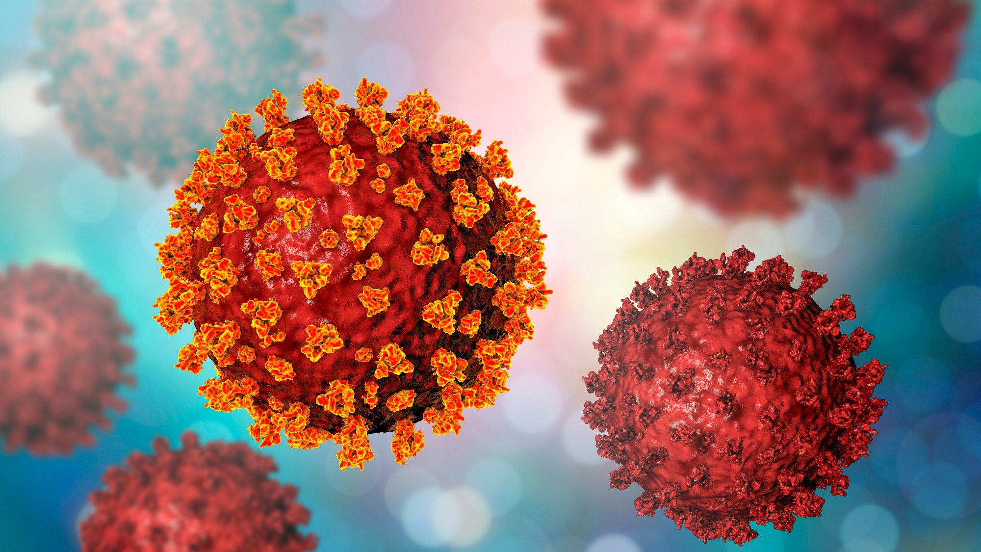
    Study: The Design, Synthesis and Mechanism of Action of Paxlovid, a Protease Inhibitor Drug Combination for the Treatment of COVID-19. Image Credit: Tobias Arhelger/Shutterstock.com

    Background 

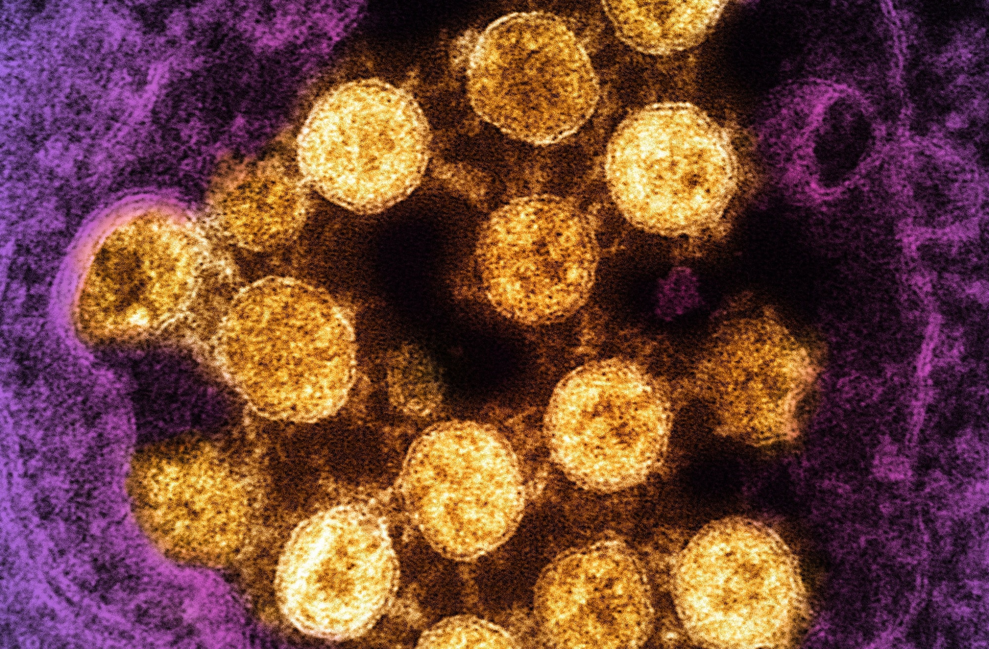
    The COVID-19 pandemic, caused by the severe acute respiratory syndrome coronavirus 2 (SARS-CoV-2) virus, significantly challenged global healthcare systems and medical science.

    In response, researchers worldwide developed vaccines with innovative mechanisms and small-molecule antivirals targeting crucial viral proteins.

    Among these, PaxlovidTM, a blend of nirmatrelvir and ritonavir PIs, stands out for its effectiveness in treating COVID-19.

    Nirmatrelvir inhibits SARS-CoV-2’s main protease, vital for viral replication, while ritonavir boosts nirmatrelvir’s effectiveness by inhibiting Cytochrome P450 3A4 (CYP3A4), an enzyme that would otherwise degrade nirmatrelvir quickly.

    Further research is needed to develop alternative main protease (MPro) inhibitors despite the success of the nirmatrelvir-ritonavir combination, ensuring continued effectiveness against COVID-19.

    PIs as antivirals for Hepatitis C virus (HCV) and Human immunodeficiency virus (HIV) 

    PI Drugs for HCV and HIV Infections

    PIs are key in treating HCV and HIV infections. HCV, a small ribonucleic acid (RNA) virus causing hepatic diseases, is targeted by PIs like asunaporevir, telaprevir, and boceprevir, focusing on the nonstructural (NS)3/4A serine protease.

    These inhibitors are peptidomimetics, containing peptide bonds and a ‘warhead’ group that binds covalently but reversibly to the enzyme’s active site.

    HIV PIs target the virus’s aspartic acid protease, which is crucial for viral replication. They are used in antiretroviral therapy, transforming HIV from fatal to chronic.

    Development and mechanism of Nirmatrelvir

    Nirmatrelvir, developed from Pfizer’s earlier SARS-CoV-1 PI .. PF-00835231, faced challenges in oral absorption.

    Modifications like altering the warhead and substituting various molecular components enhanced its binding affinity and antiviral activity, eventually leading to nirmatrelvir with a nitrile warhead, improving solubility and synthesis.

    Despite different warheads, its structural similarity to boceprevir, and its role as a covalent inhibitor of SARS-CoV-2 Mpro makes it significant in COVID-19 treatment.

    Synthesis of nirmatrelvir

    Nirmatrelvir’s synthesis involves coupling the P1 building block and the P2-P3 dipeptide, with the final step being the formation of the nitrile warhead.

    The process starts with protected amino acid derivatives, proceeding through stages like Boc-deprotection, ester cleavage, and dipeptide formation.

    The synthesis yields nirmatrelvir with high efficiency and introduces a new approach involving a Ugi-type three-component reaction for higher diastereoselectivity.

    Synthesis and structure-activity relationship (SAR) study of nirmatrelvir analogs

    Research by Chia and co-workers led to the synthesizing nirmatrelvir analogs with different P1′ moieties, examining the role of the warhead in antiviral activity.

    These studies revealed varying levels of effectiveness in protease inhibition and antiviral activity, with some derivatives showing similar or superior effects to nirmatrelvir. However, challenges in cell penetration and specificity to SARS-CoV-2 limited the broader application of these analogs.

    Novel covalent and non-covalent inhibitors of SARS-CoV-2 Mpro

    Recent developments in SARS-CoV-2 Mpro inhibitors have introduced both peptidomimetic and non-peptidic inhibitors.

    These include warheads, such as epoxide rings and fluoromethyl groups, offering alternative mechanisms of covalent binding to the enzyme.

    Non-covalent inhibitors, like ensitrelvir, show lower reactivity but better selectivity due to their secondary interaction nature. These developments represent crucial steps in diversifying therapeutic options against COVID-19 and its evolving strains.

    Ritonavir as a pharmacokinetic enhancer

    Structure, activity, and interactions of ritonavir

    Originally an HIV protease inhibitor, Ritonavir is known for its efficacy at low doses (~100 mg) in inhibiting the CYP3A4 enzyme, a crucial element in drug metabolism.

    While high doses of Ritonavir are poorly tolerated, its low-dose effectiveness is leveraged in combination therapies with other HIV protease inhibitors, enhancing their half-lives and thus reducing required dosages.

    This unique use of Ritonavir has been explored even in early COVID-19 treatments. However, its use poses risks of significant drug–drug interactions, especially with medications metabolized by CYP3A4, potentially elevating their levels to toxic concentrations.

    Additionally, Ritonavir’s effect on other enzymes and transport proteins is noted, albeit of lesser importance in Paxlovid treatment.

    Synthesis of ritonavir

    developed at Abbott Laboratories, Ritonavir’s synthesis involves complex chemical processes, combining chiral amine and carboxylic acid building blocks.

    The synthesis starts with a cyclocondensation reaction involving thioformamide and ethyl 2-chloroacetate, followed by a series of steps leading to the formation of ritonavir.

    This intricate process involves various intermediate compounds and chemical reactions, including triethylamine and 4-dimethylaminopyridine, highlighting the sophistication required in pharmaceutical synthesis.

    The production of Ritonavir demonstrates the intricate chemical engineering necessary to develop effective pharmaceutical agents.

    Paxlovid—application and activity against mutant variants

    Paxlovid, combining nirmatrelvir and ritonavir, has shown significant efficacy in reducing COVID-19-related hospitalizations and mortality.

    While it has gained emergency use authorization in various regions, its effectiveness against emerging strains and mutant variants is under continuous scrutiny.

    The evolving landscape of SARS-CoV-2 mutations necessitates ongoing monitoring to ensure the sustained efficacy of treatments like Paxlovid.

    [ad_2]

    Source link

  • SARS-CoV-2 fragments found to mimic immune system peptides, fueling inflammation

    SARS-CoV-2 fragments found to mimic immune system peptides, fueling inflammation

    [ad_1]

    In a recent study published in the journal Proceedings of the National Academy of Sciences, researchers analyzed the inflammatory capacity of fragmented components of severe acute respiratory syndrome coronavirus 2 (SARS-CoV-2).

    The intensive research during the coronavirus disease 2019 (COVID-19) pandemic has helped understand SARS-CoV-2 infection. Nevertheless, what makes the virus capable of causing a dangerous inflammatory response remains unclear. Research has suggested that amphiphilic, cationic peptides from the innate immune system undergo amyloid-like assembly with anionic nucleic acids and form proinflammatory complexes.

    Study: Viral afterlife: SARS-CoV-2 as a reservoir of immunomimetic peptides that reassemble into proinflammatory supramolecular complexes. Image Credit: NIAIDStudy: Viral afterlife: SARS-CoV-2 as a reservoir of immunomimetic peptides that reassemble into proinflammatory supramolecular complexes. ​​​​​​​Image Credit: NIAID

    The study and findings

    The present study investigated whether fragmented SARS-CoV-2 peptides assemble with anionic double-stranded RNA (dsRNA) into supramolecular complexes. The viral proteome was considered a reservoir of peptide fragments liberating after the proteolytic destruction of virions. The researchers leveraged a support vector machine (SVM) classifier to recognize antimicrobial peptide (AMP)-like sequences (xenoAMPs) in the SARS-CoV-2 proteome.

    Viral protein sequences were scanned via a moving window of 24–34 amino acids to identify potential xenoAMPs and test whether they behave like AMPs if cleaved at different positions. Sequences were selected based on the output provided by the classifier as a sigma (σ) score, wherein a strongly positive score implied the sequence was highly likely to be an AMP.

    Existence of exogenous mimics of pro-inflammatory host antimicrobial peptides (xenoAMPs) in SARS-CoV-2 proteins. (A) SARS-CoV-2 proteins are scanned with a machine-learning AMP classifier. Each queried sequence is given a σ score that measures its AMP-ness. Three representative high-scoring sequences are studied: xenoAMP(ORF1ab), xenoAMP(S), and xenoAMP(M). The grey bars mark the location where the corresponding sequences are selected. (B) SARS-CoV-2 sequences are aligned and compared to their homologs in a common cold human coronavirus HCoV-OC43: Control (ORF1ab), Control(S), and Control(M). Asterisks, colons, and periods indicate positions that have fully conserved residues, those that have strongly similar properties, and those that have weakly similar properties, respectively. Color is assigned to each residue using the ClustalX scheme. (C) σ score heatmaps compare the distribution of high-scoring sequences in three proteins from SARS-CoV-2 and HCoV-OC43. The first amino acid in each sequence is colored according to its average σ score; regions with negative average σ scores (non-AMPs) are colored white. “Hot spot” clusters of high-scoring sequences for SARS-CoV-2 (bright yellow regions bracketed in red boxes) have systematically higher scores and span wider regions of sequence space compared to HCoV-OC43. This trend suggests that hot spots in SARS-CoV-2 can generate higher scoring sequences for a greater diversity of enzymatic cleavage sites than those in HCoV-OC43.

    Existence of exogenous mimics of pro-inflammatory host antimicrobial peptides (xenoAMPs) in SARS-CoV-2 proteins. (A) SARS-CoV-2 proteins are scanned with a machine-learning AMP classifier. Each queried sequence is given a σ score that measures its AMP-ness. Three representative high-scoring sequences are studied: xenoAMP(ORF1ab), xenoAMP(S), and xenoAMP(M). The grey bars mark the location where the corresponding sequences are selected. (B) SARS-CoV-2 sequences are aligned and compared to their homologs in a common cold human coronavirus HCoV-OC43: Control (ORF1ab), Control(S), and Control(M). Asterisks, colons, and periods indicate positions that have fully conserved residues, those that have strongly similar properties, and those that have weakly similar properties, respectively. Color is assigned to each residue using the ClustalX scheme. (C) σ score heatmaps compare the distribution of high-scoring sequences in three proteins from SARS-CoV-2 and HCoV-OC43. The first amino acid in each sequence is colored according to its average σ score; regions with negative average σ scores (non-AMPs) are colored white. “Hot spot” clusters of high-scoring sequences for SARS-CoV-2 (bright yellow regions bracketed in red boxes) have systematically higher scores and span wider regions of sequence space compared to HCoV-OC43. This trend suggests that hot spots in SARS-CoV-2 can generate higher scoring sequences for a greater diversity of enzymatic cleavage sites than those in HCoV-OC43.

    Further, the team selected specific sequences from this population of (high-scoring) sequences with a high cationic charge. Specifically, they focused on prototypical candidates from the membrane (M) protein, spike (S) protein, and open reading frame 1ab (ORF1ab) polyprotein. In silico analyses showed that these xenoAMPs could be generated during proteasomal degradation, with matrix metalloproteinase 9 (MMP9) and neutrophil elastase (NE) capable of generating them.

    Next, the team compared SARS-CoV-2 xenoAMPs with homologous sequences from SARS-CoV-1 and non-pandemic human CoVs. This showed that sequences were partially conserved. A comparison of σ score heat maps of ORF1ab, S, and M proteins between SARS-CoV-2 and HCoV-OC43 revealed that high-scoring sequences were clustered into hotspots, with SARS-CoV-2 hotspots having higher scores and spanning wider regions than those of HCoV-OC43.

    Further, mass spectrometry was performed on tracheal aspirate samples from patients with severe COVID-19. The team detected fragments of host AMP, cathelicidin LL-37, in 20 samples (out of 29). By contrast, 28 samples contained viral peptide fragments, some of which had sufficiently high σ scores to qualify as xenoAMPs.

    The three xenoAMPs, xenoAMP(S), xenoAMP(M), and xenoAMP(ORF1ab), were experimentally observed to chaperone and assemble with dsRNA into complexes similar to LL-37. Polyinosine: polycytidylic acid (Poly(I:C) was used as a synthetic analog to mimic the viral dsRNA generated during replication. The structures of xenoAMPs-poly(I:C) complexes were cognate to host AMPs-dsRNA complexes.

    Next, the team investigated the robustness of these self-assembled proinflammatory complexes under non-optimal conditions. They found that the nanocrystalline structures were preserved when participating xenoAMPs were shortened. Besides, SARS-CoV-2 xenoAMPs were found to co-crystallize with LL-37, suggesting that host AMPs and xenoAMPs could synergistically activate inflammatory responses.

    The immune activation capacity of xenoAMPs from SARS-CoV-2 was compared with that of homolog peptides from HCoV-OC43 using human monocytes. XenoAMP-poly(I:C)-treated monocytes released 1.7-fold more interleukin (IL)-8 than poly(I:C) treated controls. By contrast, complexes formed with homologous peptides from HCoV-OC43 induced much lower IL-8 levels.

    In addition, xenoAMP-poly(I:C) stimulation of primary human dermal microvascular endothelial cells (HDMVECs) triggered robust production of IL-6, which was not observed with complexes formed from HCoV-OC43 peptides. Notably, xenoAMP-poly(I:C)-treated HDMVECs showed significant upregulation of several proinflammatory chemokine and cytokine genes.

    Finally, the researchers measured the immune activation capacity in mice. C57BL/6 mice unexposed to infection were treated with xenoAMP(ORF1ab)-poly(I:C) complexes or poly(I:C)-alone (control). XenoAMP(ORF1ab)-poly(I:C) treatment increased plasma levels of IL-6 and C-X-C motif chemokine ligand 1 (CXCL1) by 1.6 and 2.2 times, respectively, compared to poly(I:C)-alone. Moreover, IL-6 and CXCL1 levels increased 1.2 times in the lung compared to the control treatment.

    Conclusions

    In sum, the study has illustrated an unexpected mechanism of inflammation propagating through uninfected cells in COVID-19, wherein viral fragments mimic AMPs like LL-37. This could be salient to understand why the host immune system in COVID-19 resembles that of individuals with autoimmune conditions like rheumatoid arthritis and lupus.

    The researchers found that host proteases could generate xenoAMPs, suggesting that protease inhibitors suppressing xenoAMP generation could have a clinical impact on viral-induced inflammation. The proteolytic degradation of SARS-CoV-2 could differ across host individuals, possibly explaining the heterogeneity of infection outcomes, e.g., asymptomatic and fatal.

    Journal reference:

    • Zhang Y, Bharathi V, Dokoshi T, et al. Viral afterlife: SARS-CoV-2 as a reservoir of immunomimetic peptides that reassemble into proinflammatory supramolecular complexes. Proc Natl Acad Sci USA, 2024, DOI: 10.1073/pnas.2300644120, https://www.pnas.org/doi/10.1073/pnas.2300644120

    [ad_2]

    Source link

  • Fatigue and cognitive deficits improve over two years

    Fatigue and cognitive deficits improve over two years

    [ad_1]

    In a recent study published in the journal EClinicalMedicine, a team of scientists from Germany assessed the long-term trajectories of sequelae of severe acute respiratory syndrome coronavirus 2 (SARS-CoV-2) infections such as cognitive deficits and fatigue and attempted to identify the risk factors that could predict non-recovery from these sequelae.

    Study: Predictors of non-recovery from fatigue and cognitive deficits after COVID-19: a prospective, longitudinal, population-based study. Image Credit: p.ill.i / ShutterstockStudy: Predictors of non-recovery from fatigue and cognitive deficits after COVID-19: a prospective, longitudinal, population-based study. Image Credit: p.ill.i / Shutterstock

    Background

    Although worldwide vaccination efforts have successfully limited the transmission and severity of SARS-CoV-2 infections and lowered the morbidity and mortality associated with the coronavirus disease 2019 (COVID-19) pandemic, long coronavirus disease (long-COVID) has emerged as a serious consequential health concern. Over 60 million COVID-19 patients are believed to suffer from long-COVID, with cognitive impairments and fatigue being the most common symptoms.

    Approximately 26% of the long-COVID patients suffer from cognitive deficits, while fatigue impacts 19% of the patients, with both symptoms significantly affecting their overall quality of life and preventing the resumption of everyday activities such as work and exercise.

    Furthermore, while electronic health records of long-COVID patients indicate that cognitive deficits are observed throughout the first two years following a SARS-CoV-2 infection, the longitudinal information on fatigue is sparse. The few existing studies are primarily on older patients with preexisting comorbidities, and the results are conflicting, making it difficult to extrapolate these findings to the general population.

    About the study

    In the present study, the researchers used data from the German National Pandemic Cohort Network to evaluate the trajectories of the two most prevalent long-COVID symptoms — cognitive deficits and fatigue — over a period of 18 months in 3,000 patients. They hypothesized that long-term follow-up would indicate a recovery from both symptoms in most patients.

    The scientists also aimed to identify the risk factors that could indicate non-recovery from cognitive deficits or fatigue following COVID-19, which could be used to predict recovery rates and make informed decisions on treating these conditions. The longitudinal, prospective, multicenter, population-based study included participants above the age of 18 years who tested positive for SARS-CoV-2 through a polymerase chain reaction (PCR) test.

    Baseline assessments were conducted six months after the first SARS-CoV-2 infection, and those with reinfections were excluded from the study. Assessments for follow-up were conducted a minimum of 18 months after the SARS-CoV-2 infection.

    All participants were required to fill out an online questionnaire about fatigue, and those with symptoms that indicated post-COVID syndrome or long-COVID were invited for on-site appointments to undergo cognitive assessments. Matched controls were selected based on the PCR test date, with 30% of the baseline participants and their matched controls being invited for in-person follow-ups.

    The FACIT-Fatigue or Functional Assessment of Chronic Illness Therapy-Fatigue scale, which assesses 13 symptoms related to fatigue on a five-point scale, was used to measure one of the primary measures. Scores below the cut-off indicated recovery from fatigue, while those above the cut-off indicated persistent fatigue. The scores were used to further characterize fatigue severity.

    The Montreal Cognitive Assessment was used to assess cognitive performance, with scores between 0 and 30 indicating severe to no cognitive deficits. Educational levels were considered while assessing these scores to account for learning deficits.

    Results

    The results showed that while cognitive deficits and fatigue were the two most prevalent long-COVID symptoms, these symptoms showed improvements over two years in close to half the patients recovering from post-COVID syndrome. Furthermore, depressive symptoms and headaches were risk factors that predicted non-recovery from fatigue in the long term, while male sex, old age, and school education levels below 12 years were predictors of non-recovery from cognitive deficits.

    Compared to the pre-COVID-19 pandemic levels of fatigue, which were around 9%, clinically relevant fatigue was reported by 21% of the participants, indicating a significant health burden due to fatigue in the post-pandemic period. However, the fatigue scores were seen to improve significantly after the follow-up period of 18 months to two years.

    Psychological distress before the SARS-CoV-2 infection was thought to be linked to the persistence of fatigue since depressive symptoms were found to be one of the significant predictors of non-recovery from fatigue. Depressive symptoms and headaches could potentially be targeted for accurate diagnosis and targeted treatment of fatigue in long-COVID patients.

    Conclusions

    To summarize, the study investigated the long-term trajectories of fatigue and cognitive impairment, the two most prevalent long-COVID symptoms, in a longitudinal cohort of long-COVID patients.

    The findings suggested that while both symptoms showed improvements over a span of two years in approximately 50% of the patients, specific risk factors such as depressive symptoms and headache predicted non-recovery from fatigue in the long term. Old age and male sex were two of the risk factors indicating non-recovery from cognitive deficits in long-COVID patients.

    Journal reference:

    • Hartung, T. J., Bahmer, T., ChaplinskayaSobol, I., Deckert, J., Endres, M., Franzpötter, K., Geritz, J., Haeusler, K. G., Hein, G., Heuschmann, P. U., Hopff, S. M., Horn, A., Keil, T., Krawczak, M., Krist, L., Lieb, W., Maetzler, C., Montellano, F. A., Morbach, C., & Neumann, C. (2024). Predictors of nonrecovery from fatigue and cognitive deficits after COVID-19: a prospective, longitudinal, population-based study. EClinicalMedicine, 69.  DOI: 10.1016/j.eclinm.2024.102456, https://www.thelancet.com/journals/eclinm/article/PIIS2589-5370(24)00035-X/fulltext

    [ad_2]

    Source link

  • Congressman off-base in ad claiming Fauci shipped covid to Montana before the pandemic

    Congressman off-base in ad claiming Fauci shipped covid to Montana before the pandemic

    [ad_1]

    A fundraising ad for U.S. Rep. Matt Rosendale (R-Mont.) shows a photo of Anthony Fauci, former director of the National Institute of Allergy and Infectious Diseases, behind bars, swarmed by flying bats.

    Rosendale, who is eyeing a challenge to incumbent Sen. Jon Tester, a Democrat, maintains that a Montana biomedical research facility, Rocky Mountain Laboratories in Hamilton, has a dangerous link to the pandemic. This claim is echoed in the ad:

    “It’s been revealed that Fauci brought COVID to the Montana one year before COVID broke out in the U.S!,” it charges in all-caps before asking readers to “Donate today and hold the D.C. bureaucracy accountable!”

    The ad, paid for by Matt Rosendale for Montana, seeks contributions through WinRed, a platform that processes donations for Republican candidates. Rosendale also shared the fundraising pitch on his X account Nov. 1, and it remained live as of early February.

    Rosendale made similar accusations on social media, during a November speech on the U.S. House floor, and through his congressional office. Sometimes his comments, like those on the House floor, are milder, saying the researchers experimented on “a coronavirus” leading up to the pandemic. Other times, as in an interview with One America News Network, he linked the lab’s work to covid-19’s spread.

    In that interview clip, Rosendale recounted pandemic-era shutdowns before saying, “And now we’re finding out that the National Institute of Health, Rocky Mountain Lab, down in Hamilton, Montana, had also played a role in this.”

    Rosendale’s statements echo broader efforts to scrutinize how research into viruses happens in the United States and is part of a continued wave of backlash against scientists who have studied coronaviruses. Rosendale is considering seeking the Republican nomination to challenge Tester, in a toss-up race that could help determine which party controls the Senate in 2025. Political newcomer Tim Sheehy is also seeking the Republican nomination for the Senate.

    Rosendale proposed amendments to a health spending bill that would ban pandemic-related pathogen research funding for Rocky Mountain Laboratories and cut the salary of one of its top researchers, virologist Vincent Munster, to $1. The House has included both amendments in the Health and Human Services budget bill that the Republican majority hopes to pass. A temporary spending bill is funding the health department until March.

    We contacted Rosendale’s congressional office multiple times — with emails, a phone call, and an online request — asking what proof he had to back up his statements that the Montana lab infected bats with covid from China before the outbreak. We got no reply.

    Kathy Donbeck, of the National Institute of Allergy and Infectious Diseases’ Office of Communications and Government Relations, said in an email that the ad’s claims are false. Interviews with virologists and a review of the research paper published shortly before Rosendale’s assertions support that position.

    Where this is coming from

    Rosendale’s statements seem to stem from a Rocky Mountain Laboratories study from 2016 that looked into how a coronavirus, WIV1-CoV, acted in Egyptian fruit bats. The work, published by the journal Viruses in 2018, showed that the specific strain didn’t cause a robust infection in the bats.

    The study did not receive widespread attention at the time. But on Oct. 30, 2023, the study was highlighted by a blog called White Coat Waste Project, which says its mission is to stop taxpayer-funded experiments on animals. Some right-wing media outlets began to connect the Montana lab with the coronavirus that causes covid.

    Rosendale’s office issued an Oct. 31 news release saying the Wuhan Institute of Virology in China “shipped a strain of coronavirus” to the Hamilton lab. “Our government helped create the Wuhan flu, then shut the country down when it escaped from the lab,” Rosendale said.

    It’s a different virus

    Rocky Mountain Laboratories is a federally funded facility as part of NIAID, the nation’s top infectious disease research agency, which Fauci led for nearly 40 years.

    According to the study and Donbeck’s email, the Montana researchers focused on a coronavirus called WIV1-CoV, not the covid-causing SARS-CoV-2. They’re different viruses.

    “The genetics of the viruses are very different, and their behavior biologically is very different,” said Troy Sutton, a virologist with Pennsylvania State University who has studied the evolution of pandemic influenza viruses.

    In a review of media reports on the Montana study, Health Feedback, a network of scientists that fact-checks health and medical media coverage, showed the virus’s lineage indicated that WIV1 “is not a direct ancestor or even a close relative of SARS-CoV-2.”

    Additionally, the description of the coronavirus strain as being “shipped” suggests that it physically traveled across the world. That’s not what happened.

    The Wuhan Institute of Virology provided the WIV1 virus’s sequence that allowed researchers to make a lab-grown copy. A separate study, published in 2013 by the journal Nature, outlines the origins of the lab-created virus.

    According to the study’s methodology, the researchers used a clone of WIV1. An NIAID statement to Lee Enterprises, a media company, said the virus “was generated using common laboratory techniques, based on genetic information that was publicly shared by Chinese scientists.”

    Stanley Perlman, a University of Iowa professor who studies coronaviruses and serves on the federal advisory committee that reviews vaccines, said Rosendale’s claim is off-base.

    He said Rosendale’s focus on where the lab got its materials is irrelevant and serves “only to make people wary and scared.”

    Rosendale’s efforts to prohibit particular research at Rocky Mountain Laboratories appear ill-informed, too. Rosendale targeted banning gain-of-function research, which involves altering a pathogen to study its spread. In her email, NIAID’s Donbeck said the Rocky Mountain Laboratories study didn’t involve gain-of-function research.

    This type of research has long been controversial, and people who study viruses have said the definition of “gain of function” is problematic and insufficient to show when research, or even work to create vaccines, could cross into that type of research.

    But both Sutton and Perlman said that, any way you look at it, the Rocky Mountain Laboratories study published in 2018 didn’t change the virus. It put a virus in bats and showed it didn’t grow.

    And it had no effect on the covid outbreak a year later, first detected in Washington state.

    Our ruling

    Rosendale’s ad said, “It’s been revealed that Fauci brought COVID to the Montana one year before COVID broke out in the U.S!” The campaign ad and Rosendale’s similar statements refer to research at the Rocky Mountain Laboratories involving WIV1, a coronavirus that researchers say is not even distantly close in genetic structure to SARS-CoV-2, the virus that caused covid-19.

    Rosendale’s claim is wrong about when the scientists began their work, what they were studying, and where they got the materials. The researchers began their work in 2016 and, although they were studying a coronavirus, it wasn’t the virus that causes covid. The Montana scientists used a lab-grown clone of WIV1 for their research. The first laboratory-confirmed case of covid was not detected in the U.S. until Jan. 20, 2020. Rosendale’s ad is inaccurate and ridiculous. We rate it Pants on Fire!

    Sources:

    Viruses, “SARS-Like Coronavirus WIV1-CoV Does Not Replicate in Egyptian Fruit Bats (Rousettus aegyptiacus),” Dec. 19, 2018

    White Coat Waste Project, “Horror Show: Shady Zoo Sent Bats to NIH to Be Infected With a Wuhan Lab Coronavirus,” Oct. 30, 2023

    MattForMontana X post, Nov. 1, 2023

    Campaign ad, accessed Dec. 14, 2023

    Rep. Matt Rosendale, House floor speech, Nov. 14, 2023

    One America News Network, interview, accessed Dec. 14, 2023

    Rosendale congressional office, “Rep. Rosendale Reacts to Reports That Wuhan Lab Shipped Coronavirus to Fauci-Run Lab in Hamilton Prior to Pandemic,” Oct. 31, 2023

    National Institute of Allergy and Infectious Diseases, “History of Rocky Mountain Labs (RML),” accessed Dec. 14, 2023

    Email exchange with NIAID, beginning Dec. 14, 2023

    Statement from NIAID provided to Lee Enterprises, accessed Jan. 2, 2024

    Nature, “Isolation and Characterization of a Bat SARS-Like Coronavirus That Uses the ACE2 Receptor,” Oct. 30, 2013

    Ravalli Republic, “Rosendale Moves to Strip Rocky Mountain Lab Research Funding,” Nov. 17, 2023

    Interview, Troy Sutton, assistant professor of veterinary and biomedical sciences at Pennsylvania State University, Jan. 5, 2024

    Interview, Stanley Perlman, professor of microbiology and immunology and professor of pediatrics at the University of Iowa, Jan. 13, 2024

    FDA, “Roster of the Vaccines and Related Biological Products Advisory Committee,” accessed Jan. 16, 2024

    Health Feedback, “2018 Coronavirus Research in NIAID Montana Lab Is Unrelated to the COVID-19 Pandemic, Contrary to Claim by Fox News’s Jesse Watters,” last accessed Jan. 17, 2024

    Email exchange with OpenSecrets, an independent research group tracking money in politics, beginning Jan. 30, 2024

    CDC Museum COVID-19 Timeline, accessed Feb. 2, 2024


    Kaiser Health NewsThis article was reprinted from khn.org, a national newsroom that produces in-depth journalism about health issues and is one of the core operating programs at KFF – the independent source for health policy research, polling, and journalism.


    [ad_2]

    Source link

  • COVID-19 recovery disparities uncovered among racial and ethnic groups

    COVID-19 recovery disparities uncovered among racial and ethnic groups

    [ad_1]

    In a recent study published in Frontiers in Public Health, researchers from the United States of America (US) investigated the racial and ethnic variation in symptoms, activity level, health status, and missed work.

    Depiction of the SARS-CoV-2 virus
    Study: Ethnic and racial differences in self-reported symptoms, health status, activity level, and missed work at 3 and 6 months following SARS-CoV-2 infection. Image Credit: Kateryna Kon/Shutterstock.com

    They assessed this via follow-ups post-initial infection with severe acute respiratory syndrome coronavirus 2 (SARS-CoV-2). Although the symptoms were equally prevalent among the groups, they found that three and six months post-infection, Hispanic participants reported poorer health and reduced activity compared to non-Hispanic participants.

    Further, racial minority participants reported more negative impacts on health status, activity, and absence from work as compared to the White population.

    Background

    The coronavirus disease 2019 (COVID-19) pandemic highlighted disparities, wherein ethnic and racial minoritized populations were observed to face greater infection risks due to the essential nature of their work, limited remote work options, and challenges in practicing social distancing.

    The infected individuals faced barriers to care, including underinsurance and lack of primary care, with economic consequences. Disparities persisted in health outcomes, for example, higher hospitalization and mortality rates among Black and Hispanic populations.

    Despite these challenges, recovery-related differences after SARS-CoV-2 infection remained understudied, with existing studies having limitations like varied follow-up durations, inconsistent findings, and insufficient consideration of social health determinants.

    Researchers in the present study aimed to address this gap. The study assessed symptoms and health-related effects following SARS-CoV-2 infection across ethnicities and races, aiming to guide equitable health interventions effectively.

    About the study

    In the present study, a secondary analysis was performed using data from a US-based, prospective, multicenter, longitudinal cohort study named Innovative Support for Patients with SARS-CoV-2 Infections Registry (INSPIRE). The primary cohort involved adults positive for SARS-CoV-2 infection enrolled from December 2020 to July 2022, along with a SARS-CoV-2-negative group for considering non-SARS-CoV-2-related effects.

    A total of 3,161 participants completed enrollment and reported symptoms and other outcomes every three months via surveys. Out of these participants, 2,402 were SARS-CoV-2-positive and 759 SARS-CoV-2-negative.

    Among the SARS-CoV-2-positive participants, 14.0% were Hispanic, 11.0% were Asian, 7.9% were Black, 9.9% were categorized as Other/Multiple races, and 71.1% were White. Among the SARS-CoV-2-negative participants, 16.5% were Hispanic, 14.8% were Asian, 13.1% were Black, 8.1% were categorized as Other/Multiple races, and 64% were White.

    The researchers evaluated 21 COVID-19-like symptoms and “other symptoms” at enrollment and three and six months post-infection using the Centers for Disease Control and Prevention’s symptom list.

    During the follow-ups, health status (rated on a 5-point scale from excellent to poor), activity level compared to pre-SARS-CoV-2 symptoms (same, somewhat less, much less), and missed work in the past three months (categorized into workdays) were assessed.

    Data were collected on ethnicity and race. Interactions between ethnicity or race and SARS-CoV-2 infection status were considered.

    Generalized estimating equations (GEE) logistic regression was used to estimate marginal odds ratios (ORs) for various outcomes, adjusting for SARS-CoV-2 infection status, demographic factors, substance use, social determinants of health, pre-existing health conditions, COVID-19 vaccination status, and survey time point.

    Results and discussion

    Post SARS-CoV-2 infection, symptoms were found to be mostly similar across ethnic and racial groups over time. At three months, Hispanic individuals had higher odds of reporting fair/poor health (OR = 1.94) and reduced activity compared to their non-Hispanic counterparts. No significant differences by ethnicity were observed at six months.

    At three months, participants of Other/Multiple races had higher odds of reporting fair/poor health (OR = 1.9) and reduced activity compared to White participants. At six months, Asian participants had a greater probability of reporting fair/poor health (OR = 1.88), Black individuals reported more missed work (OR = 2.83), and Other/Multiple race participants reported more health issues (OR = 1.83), reduced activity, and missed work (OR = 2.25).

    The findings help to improve our understanding of the ethnic and racial disparities in outcomes after SARS-CoV-2 infection and could be used to inform clinical and public health initiatives and policy.

    However, the study is limited by small sample sizes in ethnic and racial subgroups, lack of adjustment for insurance and frontline worker status, potential participant representativeness issues, variations in response rates, lack of exploration of neurological and mental health sequelae, recruitment at different pandemic stages, and the absence of adjustments for multiple comparisons.

    Conclusion

    In conclusion, the findings suggest that while the symptom prevalence was similar among the groups, the ethnic and racial minority groups suffered adverse effects on health status, activity level, and absence from work as compared to non-Hispanic and White populations, respectively.

    Examining the underlying factors contributing to these differences could aid the efforts to promote health equity and improve our preparedness for future pandemics.

    Journal reference:

    • O’Laughlin KN, Klabbers RE, Mannan IE, et al. (2024). Ethnic and racial differences in self-reported symptoms, health status, activity level, and missed work at 3 and 6 months following SARS-CoV-2 infection. Frontiers in Public Health. doi: 10.3389/fpubh.2023.1324636.  https://www.frontiersin.org/articles/10.3389/fpubh.2023.1324636/full

    [ad_2]

    Source link

  • Researchers assess effectiveness of contact tracing in controlling COVID-19 pandemic

    Researchers assess effectiveness of contact tracing in controlling COVID-19 pandemic

    [ad_1]

    During the COVID-19 pandemic, Switzerland, like many other countries, relied on contact tracing to identify people likely to have been contaminated by an infected acquaintance. Has this strategy, which is standard in epidemic outbreaks, proved effective in interrupting viral transmission? Epidemiologists from the University of Geneva (UNIGE) and the Geneva University Hospitals (HUG) have analyzed the data collected in Geneva. Overall, 40% of people infected were identified via sick acquaintances. This rate fluctuated, however, depending on the variant involved, the type of housing, and neighbourhood wealth. These results, published in the journal EuroSurveillance, suggest that monitoring contacts alone is not enough to control certain epidemics. It needs to be supplemented by a range of measures that take into account the specific features of each disease.

    Contact tracing aims to identify people who have been in contact with an infected person so that they can be treated before they further transmit the disease.

    “The effectiveness of this strategy depends mainly on the characteristics of the disease in terms of symptoms, contagiousness and modes of transmission,” explains Delphine Courvoisier, assistant professor in the Department of Medicine at the UNIGE Faculty of Medicine, epidemiologist at the HUG Division of Quality of Care and delegated by the HUG as head of the ‘data’ unit at the Cantonal Medical Service during the COVID-19 pandemic, who directed this work. “In the case of Ebola, for example, where patients are only contagious after the onset of symptoms, or closer to us in the case of measles, contact tracing has proved its effectiveness in cutting transmission chains.”

    To assess the effectiveness of contact tracing for COVID-19, Delphine Courvoisier and her team analysed data of more than 140,000 cases and 185,000 contacts recorded in the canton of Geneva between June 2020 and March 2022.

    Voluntary or involuntary non-declaration?

    To determine the number of people identified by contact tracing, we first need to determine the number of people who infect each other. To do this, we looked at how many people living at the same address tested positive for SARS-Cov2 within a ten-day period.”

    Denis Mongin, research fellow in the Department of Medicine at the UNIGE Faculty of Medicine, statistician at the HUG, and expert delegated to data processing

    “Then, to remove the element of chance, we carried out a permutation test by randomly assigning an address to people. The difference between the number of people testing positive within a ten-day interval at the same address before and after permutation indicates the number of people infected at home, which is then compared with the people who had been declared as contacts. In this way, we were able to estimate the overall rate of reporting of contacts, as well as its evolution over time and its dependence on the socio-economic profile of neighbourhoods, the type of buildings, and population density.”

    On average, around 40% of infected people could be identified by contact tracing, with variations ranging from 25% at the height of epidemic waves to 60% during calmer periods. Socio-economic factors also play a major role. For example, the larger the building and the more communal areas (ground-floor shops, for example), the more likely people were not to report their contacts. “This is probably due to unintentional omissions: people bump into each other without thinking about it, they don’t necessarily know their neighbours, or the virus remains suspended in the air, for instance in the lift,” explains Denis Mongin. “What’s more, this effect disappears during the phases of restricted gatherings and compulsory masking, which also enables us to assess the effectiveness of these measures.”

    On the other hand, the higher the socio-economic status of the neighbourhoods, the fewer people reported their contacts. “Many hypotheses have been put forward: less compliance with government directives, but also greater possibility of self-isolation due to the size of housing and professions that allow people to work from home, without the need for a medical certificate,” points out Delphine Courvoisier. “In any case, this demonstrates the importance of involving sociologists and anthropologists in the development and evaluation of health policies, in order to understand the human factors involved in their success or failure.”

    One measure among many

    COVID-19 is a highly contagious disease, transmitted by aerosols and infectious before the onset of symptoms. These characteristics make contact tracing particularly complex. In the light of these results, was this strategy the best solution for reducing transmission chains? «Contact tracing alone had only a relative effect on the dynamics of the epidemic. But its importance as a psychological support for the population should not be overlooked, to reassure and listen to people during this anxiety-inducing period. The idea is not to rewrite history and call into question decisions that made sense at the time, but to use these experiences to build a more solid, multimodal response when we are faced with another large-scale epidemic”, conclude the authors.

    Source:

    Journal reference:

    Mongin, D., et al. (2024). Time trends and modifiable factors of COVID-19 contact tracing coverage, Geneva, Switzerland, June 2020 to February 2022. Eurosurveillance. doi.org/10.2807/1560-7917.es.2024.29.3.2300228.

    [ad_2]

    Source link

  • Scientists coax a deadly bacterium to destroy itself from the inside out

    Scientists coax a deadly bacterium to destroy itself from the inside out

    [ad_1]

    Northwestern University researchers have successfully coaxed a deadly pathogen to destroy itself from the inside out.

    In the new study, researchers modified DNA from a bacteriophage or “phage,” a type of virus that infects and replicates inside of bacteria. Then, the research team put the DNA inside Pseudomonas aeruginosa (P. aeruginosa), a deadly bacterium that is also highly resistant to antibiotics. Once inside the bacterium, the DNA bypassed the pathogen’s defense mechanisms to assemble into virions, which sliced through the bacterium’s cell to kill it.

    Building on a growing interest in “phage therapies,” the experimental work represents a critical step toward engineering designer viruses as new therapeutics to kill antibiotic-resistant bacteria. It also reveals vital information about the innerworkings of phages, a little-studied area of biology.

    The study will be published on Wednesday (Jan. 24) in the journal Microbiology Spectrum.

    “Antimicrobial resistance is sometimes referred to as the ‘silent pandemic,’” said Northwestern’s Erica Hartmann, who led the work. “The numbers of infections and deaths from infections are increasing worldwide. It’s a massive problem. Phage therapy has emerged as an untapped alternative to our reliance on using antimicrobials. But, in many ways, phages are microbiology’s ‘final frontier.’ We don’t know much about them. The more we can learn about how phage work, the more likely we can engineer more effective therapeutics. Our project is cutting-edge in that we are learning about phage biology in real time as we engineer them.”

    An indoor microbiologist, Hartmann is an associate professor of civil and environmental engineering at Northwestern’s McCormick School of Engineering and a member of the Center for Synthetic Biology.

    Desperate need for antibiotic alternatives

    Associated with an increase in antimicrobial use, the rise of antibacterial resistance is an urgent and growing threat to the global population. According to the Centers for Disease Control and Prevention (CDC), nearly 3 million antimicrobial-resistant infections occur each year in the United States alone, with more than 35,000 people dying as a result.

    The growing crisis has motivated researchers to look for alternatives to antibiotics, which are continually losing effectiveness. In recent years, researchers have started to explore phage therapies. But even though billions of phages exist, scientists know very little about them.

    For every bacterium that exists, there are dozens of phages. So, there is an astronomically large number of phages on Earth, but we only understand a handful of them. We haven’t necessarily had the motivation to really study them. Now, the motivation is there, and we are increasing the number of tools we have to dedicate to their study.”


    Erica Hartmann, Northwestern University

    Treatment without side effects

    To explore potential phage therapies, researchers either pinpoint or modify an existing virus to selectively target a bacterial infection without disrupting the rest of body. Ideally, scientists one day could tailor a phage therapeutic to infect a specific bacterium and design “a la carte” therapeutics with precise traits and characteristics to treat individual infections.

    “What’s powerful about phage is it can be very specific in the way that antibiotics are not,” Hartmann said. “If you take an antibiotic for a sinus infection, for example, it disrupts your entire gastrointestinal tract. A phage therapy can be designed to affect only the infection.”

    While other researchers have investigated phages therapies, almost all of those studied have focused on using phages to infect Escherichia coli. Hartmann, however, decided to focus on P. aeruginosa, one of the five most deadly human pathogens. Particularly dangerous for people with compromised immune systems, P. aeruginosa is a leading cause of hospital infections, often infecting patients with burn or surgery wounds as well as lungs in people with cystic fibrosis.

    “It is one of the highest priority, multi-drug resistant pathogens that many people are really concerned about,” Hartmann said. “It is extremely drug resistant, so there is an urgent need to develop alternative therapeutics for it.”

    Mimicking infection, bypassing defenses

    In the study, Hartmann and her team started with P. aeruginosa bacteria and purified DNA from several phages. Then, they used electroporation -; a technique that delivers short, high-voltage pulses of electricity -; to poke temporary holes in the bacteria’s outer cell. Through these holes, phage DNA entered the bacteria to mimic the process of infection.

    In some cases, the bacteria recognized the DNA as a foreign object and shredded the DNA to protect itself. But after using synthetic biology to optimize the process, Hartmann’s team was able to knock out the bacteria’s antiviral self-defense mechanisms. In these cases, the DNA successfully carried information into the cell, resulting in virions that killed the bacteria.

    “Where we were successful, you can see dark spots on the bacteria,” Hartmann said. “This is where the viruses burst out of the cells and killed all the bacteria.”

    After this success, Hartmann’s team introduced DNA from two more phages that are naturally unable to infect their strain of P. aeruginosa. Yet again, the process worked.

    Phage manufacturing in a cell

    Not only did the phage kill the bacteria, the bacteria also ejected billions more phages. These phages can then be used to kill other bacteria, like those causing an infection.

    Next, Hartmann plans to continue modifying phage DNA to optimize potential therapies. For now, her team is studying the phages expelled from the P. aeruginosa.

    “This is an important piece in making phage therapies,” she said. “We can study our phage in order to decide which ones to develop and eventually mass produce them as a therapeutic.”

    The study, “A synthetic biology approach to assemble and reboot clinically relevant Pseudomonas aeruginosa tailed phages,” was supported by the Walder Foundation, the National Science Foundation and the National Institutes of Health.

    Source:

    Journal reference:

    Ipoutcha, T., et al. (2024) A synthetic biology approach to assemble and reboot clinically relevant Pseudomonas aeruginosa tailed phages. Microbiology Spectrum. doi.org/10.1128/spectrum.02897-23.

    [ad_2]

    Source link