Like humans struggling to get through the COVID-19 pandemic, bacterial cells need social distancing to thwart viruses. But in some situations, such as inside elevators or within the candy-colored bacterial structures known as “pink berries,” staying apart just isn’t feasible.
Looking like spilled Nerds or Pop Rocks, the communal, multicellular pink berries litter the submerged surface of salt marshes in and around Woods Hole. New research conducted at the Marine Biological Laboratory (MBL) uncovers evidence that a genetic mechanism may help the berry-building bacteria -; and others like them -; protect against disease. The study, published this week in Proceedings of the National Academy of Sciences, also has implications for understanding the evolution of single-celled organisms, like bacteria, into complex multicellular ones, including humans.
It tells us about the challenges we faced back when we were little balls of cells. If you’re forming multicellular structures, you’ve got to evolve some pretty fancy immune defenses in order to stay alive.”
Lizzy Wilbanks, MBL Whitman Fellow and microbiologist at the University of California, Santa Barbara
Mysterious, mutation-generating systems
Wilbanks first encountered the pink berries as a graduate student enrolled in MBL’s Microbial Diversity course. These spherical aggregates are among the structures bacteria form when genetically similar individuals stick close together and coordinate their activity. The pink berries are populated by a species of bacteria called Thiohalocapsa PSB1, which feeds itself using sulfur and light, plus a relatively small number of other symbiotic bacteria. By working together, these cells create pockets free of oxygen, which could poison them, and acquire the weight necessary to settle safely into their ideal habitat.
Like all organisms, these cooperative microbes risk contracting viruses from their environment. Pink berries and other multicellular bacteria have a heightened need for protection, since -; like us -; they are composed of genetically similar cells packed tightly together, with no social distancing possible.
“It’s a perfect cocktail for an epidemic to blow through and wipe out everything,” Wilbanks says.
Through her collaborator Blair Paul, assistant scientist at MBL, Wilbanks learned about an unusual genetic mechanism that they found to be abundant within Thiohalocapsa. Known as diversity-generating retroelements (DGRs), this system contains sections of DNA that are transcribed into RNA and back into DNA through an error-prone process, then inserted into a target gene for mutation.
In this way, DGRs introduce lots of new genetic variation, the raw material for adaptation, into specific spots within the genomes. Scientists have found these systems in viruses, bacteria, and other microbes called archaea, yet they don’t fully understand how the microbes use them.
Wilbanks and Hugo Doré, then a postdoctoral scientist in her lab and the study’s first author, began discussing what DGRs might accomplish for Thiohalocapsa. Through their research, they learned the DGRs’ target genes include components related to those found in the immune systems of multicellular organisms, including humans, plants and even some fungi. The similarity to pieces of other organisms’ immune systems prompted the researchers to suspect the DGRs might diversify the sensor proteins Thiohalocapsa uses to defend against pathogens, analogous to the antibodies in our own immune systems.
All living organisms need to detect threats they have never encountered before. Humans and other vertebrates solve this problem by shuffling and mutating genes for their sensor proteins (antibodies) to generate a diverse army of sentinels. Though recent research has shown many components of our innate immune systems evolved from bacterial ancestors, scientists have never before seen in bacteria anything like our hyper-diverse antibodies.
A widespread immunological connection
The team first looked broadly at DGRs found in bacteria and archaea, focusing on the gene responsible for turning RNA back into DNA. This method divides the DGRs from bacteria and archaea into two groups. Within the group to which Thiohalocapsa belongs, they found that 82 percent of DGRs belong to microbes that form many-celled, cooperative structures, akin to the pink berries. Even though they belonged to distantly related microbes, the DGRs’ alterations tend to affect the same kind of immune system genes as they do in Thiohalocapsa.
Examining hundreds of individual pink berries, they found that DGRs had been actively diversifying 14 of the 15 total target genes in Thiohalocapsa. The amount of the variation found for these genes changed, however, depending on the site from which the pink berries had been collected. The viruses in pools in the same marsh may vary -; perhaps driving the differences the team saw.
“The next frontier is showing what Thiohalocapsa is actually doing with its DGRs in the environment,” Wilbanks says.
In addition to offering a peek at the evolution of life, this research has practical implications. Wastewater treatment plants use multicellular bacteria to remove nutrients that can harm local ecosystems, and federal and industrial researchers are exploring a host of other applications for engineered clumps of microbes. These microbial structures face the same challenge -; viral epidemics -; as the pink berries. When engineering these microbial systems, Wilbanks says, it makes sense to mimic the DGR-based immunity of wild communal bacteria.
H. Doré, H., et al. (2024). Targeted hypermutation of putative antigen sensors in multicellular bacteria. Proceedings of the National Academy of Sciences. doi.org/10.1073/pnas.2316469121.
Figures published today by the UK Health Security Agency (UKHSA) in its TB annual report, show that tuberculosis cases in England in 2022 were stable compared to 2021 (4,380 in 2022 compared to 4,411 in 2021).
However, additional provisional data indicate that cases of tuberculosis (TB) in England rose by 10.7% in 2023 compared to 2022 (4,850 compared to 4,380). The rise signals a rebound of TB cases to above the pre-COVID-19-pandemic numbers.
While England remains a low incidence country for TB, the current trajectory takes the UK further from the pathway to meet World Health Organization (WHO) 2035 elimination targets. UKHSA is working with partners to investigate the reasons behind the increase in TB.
TB is a bacterial infection that most frequently affects the lungs, which is when it is infectious. Symptoms include:
a cough that lasts more than 3 weeks
high temperature
drenching night sweats
loss of appetite
weight loss
It can be treated with a prolonged course of antibiotics but can be serious, particularly if not treated.
The proportion of TB notifications accounted for by people born outside the UK has been steadily rising for a number of years. However, the increase in TB in 2023 has now been seen in both UK born and non-UK born populations in England. The largest rises in cases have been in the urban centres of London, the North West and West Midlands. However, there has also been increases in the South West and North East regions where TB incidence is low.
Tuberculosis continues to be associated with deprivation and is more common in large urban areas. People born outside the UK, especially in countries in South Asia (India, Pakistan, Bangladesh), Africa (Eritrea, Nigeria) and Eastern Europe (Romania) experience the highest number of cases. For those born in the UK, TB is more common among those who experience homelessness, drug and alcohol dependence and have had contact with the criminal justice system. TB rates are much higher in UK born individuals from ethnic groups other than white.
We need collective action to tackle TB and we are working with partners across the health system to understand how we can best refocus efforts to stamp out this preventable and treatable infection.
Not every persistent cough, along with a fever, is caused by flu or COVID-19. A cough that usually has mucus and lasts longer than 3 weeks can be caused by a range of other issues, including TB. Please speak to your GP if you think you could be at risk.
Dr Esther Robinson, Head of the TB Unit at UKHSA
UKHSA continues to work with NHSE and other partners on the TB action plan, which sets out steps to improve the prevention and detection of TB, along with increasing capacity in the TB workforce.
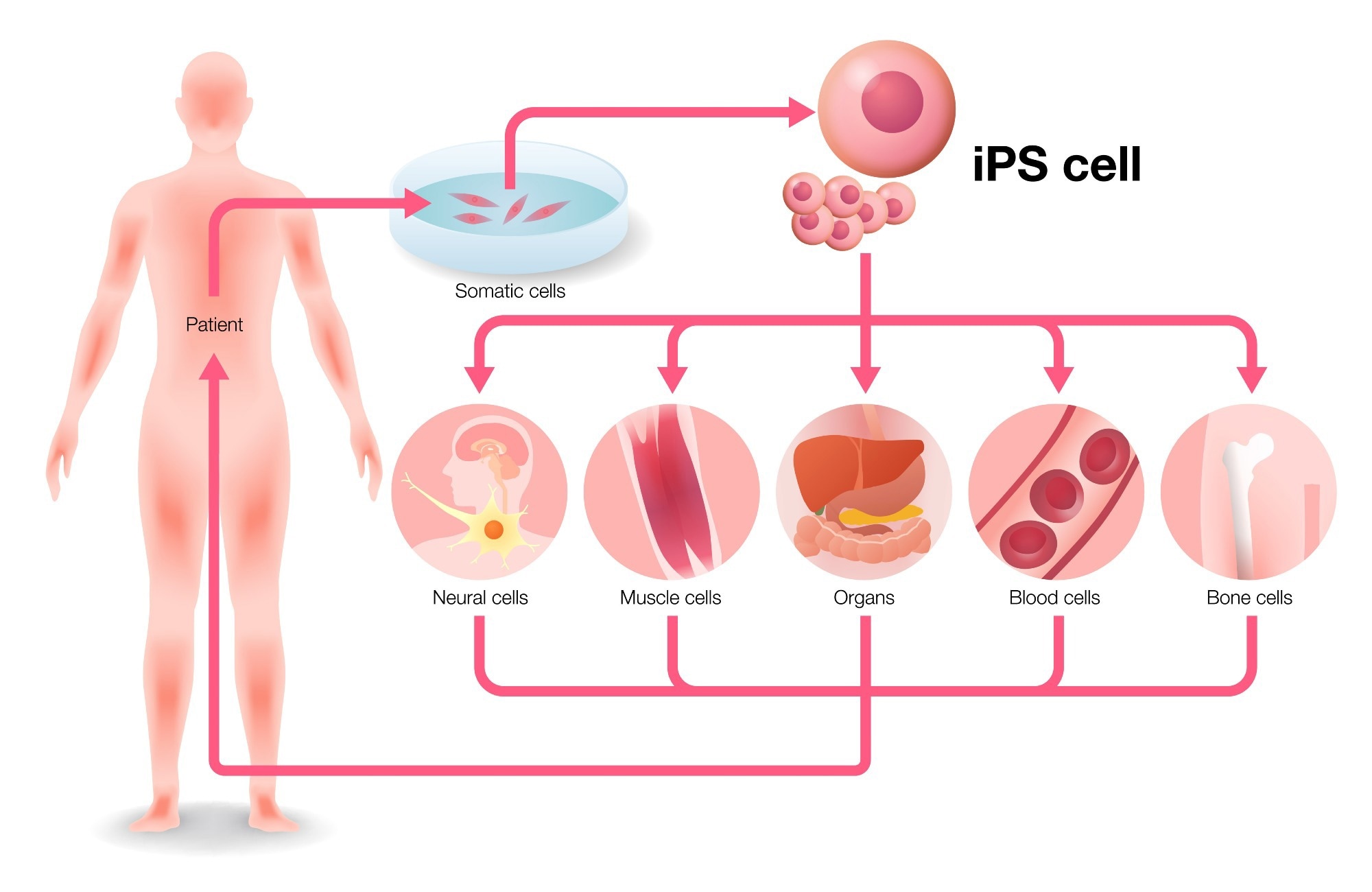
In this interview, we speak to Keith Olson and Coby Carlson from FUJIFILM Cellular Dynamics about their iPSC technology and how it is making breakthroughs in blood-brain barrier research.
Please can you introduce yourself and tell us about your role at FUJIFILM Cellular Dynamics?
Coby Carlson: My name is Coby Carlson, and I lead the Applications Team at FUJIFILM Cellular Dynamics. Our team is separate from the R&D scientists, staff, and experts focusing on stem cell culture and differentiations to create unique, specialized cell types. Our role is to test the functionality of these cells, demonstrate their performance on various platform technologies, and develop applications.
Keith Olson: My name is Keith Olson, and I am the Executive Vice President for Commercial Operations at FUJIFILM Cellular Dynamics.
FUJIFILM Cellular Dynamics is a global leader in developing and manufacturing human induced pluripotent stem cells (iPSC). Please can you tell us more about some of your core aims and missions as a company?
Coby Carlson: Fujifilm’s core technologies include reprogramming, engineering, and iPSC culture and differentiation to create unique cell types. Our mission is to bring these technologies to an industrial scale. The technology was first invented over 15 years ago, and since then, we have continuously improved and optimized it to launch it on a large scale. Our goal is to make different cell types accessible all over the world.
Keith Olson: I believe that we were pioneers of iPSC technology in the United States. We successfully brought it to market, commercialized it, and built a business around it. Our reputation is now based on our core expertise, quality, and ability to manufacture at scale for our clients in the pharmaceutical, biotech, and academic sectors.
Image Credit: metamorworks/Shutterstock.com
iPSC technology can be used to revolutionize scientific research and cell therapy. Why is this, and in what application areas within scientific research does this technology benefit the most?
Coby Carlson: The primary focus of people working with iPSC technology is toxicology/safety pharm, disease modeling, and cell therapy. Safety is crucial because we can generate cardiomyocytes from iPS cells, and these cells spontaneously beat in a dish while responding to known cardiotoxic molecules.
To measure the safety and effectiveness of drugs in a dish, we use iPSC-derived cardiomyocytes in safety toxicology. We have also participated in the CiPA (Comprehensive in Vitro Proarrhythmia Aassay) initiative to standardize this process across various labs globally. The recent FDA Modernization Act has suggested using alternatives to animal testing, making iPSC-derived cardiomyocytes a crucial component of measuring the safety and effectiveness of drugs going forward.
Keith Olson: iPSC delivers a specific gap in the market by providing customers with high-quality human cells. Instead of researching animal models or transformed cell lines, researchers can now use real human cardiomyocytes or neurons for more biologically relevant results.
At FUJIFILM Cellular Dynamics, you offer a range of iPSC cells, including neural and cardiac cells. Can you tell us more about some of the products you offer and how they are generated?
Coby Carlson: The uniqueness of iPSC technology lies in the fact that there are not many human donors who can offer their brain cells. This technology provides access to different cell types, and we can differentiate highly specialized cells to obtain unique features such as excitatory neurons and microglia. These unique cells are essential for in vitro assays, where we can test them differently than we would in an animal model. For instance, mouse models are different from human models.
Keith Olson: Everything starts from a core iPS cell bank, and we have developed specific differentiation protocols for each terminal phenotype of the cell. Whether it is a cardiomyocyte, neuron, or hepatocyte, we have a differentiation protocol that takes us from the core stem cell bank to a finished product that customers can purchase and use as an assay-ready sample.
Image Credit:Magic mine/Shutterstock.com
The blood-brain barrier (BBB) is vital in understanding how the brain functions, and its dysfunction can lead to neurological conditions such as MS, stroke, and epilepsy. Why did you choose to focus on this area, and how can your iPSC technology help to accelerate breakthroughs?
Coby Carlson: Researchers and drug developers have long struggled with the blood-brain barrier, a critical cell structure separating the blood from the brain. The barrier keeps out harmful pathogens and toxins while allowing vital nutrients to enter the brain. However, it also prevents drugs from crossing over and treating neurological diseases such as neurodegenerative disorders.
The main challenge has been replicating this barrier in vitro. One of the reasons we pursued iPSC cell technology is that we developed ways to distinguish specialized cell types from the same donor to create the blood-brain barrier. Additionally, we can cryopreserve these cells and generate media that allows them to be functional and reanimated from the thaw, enabling us to test them in an in vitro assay.
Keith Olson: We chose to develop a highly relevant human model for studying the blood-brain barrier for several reasons. The industry bottleneck and challenge in understanding how some large molecules cannot pass through the barrier made it necessary. We needed a system that truly replicated what happens in the human brain. We helped revolutionize the market by making the cell type, which was particularly difficult to cryopreserve and develop as an assay-ready product. We were able to deliver a fully isogenic solution with all cell types, and now customers can build their own real human BBB in an assay plate.
At Neuroscience 2022, you showcased research surrounding the establishment of a BBB model using iPSC-derived human cells. Can you tell us more about this research and the potential to integrate this BBB system with emerging organ-on-a-chip technologies to advance the field of drug discovery?
Coby Carlson: Generating the BBB is a significant challenge that requires assembling three different cell types. We validated our model using a Transwell assay, which measures the function of the cells on both the top and bottom of the barrier. However, we recognize that the BBB is a complex system, and microfluidic and organ-on-a-chip technologies offer a unique environment for the cells to function. Combining iPSC technology and OoC technologies will enhance cell function in these systems by providing unique environments with microfluidics and dynamic flow similar to the bloodstream. This will allow us to demonstrate the advantages of these models in a more comprehensive manner.
Keith Olson: The system is perfect for 3D modeling because it is a 3D system. It is a multi-layer, multi-cell-type organ or part of an organ, and using it as an organoid or spheroid in a complex model brings you closer to that model system. Building the barrier is crucial, and 3D modeling is the best way to achieve this. Our released product is flexible enough for clients to conduct assays in a Transwell plate or as a spheroid. They can even use more sophisticated organ-on-a-chip systems to create a comprehensive model for this biology.
What challenges are faced when generating new leads in neurological drug discovery? How can your 3D neural spheroids help to tackle these?
Coby Carlson: When we started the company, we differentiated cells from iPS lines to create highly purified neurons. While they were unique and visually pleasing, we discovered that some of their functionality was lacking. Ten years later, we have developed various products, intending to combine them to create multicellular cultures, not just in 2D systems but also in 3D systems.
Our approach involves mixing different cell types to increase complexity and biological relevance, which we believe will help improve testing and facilitate the development of new therapies and the identification of new compounds.
Keith Olson: In neurobiology, it is well-established that a human system is essential. Animal model systems simply do not replicate human biology, and studying true human biology requires a 3D system. We are not flat figures but 3D beings, and building a real neural system allows us to observe neural connections, communication, and the impact of glia on neurons, studying how cells interact as they would in the body. This is where the true value lies in what we are delivering.
Image Credit: Naumova Marina/Shutterstock.com
Are you hopeful that with continued research surrounding the BBB, we will see new advancements being made surrounding health and disease? What would this mean for both patients suffering from neurological diseases as well as clinical settings?
Coby Carlson: As a technology, the use of human iPS drive cell types is still in its infancy, with significant advances still being made. We believe that some major discoveries will be made by using this technology, whether that be by us or others. This technology has the potential to facilitate the discovery of new treatments, and we are excited about its future potential.
Keith Olson: I think anything that any of our competitors, colleagues, friends, and neighbors are doing in this space will help push the field forward. The more vendors and researchers working on creating relevant model systems for humans, the better. With the FDA Modernization Act and the desire for better test systems and models, everyone working together will likely result in better systems and, hopefully, faster, cheaper, and easier drug discovery for companies.
You were exhibiting at SLAS US 2024, an international exhibition and tradeshow bringing together both world-leading researchers and industry professionals. Can you tell us what you were exhibiting at SLAS and the importance of attending these in-person conferences?
Coby Carlson: The SLAS conference is significant as it brings together various customers, collaborators, friends, and former colleagues operating in this field. The past years have seen challenges due to the pandemic and remote work, with limited ability to collaborate effectively. The opportunity to meet face-to-face is essential when pushing these models forward and persuading companies and groups to invest significant time and resources. Having these conversations in person allows for quick decision-making and improves the efficiency of the process.
Keith Olson: The SLAS conference seems to have finally returned to normality after the pandemic. The number of attendees and vendors is impressive, and the human-to-human interaction is just unbeatable, especially when it comes to answering queries and gaining an understanding of something first-hand. Our focus remains on highlighting our entire portfolio, but we also showcased some new systems, such as the iCell® Macrophage, which is the first of its kind.
Additionally, we continued to promote our BBB system and highlighted other 3D model systems we have developed, including in the neuro and cardio spheres. Overall, it was an opportunity to build awareness for our company and its offerings.
On your website, you also offer a variety of resources for your customers, including research posters, webinars, and videos. How important are these resources in building customer relationships?
Coby Carlson: Reviews and testimonials are popular among our customers, who are primarily scientists requiring supporting evidence. We understand that content is essential, so we offer it in various formats, including small bites, digital graphical abstracts, and detailed protocols. We aim to cater to the diverse needs of our customers, providing them with the relevant information to make informed decisions about our products.
Keith Olson: We all learned a valuable lesson during the pandemic that content is king. As a result, we had to adapt and utilize various means such as websites, LinkedIn, and other portals to disseminate information to our customers and clients. Due to the absence of trade shows, these platforms were our only options. However, now that trade shows are seemingly back in full swing and everyone is attending, it presents a more effective way to communicate to a large audience at once, which is always more enjoyable.
With so many breakthroughs and technological advancements being made within the life sciences, what are you personally looking forward to the most in the coming years?
Coby Carlson: While we did not discuss it much, our cell therapy is where the truly exciting developments will take place and capture the general public’s attention. For instance, curing blindness by using retinal cells is something that could happen in our lifetime. Although I cannot speculate on the exact timeline, I believe it is inevitable. Our in vitro work behind the scenes in drug discovery and other aspects aligns well with this goal, as the same cells used in our research could be used as drugs in the future.
Keith Olson: An exciting prospect on the horizon is the potential replacement of animal model systems with validated human models within the next year or five years. If this were to happen, and the FDA, pharma companies, and biotechs were on board, it would be a major advancement for everyone. This could result in faster, cheaper, and more efficient processes. One of the main challenges in this field is the high rate of failure, and any improvements made in this area could potentially save billions of dollars for the industry.
What is next for FUJIFILM Cellular Dynamics? Are you involved in any exciting upcoming projects?
Coby Carlson: We always strive to advance our field, evaluating new technologies and creating new products. We want to expand our collaborations and work with others to achieve our scientific goals. While we can conduct R&D, we recognize that partnering with other experts, sharing information, and collaborating can lead to even greater success.
Keith Olson: Our company is constantly exploring new cell backgrounds, and we are committed to launching two to three new cell types each year. Currently, we are focused on introducing three new cell types, all of which will be launched by June or July of this year. Simultaneously, we have begun developing the next three cell types for 2024. Our goal is to continue expanding our offerings, and we hope to return next year with news of three entirely new cell lines.
About Dr. Keith R. Olson
Keith Olson is the senior commercial executive for FUJIFILM Cellular Dynamics, where we have developed the market-leading portfolio of differentiated human cells derived from iPSC.
Keith has previously served in leadership roles for Life Science products at Corning Inc, Life Technologies, DiscoveRx and Cellomics, and has launched over 700 Life Science products in his career. Keith received his bachelor’s degree in Molecular Cell Biology from Carnegie-Mellon University and his Ph.D. in the same from the University of Rochester.
About Dr. Coby Carlson
Coby Carlson is the Director of Applications Development at FUJIFILM Cellular Dynamics where his team focuses on advancing the use of human iPSC-derived cell types for drug screening, toxicity testing, and disease modeling.
He joined the company in 2012 and has extensive experience developing applications with iCell products, characterizing their functional performance on various technology platforms, and building co-culture systems in both 2D and 3D format. Coby received his PhD from the Univ. of Utah followed by a postdoctoral fellowship at UW-Madison.
Up to 80,000 people in Austria are estimated to suffer from chronic fatigue syndrome, also known as ME/CFS or myalgic encephalomyelitis/chronic fatigue syndrome. The number of ME/CFS patients is expected to rise drastically due to long-term effects of the COVID-19 pandemic. However, research in the field has neither identified mechanisms of disease onset nor causal treatment approaches. Scientists at MedUni Vienna have now identified possible biomarkers that could improve the diagnosis and treatment of long-lasting and debilitating fatigue. The study has recently been published in the Journal of Clinical Medicine.
The study by Eva Untersmayr-Elsenhuber and her team from MedUni Vienna’s Center for Pathophysiology, Infectiology and Immunology builds on earlier research on immune disorders and the intestinal barrier function in patients with ME/CFS. It is well known that ME/CFS patients often differ greatly in the clinical manifestations of their disease. However, despite intensive research, there is still no measurable parameter (biomarker) that clearly indicates the disease.
As the MedUni Vienna research team shows, ME/CFS patients can be divided into subgroups based on the function of their immune system. The study was able to identify various biomarkers in the patients that indicate immune system disorders or reduced intestinal barrier function. As a result, differences relevant to clinical care were identified in ME/CFS patients that would have remained undetected without the previous immunological stratification of the ME/CFS patient group.
In our study, we see that the immunological evaluation of ME/CFS patients is of crucial importance. Patients suffering from immunodeficiencies are characterized by an altered innate immune function. In ME/CFS patients with an intact immune system, the intestinal barrier function was reduced.”
Eva Untersmayr-Elsenhuber
According to the researchers, this not only provides a more detailed insight in different disease mechanisms, but also indicates that depending on the patient’s immune competence, some treatment approaches might be more suitable than others.
The next step will be to review the study results on a larger scale. In order to advance research in the field, the first ME/CFS Biobank in Austria is currently being set up at MedUni Vienna with the support of the WE&ME Foundation. “ME/CFS Biobank Austria” collects human samples, which will be made available for future research projects. Untersmayr-Elsenhuber: “To ensure that ME/CFS research can take place quickly and transnationally in the future, we have been coordinating with research groups in the UK, the Netherlands and Germany from the outset.”
25 per cent of those affected are bedridden
ME/CFS is a severe multisystemic disease that often leads to a high degree of disability. 60 per cent of patients are unable to work full-time and 25 per cent are bedridden. The exact causes of the disease are still unclear. As diagnosis is difficult due to the lack of biomarkers, the number of people affected cannot be precisely quantified. According to current studies, between 26,000 and 80,000 people in Austria suffer from chronic fatigue. Due to Covid-19, this number could double in the next few years. The links between infection with SARS-CoV-2 and ME/CFS are also the subject of intensive research.
In a recent study published in the journal Obesity, researchers evaluated the impact of maternal dietary protein and carbohydrate balance on offspring’s appetite and metabolic health.
Animals have nutrient-specific hunger mechanisms for both macronutrients and micronutrients. Protein prioritizing, regulating calorie intake more closely than non-protein consumption, is associated with the human obesity pandemic. Lean growth, reduced protein efficiency, senescence, insulin resistance, physiological adaptation to higher-protein diets, and genetic adaptation to ancestral high-protein diets all contribute to this problem. The impact of maternal macronutrient balance on offspring behavior and health is uncertain.
About the study
In the present study, researchers evaluated the impact of the maternal high-protein diet on offspring. They placed dams on LP or HP diets and their offspring on a food choice experiment post-weaning, subsequently constraining them to no-choice standard or Western diets (WD).
The researchers used C57BL6/Jarc mice for the experiments. They started 30 dams on the study diets at 11 weeks of age and continued them for four weeks before mating. They measured dam food consumption and body weights weekly, while 30 studs arriving at four weeks of age were housed separately outside the mating period to avoid fighting.
The team manufactured experimental diets as dry pellets, matched for minerals and vitamins. The LP diet had 10% protein, 20% fat, and 70% carbohydrate, while the HP diet had 35% protein, 20% fat, and 45% carbohydrate. The stud diets had 19% protein, 18% fat, and 63% carbohydrate, with a total calorie intake of 14 kJ/g. The team fed adult offspring two diets: the standard diet, comprising 19% protein, 18% fat, and 63% carbohydrate, with a total calorie intake of 14 kJ/g, and the WD, including 10% protein, 40% fat, and 50% carbohydrate, with a total net calorie intake of 17 kJ/g.
The team moved 15-week-old dams to breeding cages, pairing them with studs and mating for a week. Both diets yielded comparable breeding success. They housed the dams individually during gestation and measured body weight two times per week to identify pregnancies. At three weeks of weaning, they selected 64 female and male offspring from dam pools. They housed three to eight-week-pups individually, measuring body weight weekly.
The team performed a food preference experiment to evaluate the impact of maternal diets on offspring protein nutrient targets during intrauterine and early life. They conducted another choice testing round at week 40 to investigate whether the protein-based targets programmed during early life persist in the later stages. The team obtained murine blood at weeks 16 and 46 to perform oral glucose tolerance assessments and enzyme-linked immunosorbent assays (ELISA) to measure insulin levels. They also measured cholesterol, cortisone, liver function enzymes, triglycerides, and fibroblast growth factor 21 (FGF21) levels from murine sera obtained at week 46. They performed mixed-effects modeling to assess offspring data, including maternal and pup diets and gender as fixed-type effects.
Results
Offspring of high-protein diet-group dams showed higher protein consumption and body weight during early life than those of low-protein diet-group dams. The protein leverage concept, indicating that higher protein consumption targets led to higher food consumption among offspring fed no-choice diet types, resulting in higher fat mass and body weight, could predict the finding.
Dams remained on both diets throughout pregnancy and lactation, with body weights for both groups remaining similar in before-mating and initial gestational periods. However, dams fed high-protein diets were heavier in the end-gestational period to the initial 14 days of lactation, and body weights were similar towards the termination of the lactation period.
Pups from dams fed with high-protein diets ingested more protein and energy than those fed with low-protein diets for both females and males. HP targets in young murine animals were related to increased food consumption and body weights on fixed adult diets. Mice in WD had higher food intake regardless of dam diet, while standard mice showed a difference in food consumption, with pups of high-protein diet-group dams showing increased food intake compared to those of low-protein diet-group dams.
The study also found a significant three-way interaction between dam diet, pup diet, and gender in the serum biochemistry of offspring. Female offspring from high-protein diet-group dams had slightly higher FGF21 levels, while male offspring decreased with HP maternal diets at 46 weeks. Offspring from high-protein diet-group dams had increased energy expenditure in adulthood when fed a standard diet but decreased compared to maternal low-protein diet-fed groups when fed WD.
Conclusion
Overall, the study findings showed that high-protein maternal diets during preconception, pregnancy, and lactation lead to higher protein-based targets for offspring, affecting their metabolic health in later life. High-protein maternal diets combined with adult Western diets exacerbate obesity. High-protein maternal diets increase protein intake, increasing body mass and weight. In dams, an LP diet increased food consumption during gestation but caused no significant differences in body weight. Future research must elucidate the mechanisms underlying the programming persistence of maternally-induced phenotypes.
In a recent study published in the journal Nature, researchers explored the factors influencing cytokine release, a critical component of the host immunological response.
The severe acute respiratory syndrome coronavirus 2 (SARS-CoV-2) pandemic emphasized the wide variation in immunological responses between populations, with age, sex, and genetic variables all playing vital roles. However, therapy and vaccine development often disregard immunological diversity. The Milieu Intérieur research project has contributed to understanding immune homeostasis by quantitatively evaluating the impacts of age, gender, cellular composition, and genetics on immune-related gene transcript levels and those of age, gender, smoking, and cytomegalovirus (CMV) infections on leukocyte distribution in blood. Further study might help us better understand the elements that influence immune responses and how they affect clinical outcomes.
In the present study, researchers investigated environmental variables associated with cytokine responsiveness to immunological activation.
The team measured the levels of several cytokines [C‐X‐C motif chemokine ligand 5 (CXCL5), colony-stimulating factor 2 (CSF2), interferon-gamma (IFNγ), interleukin-1 beta (IL-1β), IL-2, 6, 8, 10, 12p70, 13, 17, 23, and tumor necrosis factor (TNF)] after 22 hours of whole-blood stimulations with 11 immunological agonists for 1,000 Milieu Intérieur project donors and in an unstimulated (control) condition. They categorized the stimulations as microbial, viral, T-lymphocyte activated, and cytokines.
Heat maps and principal component analyses (PCA) of 13 cytokine molecules investigated in 12 immunological stimulations revealed the individual cytokines generated by every independent condition. The team performed hierarchical clustering evaluations of log mean variations in cytokine levels to identify groups corresponding to stimulation types.
The researchers compiled 136 environmental, socio-demographic, nutritional, and clinical variables from the digital case report forms and tested for their relationships with cytokines induced in every stimulation using likelihood ratio tests (LRTs) with age, experimental batch, and gender as covariates. They also investigated human leukocyte antigen (HLA) as a predictor of immune response variability, particularly in antigen-specific responses. The team investigated whether smoking-cytokine correlations continued when particular subsets of circulating immune cells were included in their models, as these cells are related to cytokine elevations. They evaluated the biological impact of smoking on cytokine production, calculating the effect sizes for the smoking variables in the linear models and assessing the influence of 326 soluble proteins in sera obtained from 400 donors.
The researchers investigated whether epigenetic pathways contribute to the impact of smoking on adaptive immune responses. They analyzed deoxyribonucleic acid (DNA) methylation at more than 850,000 CpG sites and investigated whether the levels may explain the association between smoking and cytokine levels following SEB stimulation. The study was especially well-suited to identifying response protein quantitative trait loci (pQTLs) since it tested 5,699,237 high-quality imputed single nucleotide polymorphisms (SNPs) for relationships with the cytokines elicited by each stimulation.
Results
The team identified smoking, CMV latent infection, and body mass index (BMI) as the most significant drivers of cytokine response variability. Smoking impacts innate and adaptive immune responses, with the influence on innate responses diminishing after quitting and associated with serum carcinoembryonic antigen-related cell adhesion molecule 6 (CEACAM6) levels. However, the impact on adaptive responses lasts long after smoking cessation and is associated with epigenetic memory.
The study highlighted eleven factors related to one or more cytokines in the immune stimulations, with BMI being the most prevalent. Smoking-related factors were related to interleukin-2 and interleukin-13 (adaptive immunity) in Staphylococcus aureus enterotoxin B superantigen (SEB), anti-cluster of differentiation 3 (anti-CD3) and anti-CD28 immune stimulations, and CXCL5 following Escherichia coli infections or innate immunological stimulations. The findings indicate that smoking causes inflammation and reduces immunity against bacterial infections.
Cytomegalovirus latent infection was associated with TNF, CSF2, and IFNγ cytokines secreted by adaptive immune cells. BMI-related factors were related to CXCL5 following Bacillus Calmette-Guérin (BCG) immune stimulation, and interleukin-2 following SEB stimulation demonstrated obesity dysregulation. The team found no significant association between major histocompatibility complex (MH) class II, DQ beta 1, and HLA.DBQ1.1P, and IL-6 in the control condition.
The study found 2,416 CpG locations related to smoking in the Milieu Intérieur sample, with 129 significantly associated with IL-2 in SEB stimulation. However, 11 CpGs abolished the relationship between smoking and IL-2 and IL-13. Current smokers had lower DNA methylation than non-smokers, but former smokers had an intermediate methylation level. The number of years smoked, total cigarettes smoked, and IL-2 levels in SEB stimulation were adversely linked with DNA methylation, although the number of years after smoking typically correlated positively.
Overall, the study findings identified three novel factors, i.e., smoking status, CMV latent infection, and BMI, associated with variability in cytokine secretion following immunological stimulation. These characteristics may have clinical consequences for the risk of contracting infections, cancer, or autoimmune diseases. Smokers have a heightened inflammatory response after bacterial activation, which promptly decreases after quitting. However, the impacts on adaptive immunity last for years after stopping. The link between smoking and long-lived B and T cell subsets and DNA methylation offers a potential for long-term consequences in the adaptive response.
New research on rural New Englanders shows that gardening, hunting, fishing and other HWFP activities are important tools for maintaining food security through extreme events, such as pandemics or climate change events.
University of Vermont and University of Maine researchers found that both food insecurity and home and wild food production (HWFP) – gardening, hunting, fishing, foraging, and having “backyard” poultry or livestock – increased significantly during the COVID-19 pandemic, and those who undertook HWFP activities exhibited improved food security 9-12 months later.
The paper, published in Scientific Reports, surveyed over 1,000 individuals in rural Vermont and Maine (the two most rural states in the country) to identify their food security and food sources.
Researchers hope that policymakers will consider how HWFP might lead to a more resilient food system. “Home and wild food production is not a silver bullet, but it is a potential solution set that has been largely overlooked,” said Meredith Niles, Associate Professor at the University of Vermont, who led the study.
Programs that support HFWP are often overlooked by policymakers, but the research suggests that these activities could bolster food security, especially during ever more frequent crisis situations.
“Even during normal times, there are many barriers to food access especially for people experiencing poverty. When the COVID-19 pandemic began, there were additional barriers including travel restrictions, stay at home orders, and disruptions to the supply chain,” said Rachel Schattman, Assistant Professor of Sustainable Agriculture at the University of Maine. “While there were a variety of food assistance programs, no one had really looked at how self-provisioning things like hunting, gardening, canning, foraging and raising backyard animals contributed to food security.”
There was anecdotal evidence in the early days of the pandemic about people starting gardens and stories about canning jar shortages, but Niles says this paper brings quantitative data to back up those stories. “We were able to actually show, at a large scale with significant data, that people who did home and wild food production, especially gardening, in the early part of the pandemic, were more likely to be food secure 9 to 12 months later,” said Niles. “It’s exciting because we haven’t really seen this scale of data before and over multiple time points to assess this issue.”
“We’ve suspected that producing some of your own food through hunting, fishing, foraging, gardening helps people’s food security. This is the best evidence yet that we have that producing your own food makes a difference,” said Sam Bliss, postdoctoral fellow at the University of Vermont who was involved in the research.
One key takeaway from this report was that individuals who were newly food insecure during the pandemic seemed to be the best at recovering from food insecurity with home and wild food production, as compared to those food insecure also before the pandemic. “Our team is really interested to understand why chronically food insecure people in particular don’t seem to be able to use home and wild food production in the same way to improve food security as other people,” said Niles. “We have some information on the barriers they face and are exploring other work to assess how to overcome these issues.”
“We need policies and programs that make producing your own food more accessible to the people who could stand to benefit the most from it,” said Bliss.
Source:
Journal reference:
Niles, M. T., et al. (2024). Home and wild food procurement were associated with improved food security during the COVID-19 pandemic in two rural US states. Scientific Reports. doi.org/10.1038/s41598-024-52320-z.
In a recent study published in the journal Pediatrics, a large team of scientists from the United States (U.S.) reviewed existing studies on post-acute sequelae of coronavirus disease 2019 (COVID-19) (PASC) to understand the long-term impact of severe acute respiratory syndrome coronavirus 2 (SARS-CoV-2) infections in the pediatric population, including factors such as prevalence, clinical characteristics, risk factors, and underlying mechanisms.
The global impact of the COVID-19 pandemic has touched multiple spheres of life, with economic and social consequences apart from the massive effect on the medical and healthcare fields. Studies have shown that the pandemic has disproportionately affected specific racial and socioeconomic groups. Furthermore, a significant portion of the population continues to struggle with persistent and debilitating aftereffects and symptoms of COVID-19, which has now been called PASC or long coronavirus disease (long COVID).
Estimates indicate that the U.S. had approximately 20% pediatric cases of COVID-19, of which 10%–20% were thought to develop into PASC, which translates to roughly 5.8 million children in the country. The present study summarizes the current understanding of the epidemiology, prevalence, underlying mechanisms, clinical characteristics, and outcomes of PASC in the pediatric population.
PASC epidemiology
The review found no consensus on the prevalence of PASC among children, with a 4% to 62% prevalence being reported across studies. The researchers believe that differences in factors such as study design, follow-up durations, diagnostic criteria, and study population are responsible for the wide range of prevalence estimates. Furthermore, the broad symptoms, affecting multiple organ systems, and overlaps with existing comorbidities also make it challenging to diagnose PASC.
There is also a paucity of studies examining the trajectory of PASC in the pediatric population, with very few studies having examined the progression of the disease beyond a year. Studies found that only 15% of asymptomatic SARS-CoV-2 infections in children progress to PASC, while 45% of the symptomatic infections were found to result in long-lasting sequelae.
Furthermore, infections with variants before the emergence of Omicron were found to increase the risk of PASC. Increasing age, severity of the infection, higher body weight, chronic underlying medical conditions, and the organ systems affected during the acute SARS-CoV-2 infection were all found to be risk factors for developing PASC.
While the contribution of environmental and psycho-social factors in the development and manifestation of PASC has not been well investigated, the scientists believe that the escalating food and housing insecurity, disruption of educational and health care resources, and lower family income could have increased the mental and physical health problems in children, lowering immunity, and exacerbating existing illnesses.
PASC in children
Based on existing information, the team formulated a conceptual model for PASC in the pediatric population. They defined PASC in children as a heterogeneous group of symptoms occurring after a SARS-CoV-2 infection, consisting of persistent COVID-19 symptoms such as cough, dyspnea, fatigue, headaches, anosmia, ageusia, and chronic pain. Furthermore, exacerbation of existing conditions such as increased cough in children with asthma, deterioration of neurodevelopmental and mental health conditions, and diabetic ketoacidosis in pediatric diabetes cases are also thought to be a part of PASC.
The review emphasizes the need to give special consideration to understanding the development of PASC in children at a higher risk of SARS-CoV-2 infections due to existing comorbidities and medical conditions. The researchers also discussed the potential development of de-novo post-acute conditions and the onset of autoimmune disorders. Studies have already reported multisystem inflammatory syndrome in children (MIS-C) as being one of the prevalent complications of COVID-19 in children.
The review also provided a comprehensive summary of the wide range of manifestations and symptoms of PASC, including constitutional symptoms such as persistent fatigue, post-exertional malaise, brain fog or difficulty concentrating, depressive symptoms, and somnolence. The researchers also discussed the respiratory, cardiac, neurological, olfactory, gastrointestinal, mental health, musculoskeletal, dermatological, and inflammatory or hematological manifestations of PASC in detail.
Furthermore, the study also examined the role of PASC in exacerbating underlying conditions in children, such as asthma, fibromyalgia, and connective tissue disorders, as well as post-infectious conditions such as MIS-C and de-novo conditions such as diabetes, autoimmune disorders, and neurological problems that could potentially develop during PASC.
Conclusions
To summarize, the review examined studies investigating the long-term consequences of SARS-CoV-2 infections in children and presented a comprehensive picture of the current understanding of PASC in children. The findings indicate that while the severity and prevalence of COVID-19 in the pediatric population were not as high as in adults, PASC does entail severe and long-lasting consequences, including the development of new autoimmune conditions and diabetes. These results highlight the need to form initiatives to further understand the susceptibility of children with underlying medical conditions to SARS-CoV-2 infections.
In a study published in the journal Nature Communications, researchers screened ReFRAME (short for repurposing, focused rescue, and accelerated Medchem), a drug-repurposing library, for drugs against respiratory syncytial virus (RSV). They identified lonafarnib as a potent inhibitor of RSV fusion protein and investigated its therapeutic potential against an RSV infection.
RSV causes severe lower respiratory tract infections in young children, immunosuppressed individuals, and older adults, with millions of annual hospital admissions and deaths. The recent coronavirus disease 2019 (COVID-19) pandemic and associated interventions have led to altered RSV epidemiology, with a transient suppression and resurgence of RSV circulation, raising concerns about increased infections.
RSV infection treatment is currently symptomatic. While ribavirin shows in vitro efficacy, it is not very efficacious in patients. Palivizumab provides prophylaxis but is costly, offers only a partial reduction in hospitalization rates, and faces challenges like rapid resistance development. Although nirsevimab was recently approved for RSV prevention in newborns, there remains a dearth of therapeutic options.
Various antiviral strategies against RSV, including immunoglobulins, are being developed. Repurposing libraries containing licensed drugs or compounds in clinical development serve as repositories with potential for accelerated therapeutic applications. Researchers in the present study screened the ReFRAME library and identified lonafarnib as an RSV fusion protein inhibitor while demonstrating its therapeutic ability.
About the study
The library (of 12,000 molecules) was screened using a recombinant RSV subtype A strain GFP (short for green fluorescent protein) reporter virus. Cell viability was determined using an MTT (short for 3-[4,5-dimethylthiazol-2-yl]-2,5 diphenyl tetrazolium bromide) assay. The primary hit criteria were RSV infection ≤ 16% and cell viability ≥ 80%. Fourteen molecules met the primary criteria, and 16 additional molecules were selected. Two farnesyl-S-transferase inhibitors, lonafarnib, and tipifarnib, were evaluated and compared for their inhibitory effects on RSV infection. To identify the potential viral target of lonafarnib, passages of the RSV reporter virus were conducted with increasing doses of lonafarnib. The resulting virus populations were sequenced, and mutations were analyzed. The study additionally involved orthogonal infection assays, plaque reduction assays, RSV lentiviral pseudotype assays, and RSV F protein cell-to-cell membrane fusion assays. Surface plasmon resonance and crystallization experiments were conducted to investigate the interaction of lonafarnib with a recombinant RSV subtype A pre-fusion F protein.
Therapeutic effects of lonafarnib were evaluated by inoculating A549 cells with HRSV-A-GFP, treating with lonafarnib or ribavirin 24 hours post-inoculation, and monitoring virus spread over time. The drug’s effect in a more natural model of RSV infection and cell entry was investigated using the immortalized human basal cell line BCi-NS1.1, which was further differentiated into the pseudostratified ciliated epithelium.
Six mice were treated with oral lonafarnib or solvent control and infected with an RSV reporter virus. The animals’ weight was monitored, and on day 4, tissues were extracted, and lung RSV copy number was measured.
A Screening and validation procedure. B HEp-2 cells were infected with rHRSV-A-GFP in presence of 5 µM compound. 48 hours later, infection and cell viability were quantified via GFP and MTT readouts. Dotted lines indicate primary hit criteria and dots represent means of two technical replicates. C HEp-2 cells were infected with HRSV-A-Luc at MOI 0.01 and treated with the indicated compound concentrations. 24 hours later, supernatant was transferred onto new cells for a second round of infection. Luminescence was quantified 24 hours post inoculation of both infection rounds. Cell viability was measured via MTT readout in treated, but uninfected cells. Mean ± SD of three independent experiments. Known RSV inhibitors (F protein: presatovir; N protein: RSV604, IMPDH inhibitors (AVN944, mycophenolic acid), HSP90 inhibitors (radiciol, HSP990). 4-Sulfocalix[6]arene Hydrate (4SC6AH, unknown target). Source data are provided as a Source Data file.
Results and discussion
Twenty-one molecules, including lonafarnib, demonstrated antiviral activity against RSV. Lonafarnib is approved for Hutchinson-Gilford progeria syndrome and is in phase III clinical trials for hepatitis delta virus infections. Lonafarnib, but not tipifarnib, demonstrated inhibition of RSV infection, as evidenced by reduced reporter virus activity, plaque reduction, and suppressed syncytia formation in infected cells. Further, lonafarnib, not tipifarnib, was found to interact with the pre-fusion F protein in a binding site that has been previously observed for other fusion inhibitors.
Lonafarnib-exposed virus populations accumulated two coding mutations (T335I and T400A) within the RSV fusion protein, leading to phenotypic resistance to lonafarnib. Further, lonafarnib was found to inhibit RSV’s entry into the cells by binding to the fusion protein and inhibiting membrane fusion. This inhibition was found to be overcome by resistance mutations in the fusion protein.
In vitro, combinations of lonafarnib and ribavirin showed minor inhibitory or slightly synergistic activity at selected doses. Lonafarnib treatment post-inoculation in A549 cells restricted the spread of the HRSV GFP virus by 30% as compared to controls. In the BCi-NS1.1 cell culture model, prophylactic lonafarnib treatment from both the apical and basolateral sides dose-dependently inhibited RSV infection, resulting in a 10- to 15-fold reduction in virus load. Therapeutic application of lonafarnib only from the basolateral side also reduced virus load by approximately 50% in a clinical RSV isolate infection.
In vivo, lonafarnib-treated animals showed a significantly reduced reporter virus signal in the lung and nose compared to controls. On day 4, a dose-dependent decline was observed in viral ribonucleic acid in the lungs of treated mice, and there was a lesser weight loss compared to controls. However, cellular infiltrates were observed in the lungs of lonafarnib-treated mice.
Conclusion
In conclusion, the study identified lonafarnib as a potential therapeutic candidate for RSV treatment, highlighting the utility of drug-repurposing studies. The findings demonstrate the promising antiviral activity of lonafarnib in cell culture as well as mice models of RSV infection. Further research is warranted to confirm the findings.
In a recent study published in the journal PLOS Pathogens, researchers from the United States of America (US) investigated the resistance profile of 4’-fluorouridine (4’-FlU) against an avian influenza pandemic A/CA/07/2009 (H1N1, short for hemagglutinin 1 neuraminidase 1) (CA09).
They found that the molecule could overcome resistance in six identified escape lineages of the virus in vitro and showed promising results in mice and ferret models.
Background
Seasonal influenza viruses significantly impact public health and the economy. Every year, around one billion people worldwide get infected with the virus, and millions require hospitalization and advanced medical care. In interpandemic years, more than 600,000 people die from the disease. When zoonotic influenza viruses spill over into the human population, they can cause large-scale pandemics with even higher case fatalities. Existing influenza vaccines offer moderate protection, but efficacy diminishes in vulnerable populations and during poorly matched or pandemic virus strains.
Food and Drugs Administration (FDA)-approved antivirals, including adamantes, neuraminidase inhibitors, and baloxavir marboxil, face challenges with low genetic barriers to viral resistance.
Several antiviral resistance challenges persist, including adamantes’ widespread M2 S31N mutation, neuraminidase inhibitors’ rapid emergence of resistance, baloxavir marboxil’s swift treatment-emergent resistance, and uncertainties surrounding favipiravir’s clinical impact, despite a high resistance barrier.
In previous studies, 4’-FlU, a broad-spectrum nucleoside analog, was found to be effective against various ribonucleic acid (RNA) viruses, including beta-coronaviruses, respiratory syncytial virus (RSV), and avian influenza viruses, and displayed broad-spectrum activity and a wide therapeutic time window.
In the present study, researchers explored the therapeutic potential of 4’-FlU via resistance profiling and assessment of the pathogenesis and fitness of resistant recombinants in vitro and in vivo.
About the study
The study involved the gradual adaptation of recombinant virus CA09 (recCA09) to 4’-FlU through dose-escalation serial passaging in vitro in six independent lineages. Progeny virus titers were determined at each passage, and whole genome sequencing was conducted on 4’-FlU-experienced virus populations and dimethyl sulfoxide (DMSO)-treated control populations to identify allele-dominant mutations in RNA-dependent RNA polymerase (RdRp).
The mutations were localized using structural models. The process aimed to assess the development of resistance and understand the genetic changes associated with tolerance to 4’-FlU. Dose-response assays were conducted against recCA09 to determine inhibitory concentrations (EC50 and EC90) of 4’-FIU.
To assess the resistance profile of 4′-FlU against influenza viruses, in vitro and in vivo models, including cells, mice, and ferrets, were employed. In addition, in silico modeling was used for mechanistic characterization. The virus was intranasally inoculated.
The viral load was periodically monitored in nasal lavages (of ferrets) and respiratory tissues of mice and ferrets, extracted 4 or 8 days after infection. Statistical analysis involved the use of Student’s t-test and analysis of variance (ANOVA) to compare parameters between the groups.
Results and discussion
The EC50 and EC90 of 4’-FIU were found to be 0.14 and 0.24 μM respectively. During the in vitro adaptation of the virus to 4’-FlU six independent escape lineages with distinct mutations in the recCA09 background were generated. A 2–25-fold increase was observed in EC99 concentrations of 4’-FlU against the different lineages, confirming moderate resistance.
No combinations of mutations conferring moderate resistance to 4’-FlU were found in available complete influenza A virus (IAV) genome sequences. The mutations’ low individual frequency validated 4’-FIU’s efficacy against circulating human and avian IAVs.
The researchers could identify specific residues crucial for shaping the central cavity of the RdRP as key determinants in moderately reducing polymerase susceptibility to 4’-FlU inhibition. Additionally, they suggest that a substantial genetic barrier may hinder the emergence of more robust resistance.
Escape was found to arise from individual, additive or synergistic mutations. All the variants showed impaired fitness and attenuation in cell culture and mice models. Although the resistant variants remained pathogenic in the mice, their moderate resistance to 4’-FlU could be overcome pharmacologically in vivo.
Oral administration of 4’-FlU at either the lowest (2 mg/kg) or elevated (10 mg/kg) dose was found to effectively overcome resistance, as indicated by the survival of the mice. The positive effect of 4’-FIU was also evidenced by reduced clinical signs and lower tissue virus burden.
Even for variants with the most robust resistance or residual pathogenicity, their transmission is compromised and/or easily controlled by the standard dose of 4’-FlU. In the ferret model, the 4’-FlU-treated variants (representing four adaptation lineages) showed impaired invasion of the lower respiratory tract, rendering them either transmission-incompetent or blocking their spread to untreated sentinels.
The study’s limitations include uncertainty about whether the observed 4’-FlU resistance profile, determined in various experimental systems, equally applies to the human host.
Conclusion
In conclusion, the present study elucidates mechanisms diminishing the susceptibility of the IAV polymerase complex to 4’-FIU. In mice and ferret models, it is shown that while escape from 4’-FlU is possible, it is associated with viral attenuation and diminished transmission competence.
Thus, 4’-FlU-resistant virus populations are found incapable of attaining clinical significance or enduring in circulation in real-world scenarios, highlighting the compound’s sustained therapeutic potential. In the future, studies exploring the potential human application and dose levels of 4’-FIU could be conducted.


![A Screening and validation procedure. B HEp-2 cells were infected with rHRSV-A-GFP in presence of 5 µM compound. 48 hours later, infection and cell viability were quantified via GFP and MTT readouts. Dotted lines indicate primary hit criteria and dots represent means of two technical replicates. C HEp-2 cells were infected with HRSV-A-Luc at MOI 0.01 and treated with the indicated compound concentrations. 24 hours later, supernatant was transferred onto new cells for a second round of infection. Luminescence was quantified 24 hours post inoculation of both infection rounds. Cell viability was measured via MTT readout in treated, but uninfected cells. Mean ± SD of three independent experiments. Known RSV inhibitors (F protein: presatovir; N protein: RSV604, IMPDH inhibitors (AVN944, mycophenolic acid), HSP90 inhibitors (radiciol, HSP990). 4-Sulfocalix[6]arene Hydrate (4SC6AH, unknown target). Source data are provided as a Source Data file.](https://d2jx2rerrg6sh3.cloudfront.net/images/news/ImageForNews_771592_17076972756075995.jpg)

