Nature, Published online: 01 May 2024; doi:10.1038/d41586-024-01233-y
Organelles called mitochondria are transferred to blood-vessel-forming cells by support cells. Unexpectedly, these mitochondria are degraded, kick-starting the production of new ones and boosting vessel formation.
Two research teams have demonstrated that adding rat neurons to mouse brains that were missing crucial cells could help the organs to recover function1,2. The experiments could help scientists to better understand how different species’ brains develop, and even aid efforts to grow ‘chimeric’ pigs with human organs that could be used for transplantation in people.
Researchers have successfully generated hybrid, or chimeric, animals in the past. Among these have been mice with rat organs, including pancreases3, and mice with human neurons in their brains4. But no one had shown clearly whether rat neurons could be incorporated fully into a mouse’s brain circuits in such a way that they would become an essential part of controlling the host animal’s behaviour.
How neurons connect with one another, and fire, makes integrating cells from two species complicated, says Kristin Baldwin, a neuroscientist at Columbia University in New York City. “Neurons are not just Legos,” she says.
Early integration
In a paper published by one of the teams on 25 April in Cell1, Baldwin, molecular biologist Jun Wu at the University of Texas Southwestern Medical Center in Dallas and their colleagues attempted to test this by mixing rat and mouse neuronal cells very early in the mice’s development.
Human brain cells implanted in rats prompt excitement — and concern
First, they engineered the genes in a group of mice in a way that destroyed some neurons in the animals’ olfactory systems. This disrupted the circuits linking olfactory neurons in the nose with higher brain regions, leaving the mice unable to use their sense of smell to find mini-cookies that the researchers had buried in various places throughout the animals’ cages.
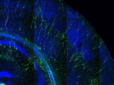
But when the researchers injected rat stem cells into blastocysts — early-stage embryos — of mice engineered in this way, the cells filled the gaps in the brain circuits. And once the mice had grown into adults, they were able to find their cookies by smell. Killing the mouse neurons created “niches” for the rat cells to take up residence in various places inside the animals’ olfactory circuits, as well as elsewhere in their bodies, Baldwin says. Her group is now working on methods for replacing specific mouse neurons with rat cells in a more targeted way.
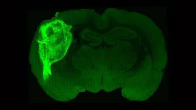
In a Cell paper published by the second team, also on 25 April2, Wu and his colleagues developed a more aggressive strategy for getting rat cells into a mouse’s brain. Using C-CRISPR, a genetic-editing tool that cuts genes in multiple places to ensure that they are fully inactivated, the researchers wiped out every trace of a gene called Hesx1 in a group of mouse blastocysts. This gene controls the development of the forebrain: a large region in the brain that coordinates much of an animal’s behaviour.
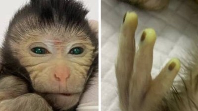
This hybrid baby monkey is made of cells from two embryos
When the researchers allowed these blastocysts to develop into mice without forebrains, the animals died shortly after birth. But when Wu and his team injected rat stem cells into the blastocysts, the forebrains that developed were made entirely of rat cells. Once the mice had grown, they were healthy and seemed to act normally, although Wu says it would be difficult to determine whether there were any subtle behavioural differences between them and normal mice.
Hiro Nakauchi, a stem cell biologist at Stanford University in California, agrees that it would be hard to establish this. Researchers in his laboratory once tried making mice ‘smarter’ by giving them rat brain cells, but they abandoned the effort when they realized that the differences between rodents with and without the cells were too minor to detect statistically without testing the behaviours of a large number of hybrid animals. Still, Nakauchi says that the new papers are meticulous analyses of chimeric animals’ brains — something he is excited about. “This is what I have been awaiting,” he says.
‘Fascinating biology’
Wu and Baldwin say that their research addresses some long-standing concerns about developing chimaeras, particularly for the purpose of transplanting tissue or organs from animals such as pigs into people. Aside from ethical considerations, there is the concern that the human body will reject a transplanted chimeric organ. But because the teams added the rat cells so early in the mice’s development — long before the embryos had formed an immune system — the animals’ bodies never learnt to recognize the cells as foreign and never attacked them.
Hybrid brains: the ethics of transplanting human neurons into animals
Another concern is a mismatch in the developmental rates of species. However, the teams found that the mouse brains developed at the same rate as they would normally, rather than at the slower pace at which a rat usually develops.
“There’s lots of fascinating biology to be learnt from this [rat–mouse] chimaera,” says Jian Feng, a physiologist at the University of Buffalo in New York. He’s not surprised that the rat cells followed the pace of the mouse’s developmental ‘clock’. In 2020, his group published a paper about a mouse embryo that it had engineered to contain up to 4% human cells5. The embryo began developing human red blood cells 17 days into gestation — much earlier than these cells develop in human embryos — suggesting that human cells, too, could follow the molecular directions of their host.
Wu says that his laboratory now plans to use the technology developed for these studies to make chimaeras by transplanting cells from wild rodent species into lab mice. It’s difficult to study wild rodents, because they are hard to maintain and breed in captivity, he says. But making stem cells from their tissue samples and inserting them into mouse blastocysts might allow researchers to study how these other species’ brains develop and function.
A person has received an experimental treatment for the first time that, if successful, will lead them to grow an additional, ‘miniature liver’. The procedure, developed by the biotechnology firm LyGenesis, marks the beginning of a clinical trial designed for people whose livers are failing, but who have not received an organ transplant.
First pig liver transplanted into a person lasts for 10 days
The approach is unusual: researchers injected healthy liver cells from a donor into a lymph node in the upper abdomen of the person with liver failure. The idea is that in several months, the cells will multiply and take over the lymph node to form a structure that can perform the blood-filtering duties of the person’s failing liver.
“It’s a very bold and incredibly innovative idea,” says Valerie Gouon-Evans, a liver-regeneration specialist at Boston University in Massachusetts, who is not involved with the company.
The person who received the treatment, on 25 March, is recovering well from the procedure and was discharged from the clinic, says Michael Hufford, chief executive of LyGenesis, which is based in Pittsburgh, Pennsylvania. But physicians will need to closely monitor them for infection because the person needs to take immunosuppressive drugs so that their body doesn’t reject the donor cells, says Stuart Forbes, a hepatologist at the University of Edinburgh, UK, who is not affiliated with LyGenesis.
Organ crisis
More than 50,000 people in the United States die each year with liver disease. In the end stage of the disease, scar tissue that has accumulated prevents the organ from filtering toxic substances in the blood, and can lead to infection or liver cancer.
A liver transplant can help, but there is a shortage of organs: about 1,000 people in the United States die every year waiting for a transplant. Thousands more aren’t eligible because they are too ill to undergo the procedure.
A person received donor liver cells on 25 March that were injected into one of their lymph nodes.Credit: LyGenesis
LyGenesis has been trialling an approach that could help people in this situation — and make use of donated livers that would otherwise go to waste because a person on the transplant waiting list with a compatible health profile hasn’t materialized in time. The company’s strategy delivers the donor cells through a tube in the throat, injecting them into a lymph node near the liver. Lymph nodes, which also filter waste in the body and are an important part of the immune system, are ideal for growing mini livers, Hufford says, because they receive a large supply of blood and there are hundreds of them throughout the body, so if a few are used to generate mini livers, plenty of others can continue to function as lymph nodes.
The treatment has so far worked in mice1, dogs and pigs2. To test the therapy in pigs, researchers restricted blood flow to the animals’ livers, causing the organs to fail, and injected donor cells into lymph nodes. Miniature livers formed within two months and had a cellular architecture resembling a healthy liver. Researchers even found cells that transport bile, a digestive fluid produced by the liver, in the mini livers of the pigs. In this case, they saw no build-up of bile acid, suggesting that the mini organs were processing the fluid.
Hufford says there’s reason to think that the organs won’t grow indefinitely in the lymph nodes. The mini organs rely on chemical distress signals from the failing liver to grow; once the new organs have stabilized blood filtering, they will stop growing because that distress signal disappears, he says. But it’s not yet clear precisely how large the mini-livers will become in humans, he adds.
The company aims to enrol 12 people into the phase II trial by mid-2025 and publish results the following year, Hufford says. The trial, which was approved by US regulators in 2020, will not only measure participant safety, survival time and quality of life post-treatment, but will also help to establish the ideal number of mini livers to stabilize someone’s health. The clinicians running the trial will inject liver cells in up to five of a person’s lymph nodes to determine whether the extra organs can boost the procedure’s success rate.
A stop-gap measure
However, mini livers might not relieve all of the complications of end-stage liver disease, says Forbes, who has formed his own company to tackle liver disease using genetically modified immune cells that stimulate repair. One of the most serious of these is portal hypertension, in which the build-up of scar tissue compresses blood vessels in the liver and can cause internal bleeding.
Pig brains kept alive outside body for hours after death
Hufford acknowledges that the mini livers are not expected to address portal hypertension, but the hope is that they can provide a stopgap until a liver becomes available for transplant, or make people healthy enough to undergo a transplant. “That would be amazing, because these patients currently have no other treatment options,” Gouon-Evans says.
LyGenesis has ambitions beyond mini livers, too. The company is now testing similar approaches to grow kidney and pancreas cells in the lymph nodes of animals, Hufford says.
If the liver trial is successful, Gouon-Evans says, it would be worth investigating whether a person’s own stem cells could be used to generate the cells that seed the lymph nodes. This technique could create personalized cells that capture the diversity of cells in the liver and don’t require immunosuppressive drugs, she says.