Tag: Stem Cells

  • Stem cells reverse woman’s diabetes — a world first

    Stem cells reverse woman’s diabetes — a world first

    [ad_1]

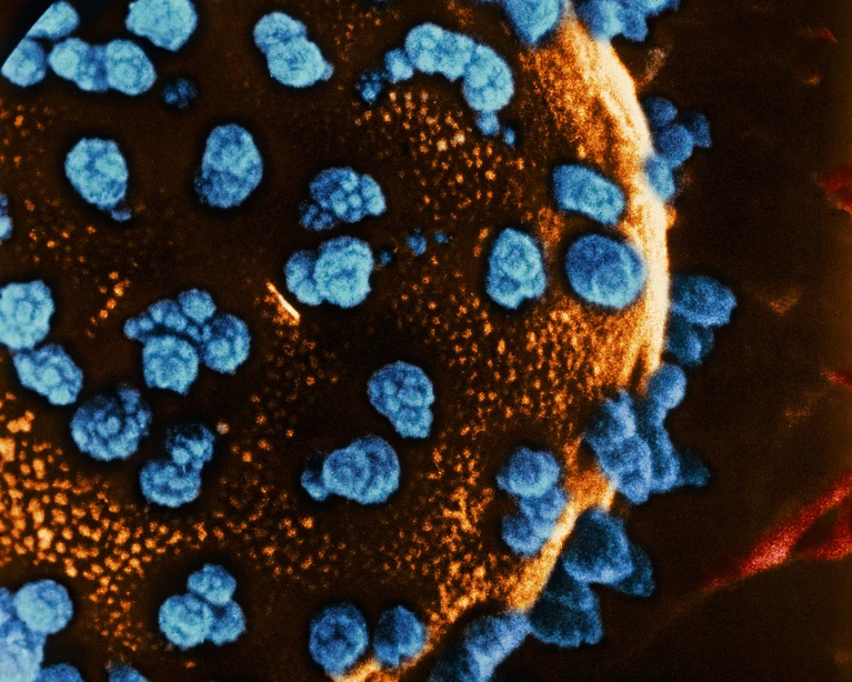
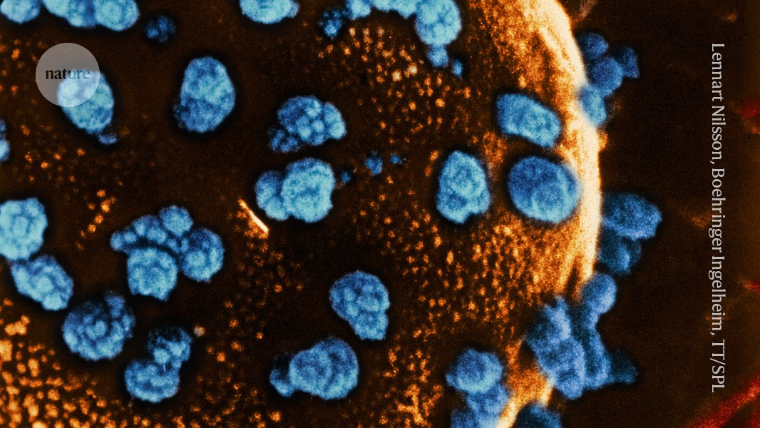
    Coloured scanning electron micrograph (SEM) of a cell in an islet of Langerhans from the pancreas.

    A woman with type 1 diabetes started producing insulin (blue) after a stem cell transplant.Credit: Lennart Nilsson, Boehringer Ingelheim International GmbH, TT/Science Photo Library

    A 25-year-old woman with type 1 diabetes started producing her own insulin less than three months after receiving a transplant of reprogrammed stem cells1. She is the first person with the disease to be treated using cells that were extracted from her own body.

    “I can eat sugar now,” said the woman, who lives in Tianjing, on a call with Nature. It has been more than a year since the transplant, and, she says, “I enjoy eating everything — especially hotpot.” The woman asked to remain anonymous to protect her privacy.

    James Shapiro, a transplant surgeon and researcher at the University of Alberta in Edmonton, Canada, says the results of the surgery are stunning. “They’ve completely reversed diabetes in the patient, who was requiring substantial amounts of insulin beforehand.”

    The study, published in Cell today, follows results from a separate group in Shanghai, China, who reported in April that they had successfully transplanted insulin-producing islets into the liver of a 59-year-old man with type 2 diabetes2. The islets were also derived from reprogrammed stem cells taken from the man’s own body and he has since stopped taking insulin.

    The studies are among a handful of pioneering trials using stem cells to treat diabetes, which affects close to half a billion people worldwide. Most of them have type 2 diabetes, in which the body doesn’t produce enough insulin or its ability to use the hormone diminishes. In type 1 diabetes, the immune system attacks islet cells in the pancreas.

    Islet transplants can treat the disease, but there aren’t enough donors to meet the growing demand, and recipients must use immune-suppressing drugs to prevent the body from rejecting the donor tissue.

    Stem cells can be used to grow any tissue in the body and can be cultured indefinitely in the laboratory, which means they potentially offer a limitless source of pancreatic tissue. By using tissue made from a person’s own cells, researchers also hope to avoid the need for immunosuppressants.

    Reprogrammed cells

    In the first trial of its kind, Deng Hongkui, a cell biologist at Peking University in Beijing, and his colleagues extracted cells from three people with type 1 diabetes and reverted them into a pluripotent state, from which they could be moulded into any cell type in the body. This reprogramming technique was first developed by Shinya Yamanaka at Kyoto University in Japan almost two decades ago. But Deng and his colleagues modified the technique3: instead of introducing proteins that trigger gene expression, as Yamanaka had done, they exposed the cells to small molecules. This offered more control over the process.

    The researchers then used the chemically induced pluripotent stem (iPS) cells to generate 3D clusters of islets. They tested the safety and efficacy of the cells in mice and non-human primates.

    In June 2023, in an operation that lasted less than half an hour, they injected the equivalent of roughly 1.5 million islets into the woman’s abdominal muscles — a new site for islet transplants. Most islet transplants are injected into the liver, where the cells cannot be observed. But by placing them in the abdomen, the researchers could monitor the cells using magnetic resonance imaging, and potentially remove them if needed.

    Insulin free

    Two-and-a-half months later, the woman was producing enough insulin to live without needing top-ups, and she has sustained that level of production for more than a year. By that time, the woman had stopped experiencing the dangerous spikes and drops in blood glucose levels, which remained within a target range for more than 98% of the day. “That’s remarkable,” says Daisuke Yabe, a diabetes researcher at Kyoto University. “If this is applicable to other patients, it’s going to be wonderful.”

    The results are intriguing, but they need to be replicated in more people, says Jay Skyler, an endocrinologist at the University of Miami, Florida, who studies type 1 diabetes. Skyler also wants to see that the woman’s cells continue to produce insulin for up to five years, before considering her ‘cured’.

    Deng says the results for the other two participants are “also very positive”, and they will reach the one-year mark in November, after which he hopes to expand the trial to another 10 or 20 individuals.

    Because the woman was already receiving immunosuppressants for a previous liver transplant, the researchers could not assess whether the iPS cells reduced the risk of rejection of the graft.

    Even if the body doesn’t reject the transplant because it doesn’t consider the cells to be ‘foreign’, in people with type 1 diabetes, because they have an autoimmune condition, there is still a risk that the body could attack the islets. Deng says they didn’t see this in the woman because of the immunosuppressants, but they are trying to develop cells that can evade this autoimmune response.

    Donor cells

    Transplants using the recipient’s own cells have advantages, but the procedures are difficult to scale up and commercialize, say researchers. Several groups have started trials of islet cells created using donor stem cells.

    Preliminary results for one trial, led by Vertex Pharmaceuticals in Boston, Massachusetts, were reported in June. A dozen participants with type 1 diabetes received islets derived from donated embryonic stem cells that were injected into the liver. They were all treated with immunosuppressants. Three months after the transplant, all the participants began producing insulin when glucose was present in their bloodstreams4. Some had become insulin independent.

    Last year, Vertex launched another trial in which islet cells derived from donated stem cells were placed in a device designed to protect them from immune-system attacks. It was transplanted into a person with type 1 diabetes, who did not receive immunosuppressants. “That trial is ongoing,” says Shapiro, who is involved in the study, which aims to enrol 17 individuals.

    Yabe is also about to start a trial using islet cells produced using donor iPS cells. He plans to develop sheets of islets and surgically place them in the abdominal tissue of three people with type 1 diabetes, who will receive immunosuppressants. The first participant should receive their transplant early next year.

    [ad_2]

    Source link

  • Temporal BMP4 effects on mouse embryonic and extraembryonic development

    [ad_1]

    Embryo recovery and documentation

    All of the animal procedures were approved by the Institutional Animal Care and Use Committee and were performed in strict adherence to Weizmann Institute guidelines. Mice were monitored for health and activity and were given ad libitum access to water and standard mouse chow with 12 h–12 h light–dark cycles. Embryos were collected from timed pregnant immune-competent C57BL/6JRccHsd or Hsd:ICR(CD-1) female mice (obtained from Envigo and mated in house with males of the same strain) between E5.5 and E9.5. Embryos were recovered from their implantation sites using fine forceps, in PBS, while carefully preserving all extraembryonic tissues. The embryos were then washed in PBS and transferred to chilled DMEM (Phenol-red free, GIBCO) supplemented with 10% FBS (Biological Industries) for imaging before dissociation. Phase-contrast images were taken using the Eclipse Ti2 inverted microscope (Nikon) and Zyla sCMOS camera (Andor). Morphological staging and analysis of embryos was conducted as previously described61.

    Cell line information

    Source of cell lines: all of the cell lines used in this study were generated in-house from a stock of validated V6.5 (C57BL/6×129) background. Authentication of cell lines: the cell lines were authenticated using genotyping PCR to ensure their identity and purity. The genotyping results confirmed the expected genetic background for each cell line. Mycoplasma contamination testing: all of the cell lines were routinely tested for mycoplasma contamination using the PCR-based Mycoplasma Detection Kit (Hylab) before use in the experiments. No mycoplasma contamination was detected in any of the cell lines.

    Trophectoderm-specific genetic manipulation

    Production of lentiviral vectors

    Lentiviral vectors were constructed to produce lentiviruses expressing gRNAs designed to selectively target either GFP expression and the third exon of Bmp4 or the third and fourth exons of the Elf5 gene locus in the trophectoderm of Cas9–GFP62 mouse blastocysts. All gRNAs were selected for minimal off-target effects using the CCTop CRISPR/Cas9 target online predictor (https://cctop.cos.uni-heidelberg.de:8043/)63. Introduction of a mega-primer (Supplementary Table 4) that includes gRNAs into a lentivector, constitutively expressing gRNAs scaffolds and a mCherry fluorescent reporter, was carried out by restriction-free cloning as previously described64. Recombinant lentiviruses were produced by transient transfection into HEK293T cells, using polyethylene imine (PEI) (PEI linear, Mr 25,000, Polyscience) as previously described65, using three envelope and packaging plasmids and one of three viral constructs: (1) pDecko-GFP/mCherry (that is, the control vector), (2) pDecko-Elf5/mCherry or (3) pDecko-Bmp4/mCherry. In brief, infectious lentiviruses were collected at 48 and 72 h after transfection, filtered through 0.45-mm-pore cellulose acetate filters and concentrated by ultracentrifugation at 20,000 rpm for 2 h. Lentiviral supernatant effective titres were determined by infection of HEK293T cells followed by fluorescence-activated cell sorting (FACS) analysis. To validate of Elf5 and Bmp4 KO, HEK293T cells were infected with the appropriate lentiviral vector expressing gRNAs targeting Elf5/Bmp4 and mCherry. Infected cells were picked by sterile sorting, subsequently transfected with px330 Cas9 targeting plasmid expressing GFP66 and sorted again prior to DNA extraction. Genomic DNA was extracted by PCR-compatible lysis buffer (10 mM Tris, pH 8, 0.45% Triton X-100, 0.45% Tween-20, 0.2 mg ml−1 proteinase K). Primers flanking the PAM sequence of each target (Supplementary Table 4) were used for amplifying the genomic segments that included the expected Cas9-mediated DNA editing, and immediately followed by Sanger sequencing (not shown).

    Mice and lentiviral transduction

    Infection of nascent blastocysts was performed using B6D2F1 (C57BL/6xDBA) (Envigo)/Cas9-GFP embryos. In brief, 3–4-week old B6D2F1 female mice were hormone primed by an intraperitoneal injection of pregnant mare serum gonadotropin (PMSG, Vetmarket) followed 46 h later by an injection of human chorionic gonadotropin (hCG, Sigma-Aldrich) and mating with homozygote Cas9-GFP males62. Embryos were collected at the zygote stage, and cultured in KSOM medium until the blastocyst stage. For efficient infection of the trophectoderm, zona pellucida was removed in acidic Tyrode’s solution (Sigma-Aldrich)27. Next, 15–20 embryos were incubated with lentiviruses, described above, in KSOM for 4–5 h. The transduced blastocysts were washed, and then were transferred into each recipient female generated after mating with vasectomized CD1 males (Envigo); the day of injection was considered to be 2.5 days post coitum. Mice were handled in accordance with institutional guidelines and approved by the Institutional Animal Care and Use Committee (IACUC, The Weizmann Institute of Science).

    Analysis of genetically manipulated embryos

    Elf5-KO embryos were collected at E7.5, and Bmp4-KO embryos were collected in a time series from E7.5 to 8.5, and all were dissected in ice-cold 1× PBS. Individual mutants were imaged using the Eclipse Ti2 inverted microscope (Nikon) and the Zyla sCMOS camera (Andor) while being maintained in DMEM supplemented with 10% FBS. Embryos positive for mCherry were selected for further morphological and transcriptome analysis (only in Elf5-KO embryos).

    Tetraploid complementation assay for generating embryonic Bmp4 KO

    Bmp4LoxP/LoxP embryonic stem cells were derived from Bmp4LoxP/LoxP mice67 (C57BL/6×129; Extended Data Fig. 11a) using standard embryonic stem cell derivation method. The cells were then treated with recombinant His-TAT-NLS-Cre (HTNC) protein (Addgene plasmid, 13763). For genotyping, individual clones were grown for two passages on gelatin-coated plates to eliminate residual MEF, and RNA was extracted using Direct-zol (Zymo RNA miniPrep, R2052) followed by cDNA production and quantitative PCR (qPCR; Supplementary Table 4). For this study, we used two validated Bmp4Δ/Δ clones and one isogenic control clone (a Bmp4+/+ HTNC-treated clone). Blastocyst injections were performed using (C57BL/6xDBA) B6D2F1 (Envigo) host embryos. In brief, 3–4-week old B6D2F1 females were hormone primed by an intraperitoneal injection of pregnant mare serum gonadotropin (PMSG, Vetmarket) followed 46 h later by an injection of human chorionic gonadotropin (hCG, Sigma-Aldrich). Embryos were collected at the zygote stage, and cultured in a CO2 incubator until the blastocyst stage. For tetraploid complementation, two-cell embryos were fused to one cell using a CF150/F instrument (BLS), by 2 DC square pulses of 30 V 40 ms and 1–2 V AC, in 0.3 M mannitol solution with BSA. On the day of the injection, embryos were placed in M2 medium using a 16-µm-diameter injection pipet (Biomedical Instruments) and a Piezo micromanipulator (Prime Tech); approximately 15 cells were injected into the blastocoel of each embryo. Approximately 20 blastocysts were transferred to each recipient female (CD1 female mice, Envigo); the day of injection was considered to be E2.5. Mice were handled in accordance with institutional guidelines and approved by the Institutional Animal Care and Use Committee (IACUC, The Weizmann Institute of Science).

    Ex utero culture of post-implantation embryos

    Pregastrulating embryos were dissected at E5.5, their Reichert’s membrane removed and individually placed into separate wells of 8-well glass-bottom ibiTreat μ-plates (iBidi; 80827/80826) filled with 250 μl of EUCM (consisting of 25% DMEM (GIBCO 11880; includes 1 mg ml−1 d-glucose and pyruvate, without phenol red and without l-glutamine) supplemented with 1× GlutaMax (GIBCO, 35050061), 100 U ml−1 penicillin–100 μg ml−1 streptomycin (Biological industries; 030311B) and 11 mM HEPES (GIBCO, 15630056), plus 50% rat serum (rat whole-embryo culture serum, ENVIGO Bioproducts B-4520) and 25% human umbilical cord blood serum (prepared in-house)). The medium was preheated for an hour in an incubator under 5% CO2 at 37 °C. Embryos were cultured statically under 5% CO2 at 37 °C. The total volume of the medium was replaced every 24 h, and the embryos were monitored by morphological assessment daily.

    BMP inhibition in ex utero and explant cultures

    For ex utero experiments, embryos were meticulously dissected at either E5.5 or E6.5 and processed for ex utero culture as described above. During the 24 h culture period, the embryos were co-cultured with 400 ng ml−1 of mouse recombinant Noggin (R&D systems, 1967-NG). Continuous morphological monitoring was conducted, and comprehensive analysis was performed using multiplexed RNA in situ HCR, as described below. In ExE explant cultures, embryos at E6.5 and 7.5 were dissected, and the ExE and EPC were precisely isolated from the embryonic compartment. Subsequent culture procedures followed an established protocol41, with the addition of 800 ng ml−1 of mouse recombinant Noggin or 1000 ng ml−1 mouse recombinant BMP4. After incubation for 24 h, bulk RNA was extracted and purified using the Micro RNA kit (Qiagen, 74004). qPCR was then used to quantify selected markers (primer details are provided in Supplementary Table 4). For temporal depletion of BMP signalling, embryos were dissected at E7.5 and were subjected to ex utero culture as described above. During the 12/24 h culture period, the embryos were co-cultured with 5 µM LDN-193189 (Sigma-Aldrich, SML0559), followed by whole-mount immunostaining.

    Immunostaining

    In this study, whole-mount immunostaining was performed as previously described24, using the following antibodies: rabbit anti-EOMES (1:100, ab23345, Abcam); rabbit monoclonal anti-Brachyury (D2Z3J) (1:100, Cell Signaling, 81694); mouse anti-KRT7 (1:100, Abcam; ab9021); goat anti-SOX2 (1:100, R&D, AF2018); rabbit anti-TFAP2C (1:100, CST, 2320); donkey anti-rabbit IgG (IgG) (H+L), Alexa 647 (1:250, Jackson ImmunoResearch, 711-605-152); donkey anti-goat IgG (H+L) Alexa 488 (1:250, Jackson ImmunoResearch, 705-545-003); goat anti-mouse IgG1 Alexa Fluor 594 (1:250, Jackson ImmunoResearch, 115-585-205).

    Spatial analysis

    Multiplex RNA in situ HCR22 was performed according to the manufacturer’s instructions (Molecular Technologies). For sample preparation, embryos were dissected and their Reichart’s membrane removed in M2 medium at consecutive timepoints after implantation until E7.5. Embryos were then washed in cold PBS and fixed in 4% PFA 4 °C overnight. After fixation, the samples were dehydrated on ice in increasing ratios of methanol (Sigma-Aldrich) and PBST (0.1% Tween-20) (Sigma-Aldrich) until freezing overnight in absolute methanol −20 °C. The embryos were then rehydrated in increasing ratios of PBST and methanol on ice, washed in PBST, treated with 10 μg ml−1 proteinase K (Thermo Fisher Scientific) and subjected to post-fixation in PFA 4% all at room temperature. For HCR staining, the samples were repeatedly washed with PBST, prehybridized with probe hybridization buffer (Molecular Technologies) for 30 min at 37 °C and then hybridized with probe sets (Molecular Technologies) for different combinations of Adm, Sox2, Eomes, Fgfr2, Ascl2, Bmp4, Lefty1, Fosl1, Chsy1, Hand1 and Pcdh12 (16 nM) at 37 °C overnight. Tissues were washed with HCR probe wash buffer (Molecular Technologies), followed by repeated washes in 5× SSCT (5× SSC with a final concentration of 0.1% Tween-20) and incubated with HCR amplifiers (Molecular Technologies) (30 pmol) in amplification buffer (Molecular Technologies) at room temperature overnight. The samples were then washed with 5× SSCT, labelled, mounted and imaged in an eight-well glass bottom/ibiTreat μ-plates (iBidi; 80827/80826). Spatial analysis was conducted using the Leica STELLARIS 8 Spectral confocal microscope and acquired using LAS-X (Leica). Fluorophores were excited by a white light laser and acousto-optical beam splitter. All embryonic specimens were visualized by maximum-intensity projection of their fluorescence signals across focal planes, and their 3D structure was assessed. The presented images were finalized using ImageJ software.

    Flow cytometry

    For isolation of single cells for scRNA-seq analysis, embryos were dissociated with 0.25% trypsin-A, 0.02% EDTA (Biological Industries) solution for 5 min at 37 °C and resuspended in DMEM without phenol red (GIBCO) supplemented with 10% FBS (Biological Industries). The samples were run on the FACS Aria-III flow cytometer (BD Biosciences, using BD FACSDiva v.9.0) using the ‘index sort’ option to retain the spectral properties of each individual sorted cell. For samples of ΔPE-Oct4-GFP44 obtained after E7.5, we further dissected areas on the basis of the localization of GFP expression. Subsequently, we used index-sorting and enriched for GFP-positive cells. The gating and sorting strategy is shown in Extended Data Fig. 13c. FlowJo v.10.7 was used to generate Extended Data Fig. 7c.

    scRNA-seq analysis

    10x Genomics

    Cultured embryos were prepared for sequencing at different developmental stages after 2 days in culture: late streak and early head fold, assessed by light microscopy. Six similar embryos were selected for each developmental stage, pooled and dissociated using trypsin-EDTA solution A 0.25% (Biological Industries, 030501B) for 5 min at 37 °C. Trypsin was neutralized with medium that included 10% FBS and cells were washed and resuspended in 1× PBS (calcium and magnesium free) with 400 μg ml−1 BSA. Cell suspension was then filtered with a 70 μm cell strainer to avoid cell clumps. Single-cell viability was determined by trypan blue staining, before being diluted to a final concentration of 1,000 cells per μl. scRNA-seq libraries were generated for each pool of embryos separately using the 10x Genomics Chromium v3 system (5,000 cell target cell recovery) and sequenced on the Illumina NovaSeq 6000 platform according to the manufacturer’s instructions.

    MARS-seq

    Single-cell cDNA plate based libraries were prepared as previously reported68,69 according to the MARS-seq protocol70, following index FACS as described above.

    scRNA-seq data processing

    10x Genomics data from ex utero cultured embryos

    Raw files were transformed into count matrices using Cell Ranger v.6.1.2 with the default parameters and with the prebuilt Cell Ranger reference package refdata-gex-mm10-2020-A (mm10 genome, GENCODE vM23/Ensemble 98). Cells with less than 2,000 counts, more than 30,000 counts or a high number of counts from mitochondrial genes relative to the number of counts from ribosomal genes were removed, resulting in 9,387 cells from the batch of late streak embryos and 3,916 cells from the batch of head fold stage embryos. For doublet removal, we ran DoubletFinder separately on the two batches, following the best practice workflow of the package. In brief, after creating a Seurat object for each batch69, Seurat principal component analysis was performed on the basis of the 2,000 most variable feature genes. For doublet detection using DoubletFinder, we calculated the fraction of artificial nearest-neighbour doublet cells (pANN) for each cell using pN = 0.25 (the relative frequency of artificial doublet cells relative to real cells) and pK = 0.02 for the batch of late streak embryos and pK = 0.01 for the batch of head fold embryos (pK is the relative neighbourhood size for the estimation of pANN). In the case of the late streak stage batch, we removed all cells with pANN > 0.2 (N = 1,753); for the batch of head fold stage embryos, we removed all cells with pANN > 0.25 (N = 291).

    MARS-seq

    MARS-seq libraries were sequenced using the NextSeq 500 or NovaSeq 6000 system. Reads were processed according to the MARS-seq2.0 protocol70 with the same specifications as previously reported12 using the STAR aligner for read alignment. Overall, we processed 129,024 wells, including the 40,868 wells of the previous version of the gastrulation atlas.

    Metacell analysis and atlas construction

    The basic idea of metacells is to partition cells into small groups of homogeneous cells, thereby removing the sparsity of RNA transcript counts associated with scRNA-seq technologies. Throughout the Article, we used metacell sizes of around 20–100 cells. For each metacell m, the absolute expression eg,m of a gene g is defined as the total number of transcript counts of this gene among all cells belonging to this metacell, normalized to the total number of counts, that is, \({e}_{g,m}={\sum }_{\{c\in m\}}{N}_{g,c}/({\sum }_{g}{\sum }_{\{c\in m\}}{N}_{g,c})\), where Ng,c is the number of the number of transcripts of gene g in the cell c.

    Embryonic and extraembryonic WT atlas

    To identify feature genes for metacell construction13, we selected all genes satisfying a minimal variance over mean (T_vm = 0.1) and coverage threshold (T_tot = 50 and T_top3 = 3). These 1,534 filtered genes were clustered into 120 clusters on the basis of their gene–gene correlation across the manifold. We manually selected and removed clusters enriched with cell-cycle- or stress-related genes, leaving 1,386 feature genes. The final metacell object (Knn = 100, minimal metacell size = 20) contained 983 metacells comprising 67,843 cells, including cells from E4.5 blastocysts.

    Metacell object of ex utero culture embryos

    The initial set of 1,017 feature genes was clustered into 60 groups and cell-cycle- and stress-related groups were removed. The metacell cover (148 metacells, 11,684 cells) was constructed on the basis of the remaining 829 feature genes and using the same parameters as above.

    Metacell2 object of Bmp4-KO embryos

    Using the framework of Metacell2 (ref. 14), all cells from the Bmp4-KO experiment were combined into a single-metacell object, including 6,705 cells from germline Bmp4-KO embryos, 11,968 cells from embryonic Bmp4-KO mutants, as well as 2,380 cells from control embryos. Metacells were constructed using the default parameters and a target size of 150,000 UMIs per metacell.

    Embryo selection and temporal ordering

    In total, cells from 287 individual embryos contributed to the embryonic and extraembryonic WT atlas. This includes cells from 153 embryos used in a previous version of the WT atlas12. Moreover, we collected 114 cells (after quality control) from two pooled samples of E4.5 blastocysts (n = 11 embryos). We selected 251 embryos with a sufficient number of embryonic cells for assigning them a developmental time (shown in Fig. 1c). The frequencies of ExE cell types were estimated on the basis of a cohort of 83 embryos (Extended Data Fig. 2b–d). This cohort included all of the embryos for which we collected the whole ExE tissue during dissection, for which we have a bright-field image to assign them a morphological stage and for which we obtained at least 10 ExE cells after quality control and filtering. Embryos with a sufficient number of embryonic cells (251 embryos) were temporally ordered as previously reported12. In brief, using the embryonic cells of an embryo, we calculate an embryo-embryo similarity matrix (Extended Data Fig. 1e) that quantifies the transcriptional similarity between two embryos. Using the similarity matrix, we compute a global goal function for each possible embryo order. Embryos are initially ordered on the basis of their morphology and then are reshuffled; each reshuffling is accepted if it improves the goal function. The final temporal ranks of each embryo are translated into developmental times (denoted as Et) by a spline interpolation of the nominal time of collection of each embryo versus its inferred transcriptional rank.

    Network flow model construction

    In a previous publication12 we introduced a network flow model that enabled us to reconstruct cellular differentiation trajectories during mouse gastrulation along the single-embryo single-cell transcriptome atlas. To infer cellular trajectories on the extended gastrulation atlas, temporally ranked embryos were grouped into 16 time bins (Fig. 1c). The network flow model for all embryonic cells was computed as previously reported. Logistic distances between metacells were calculated using the default parameters. For the estimated proliferation rate of each metacell, we interpolated between the default rate (3.5 divisions per day) and no cell division on the basis of each cell’s expression of cell-cycle-related genes. We used the same values for all additional network flow parameter as in the in the original network flow model of mouse gastrulation.

    Differential expression statistical analysis

    The expression of a gene between two groups of cells is compared using a χ2 test on the number of UMIs from that gene. If \({N}_{g}^{1}\) and \({N}_{g}^{2}\) are the number of UMIs per gene g and N1, N2 the total number of UMIs per group, we compute for each gene the χ2 statistic between the two two-dimensional vectors \(({N}_{g}^{1},{N}^{1}-{N}_{g}^{1})\) and \(\left({N}_{g}^{2},{N}^{2}-{N}_{g}^{2}\right)\). If multiple hypotheses are tested, P values are corrected using the Benjamini–Hochberg method.

    Cell cycle scores

    For each cell, its synthesis phase (S phase) and mitosis phase (M phase) score is the total number of UMIs from a list of respective marker genes, divided by the total number of UMIs of the cell. The M phase marker genes are as follows: Mki67, Cenpf, Top2a, Smc4, Ube2c, Ccnb1, Cdk1, Arl6ip1, Ankrd11, Hmmr, Cenpa, Tpx2, Aurka, AB349069, Kif4, Kif2c, Bub1b, Ccna2, Kif23, Kif20a, Sgol2a, Smc2, Kif11, Cdca2, Incenp and Cenpe. The S phase marker genes are as follows: Pcna, Rrm2, Mcm5, Mcm6, Mcm4, Ung, Mcm7, Mcm2, Uhrf1, Orc6 and Tipin.

    Single-cell scores

    Given a list of genes (referred to as cell-state markers) Gm, the single-cell score corresponding to a particular cell is calculated as the sum of counts for all genes in a given cell-state markers Gm, divided by the total sum of expression counts for all genes in the cell.

    EPC lineage analysis

    Let eg,m be the absolute expression for each gene g and all metacells m from the EPC lineage (uncommitted EPC, TGC progenitor, SpT-Gly), normalized to the total number of counts per metacell, and let leg,m = log2(eg,m + 10−5). We selected all variable genes that (1) pass a threshold of minimal expression in at least one of the metacells, that is:

    $$\mathop{\min }\limits_{m}l{e}_{g,m}\, > \,-13$$

    and that (2) pass a threshold on the difference between the highest and smallest expression in a metacell:

    $$\mathop{\max }\limits_{m}l{e}_{g,m}-\mathop{\min }\limits_{m}l{e}_{g,m} > 2.$$

    A complete list of these genes is provided in Supplementary Table 1. For further clean-up, we filtered only genes that show a minimal difference in their maximal expression among TGC progenitors and SpT-Gly metacells:

    $$|\mathop{\max }\limits_{m\in \text{TGC}}l{e}_{{gm}}-\mathop{\max }\limits_{m\in \text{SpT}-\text{Gly}}l{e}_{{gm}}| > 1.5.$$

    We observed that the large majority of the filtered genes followed one of the following behaviours: (1) high expression in TGC progenitor metacells compared to SpT-Gly and vice versa; (2) high expression in early metacells and low expression in late; or (3) low expression in early metacells and high expression in late ones. The filtered genes were therefore grouped into five clusters (arguing that this number should be enough to capture the above behaviours; Extended Data Fig. 3a) using k-means on the relative expression profiles, \(l{f}_{g,m}\,=l{e}_{g,m}-{\text{mean}}_{m}l{e}_{g,m}\). Genes from clusters 1 and 2 were used for the SpT-Gly score and genes from the clusters 4 and 5 were used for the TGC progenitor score. For pseudotime kinetics of gene expression, we fitted a principal curve to the joint distribution of Uncommitted EPC, SpT-Gly and TGC progenitor scores for all cells from the EPC lineage (Extended Data Fig. 3b) and divided the curve into 12 bins (Fig. 2a,b).

    Chorion lineage analysis

    Variable genes were filtered for all metacells from the chorion lineage (chorion progenitors, chorion) using the same parameters as for the EPC lineage (Supplementary Table 3). As in the EPC lineage, we also only filtered genes with

    $$|\mathop{\max }\limits_{m\in \text{Chorion prog.}}l{e}_{{gm}}-\mathop{\max }\limits_{m\in \text{Chorion}}l{e}_{{gm}}| > 1$$

    and clustered their relative expression into 5 clusters (Extended Data Fig. 4a). Genes from clusters 1 and 2 were used for chorion progenitor score and genes from clusters 4 and 5 for the chorion score. Gene expression kinetics (Fig. 2c,d) is shown along the principle curve fitted to the joint distribution of chorion progenitor and chorion scores for each chorion and chorion progenitor cell along 12 bins (Extended Data Fig. 4b).

    Cell type annotation of cells from embryos with genetic manipulations

    Cells from EXE Elf5-KO, all Bmp4-KO models and control embryos were annotated with a cell type from the WT atlas as previously described14,29. For each experiment, we constructed a joint metacell k-nearest neighbours similarity graph consisting of query cells from the KO embryos and WT atlas cells. For each query and each atlas cell, we sampled the empirical distribution of cell types from atlas cells among its 100 nearest neighbours. Each query cell is matched with an atlas cell (and annotated with its cell type) on the basis of matching the empirical cell type distribution of the query cell with the best-correlated cell type distribution among the atlas cells. Query cells with less than ten atlas cells among their nearest neighbours were assigned with an atlas cell type by computing the cell-metacell correlation corg(log2(ug,c + 1), log2(eg,m + 10−5)) for each atlas metacell over the list of feature genes and using the cell type of the best-correlated atlas metacell.

    Developmental timing of Elf5-KO and Bmp4-KO embryos

    Given a temporal order of embryos from the WT atlas, query embryos were assigned a best-matching WT rank as previously reported14,29: using the joint metacell k-nearest neighbours similarity graph of query cells and atlas cells, each query cell is annotated with the temporal rank of the nearest neighbour atlas cell (that is, the temporal rank of the embryo this cell belongs to). In a similar way, we resample for each atlas cell a temporal rank by assigning to it the rank of the nearest neighbour cell from a different atlas embryo. As a result, we obtain for each embryo a distribution of sampled temporal ranks of the cells belonging to the embryo. Each query embryo is mapped onto a WT rank by computing the correlation between the cumulative distributions over ranks between the query embryo and the WT embryos and using the temporal rank of best-correlated WT embryo.

    Atlas projection and cell type annotation of cells from ex utero cultured embryos

    Each cell from ex-utero-cultured embryos was projected onto the WT atlas by matching its expression profile with the best-matching atlas metacell profile: Let ug,c be the single-cell gene expression matrix for all cells c> from ex utero cultured embryos and eg,m the WT atlas gene expression per metacell m for all genes g from a set of 581 feature genes. On the basis of the cell-metacell correlation matrix

    $${\text{cor}}_{g}({\log }_{2}{(u}_{g,c}+1),{\log }_{2}({e}_{g,m}+{10}^{-5}))$$

    each cell is matched with its best-correlated metacell and annotated with the cell type of the metacell.

    Similarly, each metacell from ex utero cultured embryos is matched with its best-correlated WT atlas metacell using the correlation matrix

    $$\text{cor}({\log }_{2}{(u}_{g,k}+1),{\log }_{2}({e}_{g,m}+{10}^{-5}))$$

    where ug,k is the metacell expression matrix per gene g and metacell k from the ex utero cultured embryos.

    Cell type frequency comparison between ex utero cultured and WT embryos

    To compare the distribution of cell states in ex utero cultured embryos to the WT distribution, we first computed for each batch of ex utero cultured embryos (late streak and late head fold) a best-matching developmental time, thereby correcting for potential differences in the mean developmental time of the two groups. Ex utero cultured embryos were assigned an average developmental time using for each ex utero cell c the best-matching atlas metacell m(c). For a batch b of ex utero embryos, let \({p}_{m}^{b}\) be the number of ex utero cells for which m(c) = m normalized to the total number of cells, that is, the number of cells that project to that atlas metacell normalized by the total number of cells from that batch. Vice versa, for each WT embryo e (indexed here by their temporal rank 1, 2, …, 235), let \({p}_{m}^{e}\) be the number of cells from that embryo in the metacell m normalized to the total number of cells from that embryo. As the number of cells per metacell is low for single embryos, we use instead of \({p}_{m}^{e}\) the averaged frequency of cells \({\bar{p}}_{m}^{e}={\sum }_{f,|f-e|\le w}{p}_{m}^{f}\) for comparison with \({p}_{m}^{b}\) (over a time window w). More precisely, each ex utero batch was matched with that WT embryo e for which thedistance between the two distributions \({p}_{m}^{b}\) and \({\bar{p}}_{m}^{e}\) was minimal, that is,

    $$\text{matched WT rank}\,=\,{{\rm{argmin}}}_{e}\,|{p}_{m}^{b}-{\bar{p}}_{m}^{e}|.$$

    For the batch of late streak embryos we used a window size w = 20 and for the batch of late head fold embryos window size w = 5.

    Differential expression analysis for Bmp4-KO embryos

    Identification of batch-related genes and metacell–metacell projection

    We created a metacell object of all cells from the Bmp4 experiment. Metacells from the Bmp4 experiment were projected onto the WT atlas using the framework of MCProj14. This approach enables us to match each query metacell m and its expression profile \({e}_{g,m}^{{\rm{query}}}\,\) (g is a gene) with a corresponding matched WT atlas profile \({e}_{g,m}^{{\rm{proj}}}\). Both profiles represent the relative absolute expression, that is, \(1={\sum }_{g}{e}_{g,m}^{{\rm{query}}}={\sum }_{g}{e}_{g,m}^{{\rm{proj}}}\). To screen for batch-related genes, we computed the log fold changes between pseudo-bulk query and atlas expression profiles for all cells c from a cell typet and experimental condition b, that is:

    $${lfc}_{g,t}^{b}={\log }_{2}\left(\frac{1}{{N}_{b,t}}\sum _{c\in (b,t)}{e}_{g,m(c)}^{{\rm{query}}}+\varepsilon \right)-{\log }_{2}\left(\frac{1}{{N}_{b,t}}\sum _{c\in (b,t)}{e}_{g,m(c)}^{{\rm{proj}}}+\varepsilon \right)$$

    where m(c) is the metacell m of the cell c, Nb,t is the number of cells from cell type t and experimental condition b and ε = 5 × 10−5. There are four different experimental conditions: (1) homozygous Bmp4Δ/Δ cells from germline Bmp4Δ/+ mating (characterized on the basis of total low levels of BMP4 per embryo), (2) heterozygous Bmp4Δ/+ or Bmp4+/+ cells from germline Bmp4Δ/+ mating (normal levels of BMP4), (3) cells from embryonic Bmp4-KO mutants (tetraploid) and (4) isogenic control cells from injected (tetraploid) WT embryos (Extended Data Fig. 11a,b). We subsequently clustered all differentially expressed genes (genes g for which \(\mathop{\max }\limits_{t,b}|{{lfc}}_{g,t}^{b}| > 0.8\) and that pass a minimal expression threshold) into ten clusters. Genes from clusters displaying differential expression among the control cells were classified as lateral genes.

    Differential expression per embryo and cell type in the ExM lineage

    For each query embryo e, we computed the bulk expression per cell type t and gene g, \({f}_{g,e,t}^{q}\) and the bulk expression per cell type of time-matched WT embryos \({e}_{g,e,t}^{{\rm{WT}}}\). To analyse the effect of embryonic Bmp4-KO on the ExM lineage, we selected all of the expression profiles from the cell types early nascent mesoderm, ExM and allantois and from the control and embryonic Bmp4-KO mutants that contained at least ten cells per cell type and embryo. We then filtered all of the genes for which (1) \(\mathop{\max }\limits_{e,t}| {\log }_{2}(\,{f}_{g,e,t}^{q}+\varepsilon )\,-{\log }_{2}(\,{f}_{g,e,t}^{{\rm{WT}}}+\varepsilon )| \ge {\log }_{2}3\), that is, displaying a threefold change between embryonic Bmp4-KO and matched WT cells in at least one of the cell types and one of the embryos; and (2) passing a threshold on minimal expression in one of the conditions, that is, \(\mathop{\max }\limits_{e,t}(\,{f}_{g,e,t}^{q},{f}_{g,e,t}^{{\rm{WT}}}\,)\ge 1\times 1{0}^{-4}\). After removing lateral genes (see the previous paragraph), this resulted in 46 genes, which were subsequently clustered into three groups. Cluster 2 is shown in Extended Data Fig. 12c. Genes of clusters 1 and 3 are listed in Supplementary Table 5.

    Identification and differential gene expression of ExM PGC precursors

    Let eg,m be the absolute expression for each gene g and all metacells m from the PGCs, allantois and ExM, normalized to the total number of counts per metacell, and let leg,m = log2(eg,m + 10−5). We selected all variable genes that (1) pass a threshold of minimal expression in at least one of the metacells, that is:

    $$\mathop{\min }\limits_{m}l{e}_{g,m}\, > \,-13$$

    and that (2) pass a threshold on the difference between the highest and smallest expression in a metacell:

    $$\mathop{\max }\limits_{m}l{e}_{g,m}-\mathop{\min }\limits_{m}l{e}_{g,m} > 4.$$

    A complete list of these genes is provided in Supplementary Tables 6 and 7. For further clean-up, we filtered only genes that show a minimal difference in their maximal expression among PGCs and Allantois or ExM metacells:

    $$|\mathop{\max }\limits_{m\in \text{PGC}}l{e}_{{gm}}\,-\,\mathop{\max }\limits_{m\in \text{Allantois},\text{ExM}}l{e}_{{gm}}\,| > 1.5.$$

    As in the ectoplacental cone lineage, the filtered genes were therefore grouped into five clusters using k-means on the relative expression profiles, lfg,m = leg,m − meanmleg,m. Genes from clusters 1 were used to calculate the PGC score, and genes from clusters 3, 4 and 5 were used to calculate the allantois–ExM score. On the basis of these scores, we identified a population of 10 metacells consisting of 164 cells that are probably PGC precursors. These metacells were labelled as ExM PGC precursors on the basis of their intermediate-level PGC score and their early developmental time (Fig. 5i–k). Of the 164 cells, 32% originated from embryonic Bmp4-KO mutants, 52% from ΔPE-Oct4-GFP embryos (that is, PGC enrichment assay, see above) and the remaining 16% were from the atlas. To investigate differential gene expression, we compared PGC precursor cells from embryonic Bmp4-KO mutants with those from WT embryos (Fig. 5k) as described above. We also compared PGC cells from embryonic Bmp4-KO mutants to matched WT cells, which were a subset of cells from each group that was stratified by its PGC score (Extended Data Fig. 12d).

    Estimated and sampled number of EXE cells per age group

    The representativeness of cells in the ExE was rigorously validated using a dual-pronged approach. Initially, the ExE-to-embryonic cell ratio was computed for each age group, offering a quantitative assessment of the relative abundance of ExE cells throughout the developmental stages. Complementing the ratio calculation, nucleus count data from a previous study12,14 were used to estimate ExE cell numbers across diverse developmental stages. The datasets were harmonized by systematically pairing each embryo in the current study with a corresponding embryo from the previous dataset, aligning them on the basis of morphological stages. The integrated datasets enabled a thorough analysis of the congruence between the sampled ExE cells in the current study and the anticipated cell numbers at each timepoint. This comparison, visually depicted in Extended Data Fig. 2d, highlights the close correspondence observed between the sampled and expected ExE cell numbers, affirming the robustness of the used cell sampling methodology.

    Reporting summary

    Further information on research design is available in the Nature Portfolio Reporting Summary linked to this article.
    [ad_2]

    Source link

  • Human embryo models are getting more realistic — raising ethical questions

    Human embryo models are getting more realistic — raising ethical questions

    [ad_1]

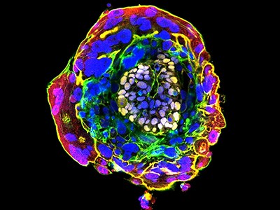
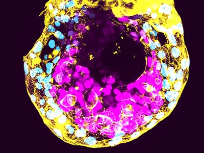
    Under his microscope, Jun Wu could see several tiny spheres, each less than 1 millimetre wide. They looked just like human embryos: a dark cluster of cells surrounded by a cavity, and then another ring of cells.

    But Wu, a stem-cell biologist at the University of Texas Southwestern Medical Center in Dallas, knew that these spheres were not what they seemed. They were laboratory-grown models of embryos, and they were far from perfect replicas.

    Entire groups of cells were absent and others were there that didn’t belong. And Wu knew that, eventually, the models would perish abruptly and chaotically.

    If embryo models were houses, then behind the facade they would have uneven floors, distorting mirrors and ghosts in their closets. Nonetheless, dozens of labs are competing to grow the best likeness of a human embryo.

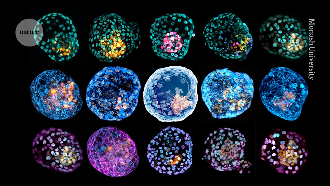
    There are as many models as there are groups making them, each recapitulating slightly different aspects of embryo development in the hope of uncovering new biology about the first weeks after conception.

    This high-stakes, high-drama period “is shrouded in mystery”, says Nicolas Rivron, a developmental biologist at the Institute of Molecular Biotechnology of the Austrian Academy of Sciences in Vienna. In the womb, these embryos are too small to be observed using ultrasound. And in the lab, there are technical, ethical — and often legal — limits to studying real embryos outside the body beyond 14 days after conception.

    Insights from embryo models could help to explain why about one-third of natural embryos don’t make it past their first weeks. This could help to address infertility, improve the success rate of in vitro fertilization and even prevent diseases that emerge early in development. Models could also be used to test the safety of drugs for embryos.

    But as the models become increasingly complex, and reach symbolic milestones, such as the first heartbeat, they raise tricky ethical questions. Ethicists, regulators and legal specialists are scrambling to keep up with the pace of research.

    Meanwhile, the field is fizzing with energy. In February, researchers organized the world’s first scientific meeting entirely dedicated to embryo models. And several scientists have launched spin-off companies to use models to develop therapeutic molecules, test drugs and improve fertility treatments. Embryo models are “pretty much the hottest topic right now”, says Insoo Hyun, a bioethics consultant for the Broad Institute of MIT and Harvard in Cambridge, Massachusetts.

    Epic show

    The meeting of egg and sperm triggers a process of rapid and precisely choreographed cell division and differentiation. In the first week, around 100 cells form a hollow circle known as the blastocyst. This is made up of three distinct groups that eventually grow into the embryo, the supportive yolk sac and the placenta.

    Then the embryo implants itself in the uterus. From about two weeks, embryos go through a process known as gastrulation, in which cells are committed to becoming one of three cell types and organize into layers. These layers differentiate further into lungs, guts, muscles and other organs, in a process known as organogenesis.

    “The embryo is never static,” says Naomi Moris, a developmental biologist at the Francis Crick Institute in London. “It undergoes these huge dramatic shifts.”

    Researchers have attempted to recreate this epic show in a dish. These efforts — often done in mice and then in humans — have typically captured snapshots of the process.

    In 2014, researchers coaxed human embryonic stem cells into three distinct rings — precursors to cells that form the embryo and the placenta1. Later models featured amniotic cavities and yolk sacs, and some were 3D. By 2020, some researchers had recapitulated an aspect of gastrulation2, in which the embryo elongates into a tube-like structure.

    But many of the early studies wouldn’t be considered models of an entire embryo by today’s standards, says Wu.

    Complete model

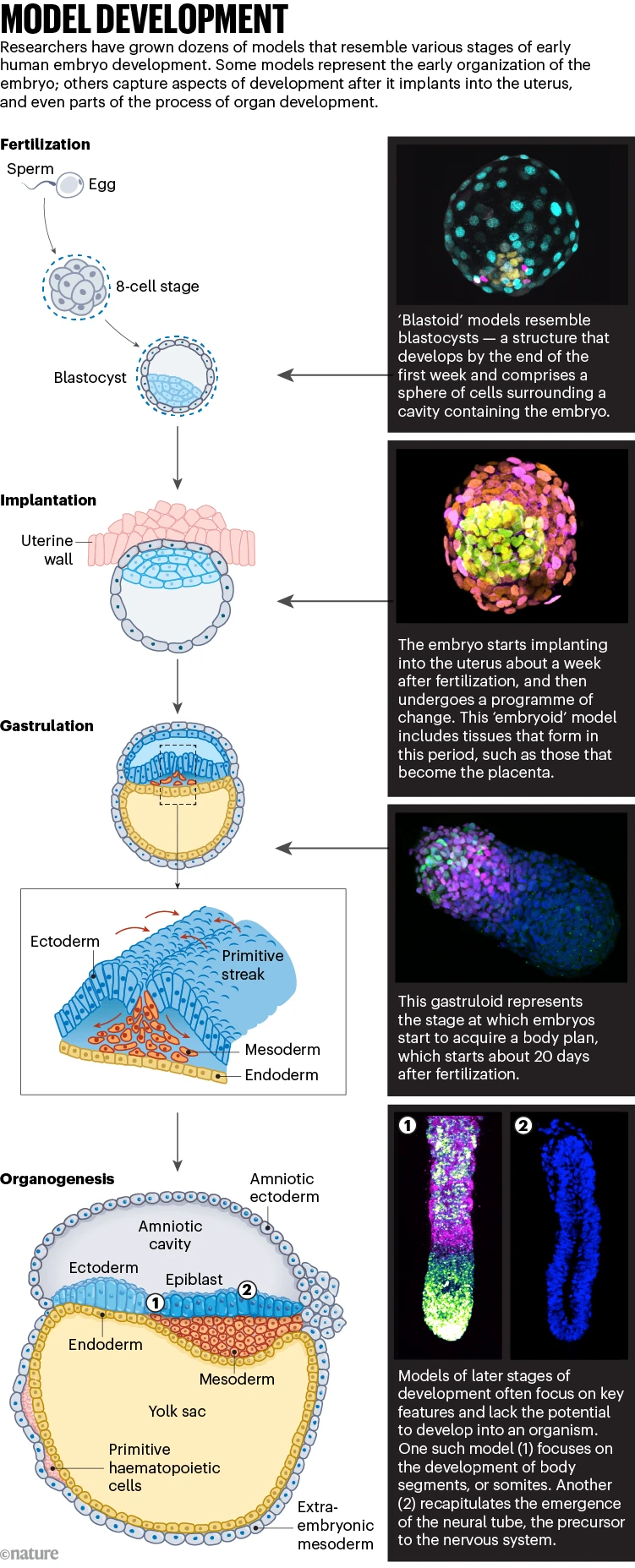
    A major milestone came in 2021, when Wu’s group3 and another team4 published models that resemble the human blastocyst (see ‘Model development’) — typically the stage at which embryos are transferred to the uterus during in vitro fertilization.

    Model development: infographic that shows stages of embryonic development, and the equivalent laboratory models that are grown to study them.

    Credit: Nik Spencer/Nature; top to bottom: ©Rivron/Nature/IMBA; Bailey A. T. Weatherbee et al./Nature (CC-BY); Naomi Moris/Univ. Cambridge; Yuchuan Miao et al./Nature; Xufeng Xue et al./Nature

    These models, called blastoids, contain cells that form the embryo and those that will support it, called extra-embryonic cells, making them the first ‘complete’ or ‘integrated’ models of the human embryo.

    “They’re not perfect,” says Marta Shahbazi, a stem-cell and developmental biologist at the MRC Laboratory of Molecular Biology in Cambridge, UK. But “they’re pretty good”.

    Some groups have tried to capture even earlier stages of development, using cells with the ability to turn into every cell type required for embryonic development (most models use cells with more limited abilities).

    In 2022, Miguel Esteban, a stem-cell biologist at the biotechnology company BGI Cell in Shenzhen, China, and his colleagues developed a model resembling the eight-cell embryo that typically forms three days after fertilization5. And this June, Du Peng, a stem-cell biologist at Peking University in Beijing, made similar blastomere mimics that eventually form blastoids without needing to be doused in chemicals6.

    Writing the rules

    Ever since the first embryo models appeared, ethicists have been striving to address the dilemmas that they pose. The International Society for Stem Cell Research (ISSCR) developed guidelines in 2021; many countries are considering their own guidelines and legislation.

    Australia’s rules are some of the most stringent in the world. In 2020, biochemist Jose Polo, who leads a team based at Monash University in Melbourne and the University of Adelaide, informed Australia’s regulatory body overseeing embryo research, the National Health and Medical Research Council, that he had developed blastoids, and was asked to put the work on hold.

    The regulator wanted to assess whether blastoids met the criteria to be considered an embryo under the current laws, which define embryos as biological entities with the potential to develop to a stage, roughly two weeks in, at which a structure called the primitive streak appears and the entity moves towards having a body plan.

    Some five months later, the answer came back: they did, said the agency, because of their theoretical potential to develop a primitive streak. As a result, the same limits that apply to research on real embryos would apply to blastoids.

    It was a devastating setback, says Polo. His team had to get a specific embryo licence, which bars the group from growing blastoids to study later stages of gastrulation and organogenesis. “I think that they made a mistake,” says Polo about the agency’s ruling. The rules also limit the number of blastoids that can be made, and require stricter consent from those donating cells to be used in them.

    Every country is charting its own course. Some points of contrast are how countries define an embryo, whether that definition extends to embryo models and how permissive the rules are to research. Regulators are often guided by rules and norms designed for research on real human embryos when thinking about embryo models, says Megan Munsie, a developmental biologist and bioethicist at Murdoch Children’s Research Institute in Melbourne.

    And these discussions often transcend the world of research, finding relevance in other domains, such as reproductive health, abortion, women’s rights and regenerative medicine, says Alfonso Martinez Arias, a developmental biologist at the University Pompeu Fabra in Barcelona, Spain. “Our echo chamber is very large,” he told a room packed with his peers at the annual ISSCR meeting in Hamburg, Germany, in July.

    Animated sequence of a 3D reconstruction of a day 8 human stem cell-based embryo model.

    Human embryo models are trying to capture aspects of the development of real embryos, such as this one at the 16-cell stage.Credit: Bernardo Oldak et al./Nature

    Because embryo models differ in lots of ways from the real structures, most countries treat the two differently. In Spain, for example, the definition of an embryo is based on fertilization, which excludes embryo models, says Nienke de Graeff, a bioethicist at Leiden University Medical Centre, the Netherlands.

    Some definitions focus on the embryo’s potential to form or become something else. The ISSCR has said that, based on their potential, embryo models cannot be considered to be embryos, and most countries take a similar view.

    Some have proposed revising regulations concerning real embryos to cover some types of embryo model. In the Netherlands, says Nienke, a scientific advisory body proposed a ban on growing the models beyond the equivalent of 28 days in real-embryo terms. France is considering the same limit. Researchers in the United Kingdom did something a little different in July: they published voluntary guidelines for embryo models that do not set fixed limits on how long they can be cultured. The guidelines could eventually lead to the passing of binding legislation — as happened with similar voluntary UK guidelines around embryo research several decades ago.

    The UK guidelines and 2021 ISSCR guidelines do, however, forbid the transfer of human embryo models into a uterus, and several other countries, including Sweden and Japan, are considering introducing similar restrictions.

    Science accelerates

    Meanwhile, the science keeps moving at such a pace that regulators have a lot to keep up with. In June 2024, the ISSCR announced that it had set up a working group to assess the state of the science and review earlier guidelines, in light of the models published since 2021.

    In 2023, around half a dozen teams described models that recapitulate the development of embryos just after implantation. Two models in particular were widely covered by the media — one by Magdalena Zernicka-Goetz, a developmental biologist at the California Institute for Technology in Pasadena, and one by Jacob Hanna, a stem-cell biologist at the Weizmann Institute of Science in Rehovot, Israel. They were described as complete post-implantation models, but that title has been hotly debated.

    “These are not complete models,” says Rivron. The one by Zernicka-Goetz’s group7 doesn’t have cells that behave like trophoblasts, which provide nutrition for the embryo — and although Hanna’s8 does contain a trophoblast-like layer, it isn’t as organized as the real thing, say researchers.

    “It’s almost like a beauty contest — whose ‘model’ looked better,” says Jianping Fu, a bioengineer at the University of Michigan in Ann Arbor. “There’s a lot of excitement, but at the same time, there’s some hype in the field right now.”

    Some researchers question the value of chasing a complete model. It’s a “pretty exquisite balancing act”, says Hyun. Researchers want models to resemble an embryo closely enough that they provide real insight into human development but not so closely that they can’t tell the difference between the two, and so risk restrictions to their work. “You want to skate as close to the edge as possible, without falling over,” he says.

    Some researchers try to avoid this ethical dilemma by intentionally introducing changes to their embryo models that would make it impossible for the model to result in an organism. For example, Hanna has started working on models in which genes involved in brain and heart development have been inactivated. He has inferred from discussions with Christian and Jewish leaders in his community that an embryo model lacking brain or heart tissue would not be considered a form of person.

    Fu has described a model9 that he says alleviates some of the ethical burdens because, although it reaches gastrulation, it gets there without first forming the primitive streak.

    Such models can be insightful and useful, say researchers. One, first published last December, has generated excitement because of how well it reflects some aspects of real embryos10, as well as its direct implications for the clinic. When Mo Ebrahimkhani, a stem-cell bioengineer at the University of Pittsburgh, Pennsylvania, and his colleagues grew 3D models on a dish, they noticed the appearance of tiny blood islands. Those islands held the first progenitors of blood cells, including immune cells known as macrophages, platelet-producing cells and cells containing haemoglobin. As Ebrahimkhani has found in unpublished work, these models could be used to produce large amounts of blood stem cells, which could be useful for transplants in people with cancer or genetic diseases.

    Organ making

    At about three weeks, the embryo starts the momentous organ-growing process that lasts more than a month. Several teams have homed in on aspects of this organogenesis, growing models that resemble only pieces of the embryo — this has potentially fewer ethical constraints than modelling the whole structure.

    One such model11 recapitulates the rhythmic process by which the body forms repeating segments known as somites, which give rise to vertebrae. And earlier this year12, Fu’s team described a model of the neural tube, the progenitor of the central nervous system, complete with a treasure trove of cells, including the precursors to some neurons.

    But even these models aren’t without controversy; those that contain nerve cells raise ethical questions around the emergence of sentience, says Hyun.

    The next frontier for embryo-like models is to nestle them in environments that more closely resemble the womb, and study how embryos interact with its lining. Researchers have shown that blastoids placed on cells that make up the lining of the uterus can burrow in and fuse correctly. Co-culturing embryo models with this maternal tissue could help models develop as natural embryos would.

    Some scientists have gone even further using cells from other animals, including non-human primates. For example, in unpublished work, Liu Zhen, a developmental biologist at the Institute of Neuroscience at the Chinese Academy of Sciences in Shanghai, has grown monkey blastoids in a dish for long enough to reach early organogenesis.

    In 2023, Liu transferred blastoids into eight monkeys, three of which experienced the hormone surge observed in early pregnancy13. The blastoids formed gestation sacs but then stopped developing. Cow and mouse blastoids transferred into their respective species also don’t survive long.

    There are currently no rules prohibiting the transfer of non-human embryo models into living animals, with the exception of humans, but Hyun worries that if such transfers do lead to a live birth, that will cause a backlash against the research and have a detrimental effect on studies using human embryo models. The intellectual leap from monkey to humans is easy to make, he says. “You don’t actually have to do the human experiment to have a pretty major concern.”

    When is it an embryo?

    Most researchers agree that today’s human embryo models are nowhere near the real thing. The key challenge for researchers who are developing guidelines is determining “when an embryo model would be considered equivalent to an embryo”, says Amander Clark, a developmental and stem-cell biologist at the University of California, Los Angeles, who co-chairs the ISSCR’s working group on embryo models.

    Given that ethical norms prohibit the transfer of embryo models into a uterus to see whether they can give rise to an organism, researchers are coming up with other tests of their potential. Some groups are developing tools to better compare embryo models with real embryos, for example by looking at the cells’ RNA profiles. But even these gene-expression exercises can miss key information, says Hanna, such as the positions of cells in an embryo.

    Assessing and improving models is important for science, as well as for ethics, says Fredrik Lanner, a stem-cell and developmental biologist at the Karolinska Institute in Stockholm. If the models aren’t good, then they’re not useful, he says. It’s “really critical now for us to not waste time on bad models”.

    That standard setting is especially important as the field starts to offer real insights into early embryonic development. There have already been some surprises, says Moris. Most notable is the ability of cells to switch personas — their plasticity — and their capacity to self-organize, often without the aid of extra-embryonic tissue.

    Some models are even revealing new phenomena. Researchers have long known that, in some mammals, embryos that are fertilized in the summer ‘hibernate’ and restart development later so that the young are born in the spring. In unpublished work, Rivron and his colleagues have been able to cajole human blastoids into this state of suspended development.

    But so far, he says, embryo models have not led to scientific discoveries of societal value, and it’s not clear which models will help researchers get there.

    “There’s been a lot of debate, arguments, especially drama, in the past”, along the lines of “my model is better than yours”, says Wu. The reality is that every model is imperfect, but every one is useful, he says. It just depends on what the question is.

    [ad_2]

    Source link

  • Lab-grown stem cells could be a ‘breakthrough’ for cancer treatment

    Lab-grown stem cells could be a ‘breakthrough’ for cancer treatment

    [ad_1]

    Stem cells are produced by bone marrow and can turn into different types of blood cells

    KATERYNA KON / SPL/ Alamy

    Human blood stem cells have been made in a laboratory for the first time, which could significantly improve how we treat certain types of cancer.

    The lab-grown cells have so far only been tested in mice, but when infused into the animals, the cells became functional bone marrow at similar levels to those seen after umbilical cord blood cell transplants.

    Treating cancers such as leukaemia and lymphoma via radiation and chemotherapy can destroy the blood-forming cells in bone marrow. A stem cell transplant means that new, healthy bone marrow and blood cells can grow. Umbilical cords are a particularly rich source of stem cells, but donations are limited and the transplant can be rejected by the body.

    The new method would allow researchers to produce stem cells from the actual patient, eliminating the supply issue and reducing the risk that their body would reject them.

    First, human blood or skin cells were turned into so-called pluripotent stem cells through a process called reprogramming. “This involves temporarily turning on four genes, with the result that the patient cells revert to an early stage of development when they can become any cell in the body,” says Andrew Elefanty at the Murdoch Children’s Research Institute in Melbourne.

    The second stage involved turning the pluripotent cells into blood stem cells. “We first make thousands of small floating balls of cells, a few hundred cells in each ball, and direct them to change from being stem cells to sequentially become blood vessels and then blood cells,” says Elefanty. This process, called differentiation, takes about two weeks and makes millions of blood cells, he says.

    These cells were then infused into mice that lacked an immune system and became functional bone marrow in up to 50 per cent of cases. This means it made the same cells that carry oxygen and fight infections as healthy human bone marrow does, says Elefanty. “It is this unique ability to make all the blood cell types for a prolonged period of time that defines the cells as blood stem cells,” he says.

    Abbas Shafiee at the University of Queensland in Brisbane says the work is a “magnificent breakthrough” towards new therapies for blood cancers. “It has not been done before and it has a lot of potential for the future.” But even once animal testing is complete, a lot of research in humans needs to be done before the approach can be used in clinics, he says.

    Simon Conn at Flinders University in Adelaide, Australia, says a key advantage of the team’s approach is that it could be scaled up to produce “an essentially never-ending supply” of blood stem cells. But he adds that the work was based on either blood or skin cells, with the rate of success and the diversity of the blood cells depending on the initial cell type.

    “This suggests the treatment, even at the preclinical stage in mice, is not consistent, which will need to be addressed prior to any clinical trials in human patients,” he says.

    Topics:

    [ad_2]

    Source link

  • Tissues stay fit by balancing clearance of dying cells with regeneration

    Tissues stay fit by balancing clearance of dying cells with regeneration

    [ad_1]

    • RESEARCH BRIEFINGS

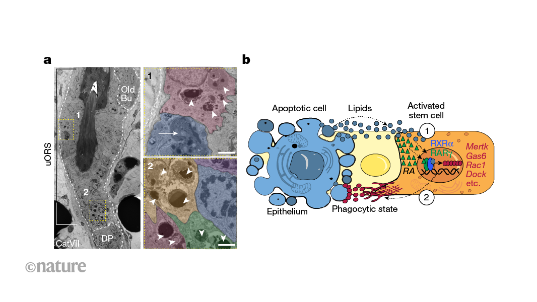
    Clearance of dying cells is essential to the health of tissues. Study of hair follicles finds that, when follicle stem cells encounter dying neighbours, they become temporarily able to engulf and digest them. The molecular underpinnings of this reveal how clearance is spatially and temporally tuned to maintain tissue fitness.

    [ad_2]

    Source link

  • ‘epigenetic’ reset in human cells paves the way

    ‘epigenetic’ reset in human cells paves the way

    [ad_1]

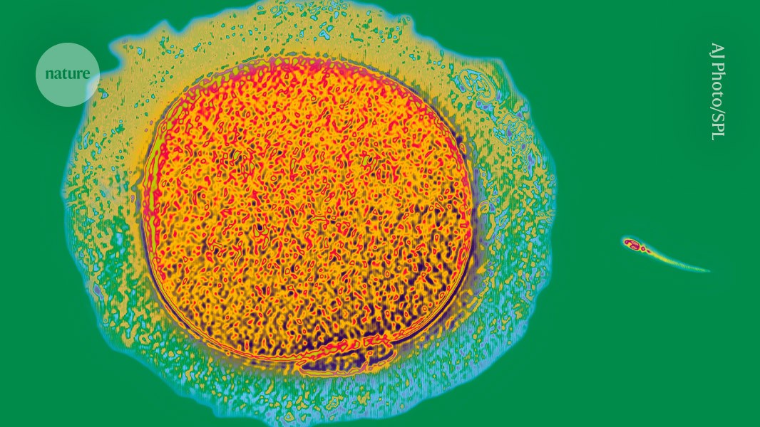
    Coloured light micrograph of a sperm cell (small, with a long tail) approaching an egg cell (much larger, roughly circular) on a green background.

    A sperm cell (right) swims towards a human egg (artificially coloured).Credit: AJ Photo/Science Photo Library

    The day when human sperm and eggs can be grown in the laboratory has inched a step closer, with the discovery of a way to recreate a crucial developmental step in a dish1.

    The advance, described 20 May in Nature, addresses a major hurdle: how to ensure that the chemical tags on the DNA and associated proteins in artificially produced sperm and eggs are placed properly. These tags are part of a cell’s ‘epigenome’ and can influence whether genes are turned on or off. The epigenome changes over a person’s lifetime; during the development of the cells that will eventually give rise to sperm or eggs, these marks must be wiped clean and then reset to their original state.

    “Epigenetic reprogramming is key to making the next generation,” says Mitinori Saitou, a stem-cell biologist at Kyoto University in Japan, and a co-author of the paper. He and his team worked out how to activate this reprogramming — something that had been one of the biggest challenges in generating human sperm and eggs in the laboratory, he says.

    But Saitou is quick to note that there are further steps left to conquer, and that the epigenetic reprogramming his lab has achieved is not perfect.

    “There is still much work to be done and considerable time required to address these challenges,” agrees Fan Guo, a reproductive epigeneticist at the Chinese Academy of Sciences Institute of Zoology in Beijing.

    Eggs in a dish

    Growing human sperm and eggs in the laboratory would offer hope to some couples struggling with infertility. It would also provide a way to edit disease-causing DNA sequences in sperm and eggs, sidestepping some of the technical complications of making such edits in embryos. And understanding how eggs and sperm develop can give researchers insight into some causes of infertility.

    But in addition to its technical difficulty, growing eggs and sperm in a dish — called in vitro gametogenesis — would carry weighty social and ethical questions. Genetic modification to prevent diseases, for example, could lead to genetic enhancement to boost traits associated with intelligence or athleticism.

    Epigenetic reprogramming is key to the formation of reproductive cells — without it, the primordial cells that would eventually give rise to sperm and eggs stop developing. Furthermore, the epigenome affects gene activity, helping cells with identical DNA sequences to take on unique identities. The epigenome helps to differentiate a brain cell, for example, from a liver cell.

    Researchers know how to grow mouse eggs and sperm using stem-cell-like cells generated from skin. But the protocols used don’t work in human cells: “There is a big gap between mice and humans,” says Saitou.

    Pressing reset on the epigenome

    So Saitou and his colleagues began an arduous search for a way to control epigenetic reprogramming in human cells. They found that a protein called BMP2 was essential for this step and that adding it to their cultures promotedepigenetic reprogramming. The cells grown in this culture were able to progress a step further in their development than were cells in cultures without added BMP2.

    After epigenetic reprogramming, the cells’ development halted again. Even so, each step towards in vitro gametogenesis holds “immense significance”, says Guo. Saitou and his colleagues are now hunting for ways to nudge the cells further along the path to becoming sperm and eggs.

    The researchers carefully analysed epigenetic marks in their laboratory-grown cells and found that although many of these imprints had been wiped away, a few remained. This means that the reprogramming might be incomplete — which could have serious consequences if such cells were used for reproduction. “If imprinting on even one gene is aberrant, that could lead to disease,” says Saitou.

    Such caveats are important to bear in mind, he says: the field of in vitro gametogenesis is advancing rapidly, and these results, along with other developments in the past few years, could fuel speculation and false claims that a solution is just around the corner. “I think in maybe five years or so, things will get more settled,” he says. “And then only the good science will remain.”

    [ad_2]

    Source link

  • Therapeutic potential of human umbilical cord-derived mesenchymal stem cells in Crohn’s disease

    Therapeutic potential of human umbilical cord-derived mesenchymal stem cells in Crohn’s disease

    [ad_1]

    In a recent study published in eBioMedicine, researchers investigated whether human umbilical cord-derived mesenchymal stem cells (hUC-MSCs) could heal luminal ulcers in Crohn’s disease (CD) patients.

    Study: hUC-MSCs therapy for Crohn’s disease: efficacy in TNBS-induced colitis in rats and pilot clinical study. Image Credit: Lobachad/Shutterstok.comStudy: hUC-MSCs therapy for Crohn’s disease: efficacy in TNBS-induced colitis in rats and pilot clinical study. Image Credit: Lobachad/Shutterstok.com

    Background

    CD is a developing global disease with a poor overall effectiveness rate, posing a considerable therapeutic challenge to health professionals. Current therapies have limitations, and recovery rates are far from expected.

    MSC-based therapy has demonstrated promise as a potential new treatment option for various disorders, including CD of the perianal fistulizing type. However, limited evidence is available on the efficacy of MSCs in treating or healing intestinal ulcers in CD. 

    Human umbilical cord MSCs have acquired popularity due to their immunomodulatory capabilities, simplicity of collection, limitless source of stem cells, low immunological rejection, and lack of tumorigenic features.

    Despite their usefulness in immunological and inflammatory illnesses, there has been no research on using hUC-MSCs locally to heal intestinal ulcers in CD patients.

    About the study

    The present study researchers explored hUC-MSC safety and effectiveness in CD management.

    The researchers used the 4,6-trinitrobenzene-sulfonic acid (TNBS)-inflicted colitis rat model to investigate the effectiveness of hUC-MSC therapy in CD. They calculated the disease activity index (DAI) by adding values for body weight loss, stool consistency, and stool blood.

    The colon macroscopic damage index (CMDI) assessed the level of colonic inflammation. They analyzed colon samples under a microscope to establish histopathological scores (HPS).

    From November 2020 to October 2023, the researchers ran a pilot, open-label clinical study with 17 refractory CD patients. Participants received a local submucosal injection of hUC-MSCs (60 × 106 cells/10 mL) by colonoscopy, followed by an intravenous drip of 1.0 × 10⁶ hUC-MSCs/kg/100 mL the following day.

    The study included individuals aged 18–75 years with moderate-severe CD for ≥3 months, baseline CDAI scores of 220-450, and endoscopic ulcers. They did not respond to conventional or advanced treatment, including immunomodulators and biologics.

    The researchers followed the patients for 24 weeks, measuring laboratory and clinical markers on days 0, week 4, week 8, week 12, and week 24.

    They performed endoscopic assessments at the start of the trial and, after 12 weeks, obtained mucosal specimens from lesion margins for ribonucleic acid (RNA) sequencing. The researchers excluded individuals with a history of pregnancy, CD complications, comorbidities, prior surgery, uncontrolled infections, or cancer in the past five years.

    The team also produced two hUC-MSC in vitro co-cultures using mucosal tissues and the human monocyte cell line THP-1-induced M1 macrophages. Reverse transcription-polymerase chain reaction (RT-PCR) measured inflammatory and intestinal barrier function-related genes.

    In addition, the researchers performed gene ontology (GO) analysis and explored the Kyoto Encyclopedia of Genes and Genomes (KEGG) pathway enrichment.

    They used enzyme-linked immunosorbent assays (ELISA) to assess the expression of tumor necrosis factor-stimulated gene-6 protein (TSG-6) in hUC-MSC culture supernatants after 48 hours of stimulation with lipopolysaccharide (LPS), interferon-gamma (IFNγ), and TNF-alpha (TNFα) for 48 hours.

    Results

    In preclinical studies, hUC-MSCs raised body weight while decreasing DAI, CMDI, and HPS scores in the TNBS-inflicted colitis murine model, with significant reductions in intestinal mucosal injury, edema, hyperemia, and ulcerations.

    Local and submucosal hUC-MSC injections resulted in intestinal ulcer regression in the study participants.

    Among the subjects, eight (47%) displayed endoscopic responses (a ≥50% improvement in SES-CD scores), and three (18%) demonstrated mucosal healing, with parallel improvements of laboratory and clinical markers without significant side effects.

    Human umbilical cord-derived MSCs increased transcripts associated with intestinal barrier integrity while decreasing those related to inflammatory intestinal mucosal pathways, including TNF-α, interleukin-17 (IL-17), and toll-like receptor (TLR) signaling.

    The treatment dramatically enhanced RNA levels of tight junction proteins [E-cadherin (CDH1), zonula occludens-1 (ZO1), and claudin-1 (CLDN1)] in the intestinal epithelium and intestinal pro-inflammatory genes (TNF-α, IL-1β, and IL-6).

    Furthermore, hUC-MSCs reduced THP1-induced M1 macrophage polarization and messenger RNA (mRNA) expressions of TNF-α, IL-1β, and IL-6.

    MSCs obtained from the human umbilical cord suppressed Janus kinase/signal transducers and activators of transcription (JAK/STAT) phosphorylation in intestinal mucosal cells from Crohn’s disease patients.

    Conclusions

    The study findings showed that submucosal injection of hUC-MSCs by colonoscopy, paired with an intravenous drip, reduced TNBS-inflicted colitis among rats. This technique may treat refractory CD with clinical effectiveness and tolerable risk.

    TSG-6 release may contribute to the therapeutic effect by inhibiting JAK/STAT phosphorylation, reducing M1 macrophage polarization, and improving intestinal barrier function.

    In individuals with moderately or highly active CD, hUC-MSC treatment resulted in clinical remission and the repair of intestinal ulcers.

    The expression of STAT1, M1 macrophage-associated specific markers, and pro-inflammatory factors in the intestinal mucosa dropped considerably following therapy. Further research is required to establish the efficacy of hUC-MSC treatment and investigate the underlying processes of the therapy.

    [ad_2]

    Source link

  • Rat neurons repair mouse brains — and restore sense of smell

    Rat neurons repair mouse brains — and restore sense of smell

    [ad_1]

    Two research teams have demonstrated that adding rat neurons to mouse brains that were missing crucial cells could help the organs to recover function1,2. The experiments could help scientists to better understand how different species’ brains develop, and even aid efforts to grow ‘chimeric’ pigs with human organs that could be used for transplantation in people.

    Researchers have successfully generated hybrid, or chimeric, animals in the past. Among these have been mice with rat organs, including pancreases3, and mice with human neurons in their brains4. But no one had shown clearly whether rat neurons could be incorporated fully into a mouse’s brain circuits in such a way that they would become an essential part of controlling the host animal’s behaviour.

    How neurons connect with one another, and fire, makes integrating cells from two species complicated, says Kristin Baldwin, a neuroscientist at Columbia University in New York City. “Neurons are not just Legos,” she says.

    Early integration

    In a paper published by one of the teams on 25 April in Cell1, Baldwin, molecular biologist Jun Wu at the University of Texas Southwestern Medical Center in Dallas and their colleagues attempted to test this by mixing rat and mouse neuronal cells very early in the mice’s development.

    First, they engineered the genes in a group of mice in a way that destroyed some neurons in the animals’ olfactory systems. This disrupted the circuits linking olfactory neurons in the nose with higher brain regions, leaving the mice unable to use their sense of smell to find mini-cookies that the researchers had buried in various places throughout the animals’ cages.

    But when the researchers injected rat stem cells into blastocysts — early-stage embryos — of mice engineered in this way, the cells filled the gaps in the brain circuits. And once the mice had grown into adults, they were able to find their cookies by smell. Killing the mouse neurons created “niches” for the rat cells to take up residence in various places inside the animals’ olfactory circuits, as well as elsewhere in their bodies, Baldwin says. Her group is now working on methods for replacing specific mouse neurons with rat cells in a more targeted way.

    In a Cell paper published by the second team, also on 25 April2, Wu and his colleagues developed a more aggressive strategy for getting rat cells into a mouse’s brain. Using C-CRISPR, a genetic-editing tool that cuts genes in multiple places to ensure that they are fully inactivated, the researchers wiped out every trace of a gene called Hesx1 in a group of mouse blastocysts. This gene controls the development of the forebrain: a large region in the brain that coordinates much of an animal’s behaviour.

    When the researchers allowed these blastocysts to develop into mice without forebrains, the animals died shortly after birth. But when Wu and his team injected rat stem cells into the blastocysts, the forebrains that developed were made entirely of rat cells. Once the mice had grown, they were healthy and seemed to act normally, although Wu says it would be difficult to determine whether there were any subtle behavioural differences between them and normal mice.

    Hiro Nakauchi, a stem cell biologist at Stanford University in California, agrees that it would be hard to establish this. Researchers in his laboratory once tried making mice ‘smarter’ by giving them rat brain cells, but they abandoned the effort when they realized that the differences between rodents with and without the cells were too minor to detect statistically without testing the behaviours of a large number of hybrid animals. Still, Nakauchi says that the new papers are meticulous analyses of chimeric animals’ brains — something he is excited about. “This is what I have been awaiting,” he says.

    ‘Fascinating biology’

    Wu and Baldwin say that their research addresses some long-standing concerns about developing chimaeras, particularly for the purpose of transplanting tissue or organs from animals such as pigs into people. Aside from ethical considerations, there is the concern that the human body will reject a transplanted chimeric organ. But because the teams added the rat cells so early in the mice’s development — long before the embryos had formed an immune system — the animals’ bodies never learnt to recognize the cells as foreign and never attacked them.

    Another concern is a mismatch in the developmental rates of species. However, the teams found that the mouse brains developed at the same rate as they would normally, rather than at the slower pace at which a rat usually develops.

    “There’s lots of fascinating biology to be learnt from this [rat–mouse] chimaera,” says Jian Feng, a physiologist at the University of Buffalo in New York. He’s not surprised that the rat cells followed the pace of the mouse’s developmental ‘clock’. In 2020, his group published a paper about a mouse embryo that it had engineered to contain up to 4% human cells5. The embryo began developing human red blood cells 17 days into gestation — much earlier than these cells develop in human embryos — suggesting that human cells, too, could follow the molecular directions of their host.

    Wu says that his laboratory now plans to use the technology developed for these studies to make chimaeras by transplanting cells from wild rodent species into lab mice. It’s difficult to study wild rodents, because they are hard to maintain and breed in captivity, he says. But making stem cells from their tissue samples and inserting them into mouse blastocysts might allow researchers to study how these other species’ brains develop and function.

    [ad_2]

    Source link

  • Targeting RNA opens therapeutic avenues for Timothy syndrome

    Targeting RNA opens therapeutic avenues for Timothy syndrome

    [ad_1]

    Nature, Published online: 24 April 2024; doi:10.1038/d41586-024-00911-1

    A therapeutic strategy that alters gene expression in a rare and severe neurodevelopmental condition has been tested in stem-cell-based models of the disease, and has been shown to correct genetic and cellular defects.

    [ad_2]

    Source link

  • Womb pressure shapes facial features, UCL study reveals

    Womb pressure shapes facial features, UCL study reveals

    [ad_1]

    Physical cues in the womb, and not just genetics, influence the normal development of neural crest cells, the embryonic stem cells that form facial features, finds a new study led by UCL researchers.

    The study published in Nature Cell Biology found that an increase in hydrostatic pressure sensed by the embryo can hinder the healthy development of facial features in mouse and frog embryos and in human embryoids (cell structures grown in the lab from human stem cells), suggesting that differences in pressure might affect the risk of facial malformations.

    The researchers found that when neural crest cells are exposed to higher-than-usual levels of pressure, key cell signaling pathways are impeded, and the risk of craniofacial malformations significantly increases.

    Lead author Professor Roberto Mayor (UCL Cell & Developmental Biology) said: “Our findings suggest that facial malformations could be influenced not only by genetics but by physical cues in the womb such as pressure.

    “When an organism is experiencing a change in pressure, all the cells – including the embryo inside the mother – are able to sense it.

    Our work shows that embryos are sensitive to pressure, but we do not know how sensitive they are; for instance, will a change in the pressure inside the uterus be able to affect the embryo? This will require further research to understand how changes inside the body as well as in environmental pressure might influence human embryo development.”


    Roberto Mayor, Professor, Cell & Developmental Biology, University College London

    The researchers say their findings could also have implications for stem cell research, as the study indicates that the development and differentiation (the process of stem cells becoming specialized cells) of stem cells are under the influence of pressure. Understanding this connection could transform how scientists manipulate stem cells for various therapeutic purposes.

    The findings add to work by Professor Mayor and colleagues at UCL into how mechanical cues in the womb can influence the development of facial features, as they have previously found that cells in the developing embryo sense the stiffness of other cells around them, which is key to them moving together to form the face and skull.

    Source:

    Journal reference:

    Alasaadi, D. N., et al. (2024). Competence for neural crest induction is controlled by hydrostatic pressure through Yap. Nature Cell Biology. doi.org/10.1038/s41556-024-01378-y

    [ad_2]

    Source link