With advances in neuroscience and the development of new technologies, new ethical considerations have emerged. This is particularly true for human brain organoids, which are three-dimensional tissues grown from stem cells that partially replicate the characteristics of the human brain. Brain organoids have emerged as important tools for studying brain development and disease, but there are concerns about the possibility of these organoids developing consciousness. This has important implications for research ethics and the need to obtain informed consent from cell donors.
To address these questions, an international team of researchers has sought to shed light on the intricate ethical landscape of brain organoid research, offering insights that will be important for researchers, ethicists, and policymakers alike. Through a comprehensive literature review and ethical analysis, they examined how the potential for consciousness in brain organoids complicates the process of obtaining informed consent from cell donors. Their study revealed uncertainties in two key aspects: the scientific understanding of consciousness in brain organoids and the moral implications of brain organoid consciousness. These uncertainties pose significant challenges for respecting donor autonomy and determining the scope of consent in human brain organoid research.
To clear these uncertainties, the researchers proposed three tentative methods for obtaining consent from donors. First, to address donor concerns and uncertainties, they advocated for project-specific consent procedures by explicitly informing cell donors that their cells will be used in brain organoid research. Second, they emphasized the importance of incorporating the abovementioned uncertainties into consent procedures by providing donors with comprehensive information about the potential for brain organoid consciousness and measures implemented to address this. Finally, they proposed the development of a risk framework for brain organoid research to guide ethical considerations and minimize potential harm.
The researchers note that some scientists may believe that such concerns are unwarranted, at least at the current stage. However, they argue that if the goal of human brain organoid research is to contribute to the advancement of science and medicine, and ultimately society as a whole, it is important to conduct research that earns public trust.
Says Dr. Sawai “Ignoring these aspects may lead to short-term success, but it’s unlikely to be sustainable in the long term. Our findings can be considered foundational research that solidifies the ethical groundwork essential for the progression of scientific and medical research.”
The findings of this study have far-reaching implications for the fields of neuroscience and research ethics, especially in terms of how future studies obtain informed consent from cell donors. As brain organoid research progresses, it is imperative to navigate these ethical complexities, particularly those regarding potential consciousness, with diligence and foresight. By tailoring informed consent procedures and prioritizing ethical oversight, scientists can uphold the principles of autonomy while advancing our understanding of the brain. This study serves as a call to action for researchers, ethicists, and policymakers to engage in thoughtful discourse and decision-making regarding brain organoid research. By confronting these ethical challenges head-on, scientists can ensure that the quest to understand the brain is guided by ethical principles.
Kataoka, M., et al. (2024) The Donation of Human Biological Material for Brain Organoid Research: The Problems of Consciousness and Consent. Science and Engineering Ethics. doi.org/10.1007/s11948-024-00471-7.
In a collaborative study, researchers from Kyushu University and Harvard Medical School have identified proteins that can turn or “reprogram” fibroblasts -; the most commonly found cells in skin and connective tissue -; into cells with similar properties to limb progenitor cells. Publishing in Developmental Cell, the researchers’ findings have enhanced our understanding of limb development and have set the stage for regenerative therapy in the future.
Globally, close to 60 million people are living with limb loss. Amputations can result from various medical conditions such as tumors, infections, and birth defects, or due to trauma from industrial accidents, traffic accidents, and natural disasters such as earthquakes. People with limb injuries often rely on synthetic materials and metal prostheses, but many researchers are studying the process of limb development, with the aim of bringing regenerative therapy, or natural tissue replacement, one step closer as a potential treatment.
“During limb development in the embryo, limb progenitor cells in the limb bud give rise to most of the different limb tissues, such as bone, muscle, cartilage and tendon. It’s therefore important to establish an easy and accessible way of making these cells,” explains Dr. Yuji Atsuta, lead researcher who began tackling this project at Harvard Medical School and continues it as a lecturer at Kyushu University’s Graduate School of Sciences.
Currently, a common way to obtain limb progenitor cells is directly from embryos, which, in the case of human embryos, raises ethical concerns. Alternatively, they can be made using induced pluripotent stem cells -; adult cells which are reprogrammed into an embryonic-like state, and which can later be coaxed into specific tissue types. The new method developed by Atsuta and colleagues, which directly reprograms fibroblast cells into limb progenitor cells and bypasses induced pluripotent stem cells, simplifies the process and reduces costs. It also mitigates the concern of cells turning cancerous, which often occurs with induced pluripotent stem cells.
In the initial phase of the study, the researchers looked at what genes were expressed in the early limb buds in mice and chicken embryos. Almost all cells in the body, including fibroblasts and limb progenitor cells, contain identical genomic DNA, but the different properties and functions of each cell type emerge during development due to changes in gene expression (in other words, which genes are active, and which proteins are produced by the cell). One way that gene expression is controlled in cells is by specific proteins, called transcription factors.
The research group identified 18 genes, mostly transcription factors, that are more highly expressed in the early limb bud compared to other tissues. They cultured fibroblasts from mouse embryos and introduced these 18 genes into the fibroblasts using viral vectors so that the cells produced these 18 protein factors. They found that the modified fibroblasts took on the properties and showed similar gene expression to naturally-occurring limb progenitor cells found in limb buds.
Next, over a series of experiments, the researchers narrowed down their selection and determined that only three protein factors were essential to reprogram mouse fibroblasts into limb progenitor-like cells: Prdm16, Zbtb16,andLin28a. A fourth protein, Lin41, helped the cultured limb progenitor cells grow and multiply more rapidly.
The researchers not only confirmed that the reprogrammed limb progenitor cells had similar gene expression to natural limb progenitor cells, but also had similar ability.
These reprogrammed cells are not only molecular mimics; we have confirmed their potential to develop into specialized limb tissues, both in laboratory dishes (in vitro) and also in living organisms (in vivo). Testing in vivo was particularly challenging, as we had to transplant the reprogrammed mouse cells into the limb buds of chicken embryos.”
Dr. Yuji Atsuta, lead researcher
In these experiments, the researchers used lentiviruses, which insert genes directly into the infected cells’ genome, raising the risk that the cells can become cancer. Instead, the team is considering other safer vectors, such as adeno-associated viruses or plasmids, which deliver genes to the cells without inserting genes into the genome.
Atsuta’s lab group is now trying to apply this method to human cells, for future therapeutic applications, and also to snakes, whose ancestors had limbs that were subsequently lost during evolution.
“Interestingly, the reprogrammed limb progenitor cells generated limb bud-like organoids, so it seems possible to generate limb tissues in species that no longer possess them. The study of limbless snakes can uncover new pathways and knowledge in developmental biology.“
Source:
Journal reference:
Atsuta, Y., et al. (2024). Direct reprogramming of non-limb fibroblasts to cells with properties of limb progenitors. Developmental Cell. doi.org/10.1016/j.devcel.2023.12.010.
An kidney organoid made from amniotic cells Credit: Giuseppe Calà, Paolo De Coppi and Mattia Gerli
Cells taken from the fluid around growing fetuses have been used to make organoids, 3D bundles of cells that mimic tissue. These organoids could help researchers to understand diseases that develop in the fetus during pregnancy.
The researchers grew organoids from lung, kidney and small intestine cells shed into amniotic fluid collected from 12 pregnancies between the 16th and 34th weeks of gestation. This is the first time that organoids have been grown directly from cells taken from ongoing pregnancies, says Mattia Gerli, a stem-cell biologist at University College London and a co-author of the study, which is published in Nature Medicine1.
Around 13,000 children in the United Kingdom were born with at least one congenital anomaly in 2020, out of nearly 600,000 births. The authors hope that the organoids could one day provide information about how congenital conditions progress, and even be used to personalize treatment for individual fetuses in future.
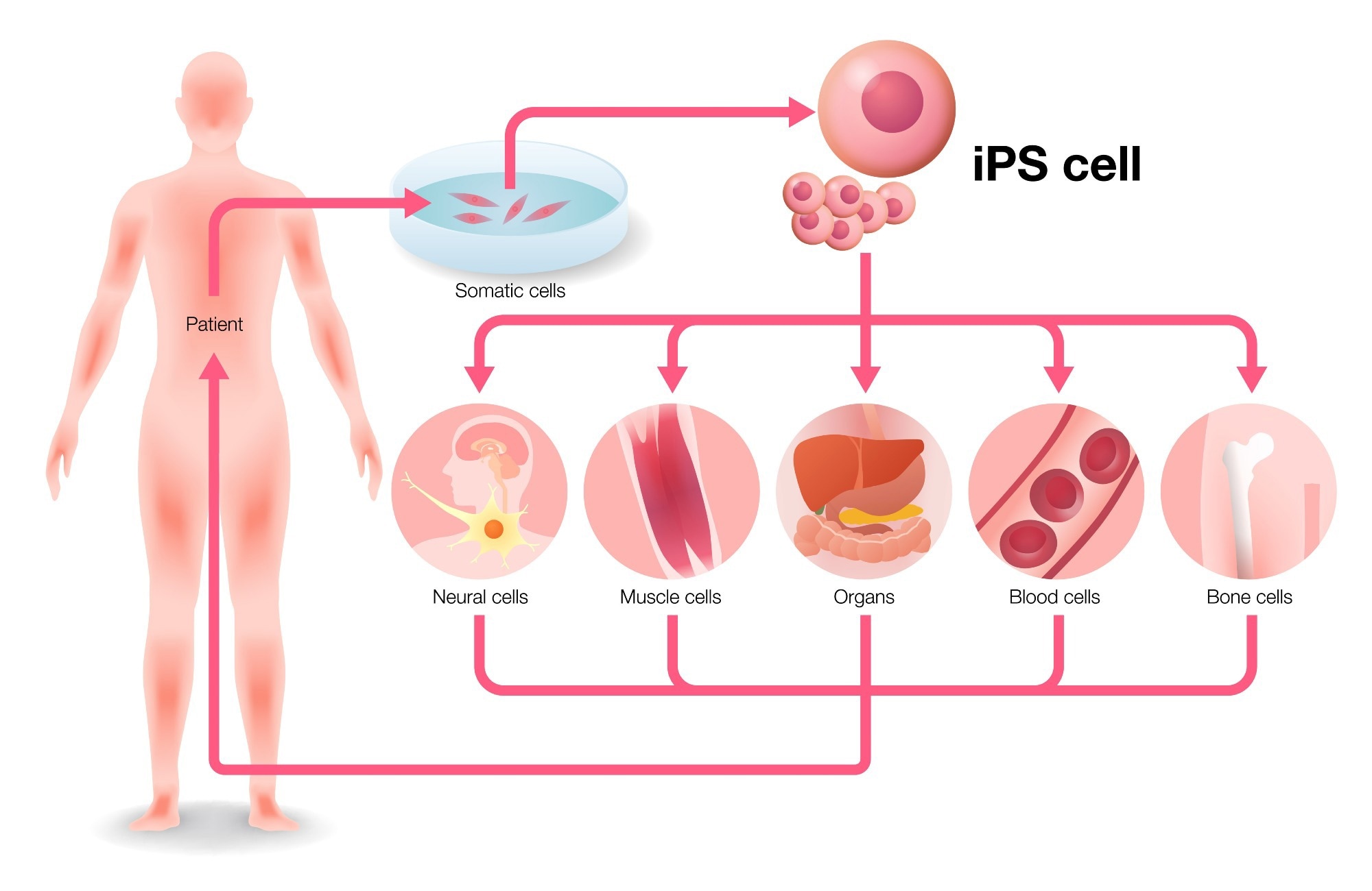
Organoids are usually grown from cells taken from biopsies, which are then programmed into induced pluripotent stem cells, which are mature cells that are reprogrammed so they can differentiate into any type of cell. The technique can produce complex structures but takes a long time. Many tissue types have now been studied by means of organoids, including the brain, heart and retina. The organoids are used to model how the tissue functions and reacts in response to drugs and diseases.
But modelling fetal tissue in this way remains challenging because researchers have limited access to the necessary cells. One option is to use tissue taken from terminated pregnancies, but this is limited to earlier stages of gestation and is accompanied by ethical issues. In the latest study, the researchers instead used amniotic fluid as a source of living cells, shed as the fluid surrounds and supports the growing fetus. This allows the researchers to study fetal tissue at later stages of development.
Fluid extraction
Samples were obtained either through amniocentesis — which involves a needle being inserted into the womb and removing amniotic fluid, and is usually performed at up to 20 weeks of gestation — or through amniotic drainage to remove excess fluid, at up to 34 weeks’ gestation. These are standard procedures during prenatal care, so “they give us the opportunity to take amniotic fluid without any additional procedure”, says study co-author Paolo De Coppi, a paediatric surgeon at Great Ormond Street Hospital in London. All samples were taken from people who were undergoing one of the procedures independent of the study.
The researchers first isolated individual cells from the samples and characterized their origins. Most were from the epithelial layer — the layer of cells that covers an organ’s surfaces. Epithelial cells “naturally come together and assemble”, making them ideal for forming organoids, says Benoit Bruneau, a researcher of paediatric heart disease at the Gladstone Institute of Cardiovascular Disease in San Francisco, California. “A lot of congenital diseases involve the epithelial tissues,” says Gerli, so the resulting organoids are relevant for studying such conditions.
The team grew organoids from three organs — the small intestines, kidneys and lungs. The cells were transferred into a gel medium to multiply and grow. Each organoid expressed the genes and proteins of the organ it originated from.
Alongside the tissue-like organoids, the researchers also modelled congenital diaphragmatic hernia (CDH), a disorder where the diaphragm fails to develop correctly, using cells from samples affected by the disorder.
Unlike organoids made from pluripotent stem cells, the amniotic fluid cells already have an organ identity. “There is no reprogramming, no manipulation,” says Gerli, “we’re just allowing the cells to express their potential.” This makes future applications in clinic more feasible, he adds. The relatively simple techniques also cut the time needed to grow the organoids to just four to six weeks, from the five to nine months typically needed when stem cells are used.
Future possibilities
The research isn’t ready to transfer to the clinic yet, the authors say. Organoids grown from amniocentesis could perhaps be used to screen treatments. At the moment, only epithelial tissue from the three organs has been successfully grown into organoids using this technique. More complex congenital disorders probably affect multiple tissue layers, such as mesenchymal cells, another type of cell found in several organs. And it might not be possible to use the method to model organs that don’t shed cells into the amniotic fluid, such as the brain or the heart, says Bruneau, who studies the most common congenital disorder — heart defects.
“The question is, how faithfully do these organoids reveal the underpinnings of the disease and how useful are they for not just modelling, but for drug testing?” says Bruneau. Their capabilities will have to be compared with those of organoids derived from biopsies or pluripotent stem cells, in terms of the level of responsiveness to drugs, says Núria Montserrat, who studies organ regeneration at the Institute for Bioengineering of Catalonia in Barcelona, Spain.
Gerli says that the next steps are to test the capabilities of the CDH organoids for modelling the disease. Comparisons to patient data will be needed to work out how characteristics of the disease are reflected in the gene and protein expression of the organoids. This will begin to answer questions about their usefulness. “We hope this just opens up to more research by us and others,” says De Coppi.
Nerve cells in the human brain take a remarkably long time to mature. This study identifies an epigenetic ‘barrier’ in neural precursor cells that determines the rate of neuronal maturation and is slowly released during the process. Inhibition of the barrier is shown to accelerate maturation in multiple human stem-cell-based models.
The first stem cell culture method that produces a full model of the early stages of the human central nervous system has been developed by a team of engineers and biologists at the University of Michigan, the Weizmann Institute of Science, and the University of Pennsylvania.
Models like this will open doors for fundamental research to understand early development of the human central nervous system and how it could go wrong in different disorders.”
Jianping Fu, U-M professor of mechanical engineering and corresponding author of the study in Nature
The system is an example of a 3D human organoid—stem cell cultures that reflect key structural and functional properties of human organ systems but are partial or otherwise imperfect copies.
“We try to understand not only the basic biology of human brain development, but also diseases—why we have brain-related diseases, their pathology, and how we can come up with effective strategies to treat them,” said Guo-Li Ming, who along with Hongjun Song, both Perelman Professors of Neuroscience at UPenn and co-authors of the study, developed protocols for growing and guiding the cells and characterized the structural and cellular characteristics of the model.
For example, organoids developed using patient-derived stem cells may be used for identifying which drugs offer the most successful treatment. Already, human brain and spinal cord organoids are used to study neurological and neuropsychiatric diseases, but they often mimic one part of the central nervous system and are disorganized. The new model, in contrast, recapitulates the development of all three sections of embryonic brain and spinal cord simultaneously, a feat that has not been achieved in previous models.
“The system itself is really groundbreaking,” said Orly Reiner, the Berstein-Mason Professorial Chair of Neurochemistry at Weizmann and co-author of the study who developed cellular tools to identify neural cell types in the model. “A model that mimics this structure and organization has not been done before, and it offers numerous possibilities for studying human brain development and especially developmental brain diseases.”
While the model is faithful to many aspects of the early development of the brain and spinal cord, the team notes several important differences. For one, neural tube formation—the very first stage of central nervous system development—is very different. The model can’t be used to simulate disorders that stem from improper closure of the neural tube such as spina bifida.
Instead, the model started with a row of stem cells roughly the size of the neural tube found in a 4-week-old embryo—about 4 millimeters long and 0.2 millimeters in width. The team stuck the cells to a chip patterned with tiny channels that the team used to introduce materials that enabled the stem cells to grow and guided them toward building a central nervous system.
The team then added a gel that allowed the cells to grow in three dimensions and chemical signals that nudged them to become the precursors of neural cells. In response, the cells formed a tubular structure. Next, the team introduced chemical signals that helped the cells identify where they were within the structure and progress to more specialized cell types. As a result, the system organized itself to mimic the forebrain, midbrain, hindbrain and spinal cord in a way that mirrors embryonic development.
“As an engineer, the challenging part is to learn neural development and stem cell biology,” said Xufeng Xue, first author of the study and a postdoctoral fellow in mechanical engineering U-M. “It was a team effort to make this happen, with amazing collaborators at UPenn and Weizmann.”
The team grew the cells for 40 days, simulating development of the central nervous system to about 11 weeks post-fertilization. In this time, the team was able to demonstrate the roles of specific genes in spinal cord development and learn how certain cell types in the early human nervous system differentiate into different cells with specialized functions.
“In many cases, animal models simply do not recapitulate either the characteristics or the degree of severity seen in human brain diseases such as microcephaly,” Song said. “Even nonhuman primates are not the same. So in the context of disease biology and treatment strategies, a human cell model is almost irreplaceable.”
The team plans to apply the model to study different human brain diseases using patient derived stem cells.
Xue hopes to continue using this model to study the interplay among different parts of the brain during development. He is also interested in studying how the brain sends instructions for movement via the spinal cord. This line of inquiry, which could shed new light on disorders like paralysis, would require the neurons to link up into working circuits—something that was not observed in this study.
Insoo Hyun, a bioethicist at the Museum of Science in Boston who was not part of the study, notes that experiments like these are closely scrutinized before they are allowed to move forward.
“Research groups must be clear about the scientific question they are trying to answer—and that the degree of development they allow in the model is the minimum to answer the question,” he said.
The model does not include peripheral nerves or functioning neural circuitry—features that are critical for humans’ ability to experience our environment and process that experience.
The study was funded by the Michigan-Cambridge Collaboration Initiative, University of Michigan, State of Michigan, Dr. Miriam and Sheldon G. Adelson Medical Research Foundation, National Science Foundation and National Institutes of Health.
The research conforms to the 2021 Guidelines for Stem Cell Research and Clinical Translation recommended by the International Society for Stem Cell Research. All protocols used in this work were approved by the Human Pluripotent Stem Cell Research Oversight Committee at the University of Michigan, Ann Arbor.
The team has applied for patent protection with the assistance of U-M Innovation Partnerships and is seeking partners to bring the technology to market.
Surveys show most men in the United States are interested in using male contraceptives, yet their options remain limited to unreliable condoms or invasive vasectomies. Recent attempts to develop drugs that block sperm production, maturation, or fertilization have had limited success, providing incomplete protection or severe side effects. New approaches to male contraception are needed, but because sperm development is so complex, researchers have struggled to identify parts of the process that can be safely and effectively tinkered with.
Now, scientists at the Salk Institute have found a new method of interrupting sperm production, which is both non-hormonal and reversible. The study, published in Proceedings of the National Academy of Sciences (PNAS)on February 20, 2024, implicates a new protein complex in regulating gene expression during sperm production. The researchers demonstrate that treating male mice with an existing class of drugs, called HDAC (histone deacetylase) inhibitors, can interrupt the function of this protein complex and block fertility without affecting libido.
Most experimental male birth control drugs use a hammer approach to blocking sperm production, but ours is much more subtle. This makes it a promising therapeutic approach, which we hope to see in development for human clinical trials soon.”
Ronald Evans, senior author, professor, director of the Gene Expression Laboratory, and March of Dimes Chair in Molecular and Developmental Biology at Salk Institute
The human body produces several million new sperm per day. To do this, sperm stem cells in the testes continuously make more of themselves, until a signal tells them it’s time to turn into sperm-;a process called spermatogenesis. This signal comes in the form of retinoic acid, a product of vitamin A. Pulses of retinoic acid bind to retinoic acid receptors in the cells, and when the system is aligned just right, this initiates a complex genetic program that turns the stem cells into mature sperm.
Salk scientists found that for this to work, retinoic acid receptors must bind with a protein called SMRT (silencing mediator of retinoid and thyroid hormone receptors). SMRT then recruits HDACs, and this complex of proteins goes on to synchronize the expression of genes that produce sperm.
Previous groups have tried to stop sperm production by directly blocking retinoic acid or its receptor. But retinoic acid is important to multiple organ systems, so interrupting it throughout the body can lead to various side effects-;a reason many previous studies and trials have failed to produce a viable drug. Evans and his colleagues instead asked whether they could modulate one of the molecules downstream of retinoic acid to produce a more targeted effect.
The researchers first looked at a line of genetically engineered mice that had previously been developed in the lab, in which the SMRT protein was mutated and could no longer bind to retinoic acid receptors. Without this SMRT-retinoic acid receptor interaction, the mice were not able to produce mature sperm. However, they displayed normal testosterone levels and mounting behavior, indicating that their desire to mate was not affected.
To see whether they could replicate these genetic results with pharmacological intervention, the researchers treated normal mice with MS-275, an oral HDAC inhibitor with FDA breakthrough status. By blocking the activity of the SMRT-retinoic acid receptor-HDAC complex, the drug successfully stopped sperm production without producing obvious side effects.
Another remarkable thing also happened once the treatment was stopped: Within 60 days of going off the pill, the animals’ fertility was completely restored, and all subsequent offspring were developmentally healthy.
The authors say their strategy of inhibiting molecules downstream of retinoic acid is key to achieving this reversibility.
Think of retinoic acid and the sperm-producing genes as two dancers in a waltz. Their rhythm and steps need to be coordinated with each other for the dance to work. But if you throw something in that makes the genes miss a step, the two are suddenly out of sync and the dance falls apart. In this case, the HDAC inhibitor causes the genes’ misstep, halting the dance of sperm production.
However, if the dancer can find its footing and get back in step with its partner, the waltz can resume. In the same way, the authors say that removing the HDAC inhibitor allows the sperm-producing genes to get back in sync with the pulses of retinoic acid, turning sperm production back on as desired.
“It’s all about timing,” says co-author Michael Downes, a senior staff scientist in Evans’ lab. “When we add the drug, the stem cells fall out of sync with the pulses of retinoic acid, and sperm production is halted, but as soon as we take the drug away, the stem cells can reestablish their coordination with retinoic acid and sperm production will start up again.”
The authors say the drug doesn’t damage the sperm stem cells or their genomic integrity. While the drug was present, the sperm stem cells simply continued regenerating as stem cells, and when the drug was later removed, the cells could regain their ability to differentiate into mature sperm.
“We weren’t necessarily looking to develop male contraceptives when we discovered SMRT and generated this mouse line, but when we saw that their fertility was interrupted, we were able to follow the science and discover a potential therapeutic,” says first author Suk-Hyun Hong, a staff researcher in Evans’ lab. “It’s a great example of how Salk’s foundational biological research can lead to major translational impact.”
Other authors include Glenda Castro, Dan Wang, Russell Nofsinger, Annette R. Atkins, and Ruth T. Yu of Salk, Maureen Kane, Alexandra Folias, and Joseph L. Napoli of UC Berkeley, Paolo Sassone-Corsiof UC Irvine, Dirk G. de Rooij of Utrecht University, and Christopher Liddle of the University of Sydney.
The work was supported by the National Institutes of Health (grants CA265762 and CA220468) and the Next Generation Sequencing and Flow Cytometry Cores at Salk, funded by the Salk Cancer Center (NCI grant NIH-NCI CCSG: P30 014195).
Source:
Journal reference:
Hong, S-H., et al. (2024) Targeting nuclear receptor corepressors for reversible male contraception.PNAS. doi.org/10.1073/pnas.2320129121.
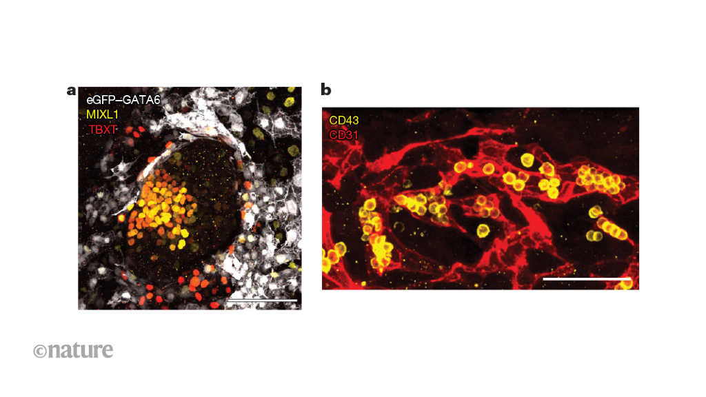
Human embryos are extremely difficult to study. This lack of samples limits our understanding of crucial developmental stages, such as the early formation of blood cells. A stem-cell-based model closely captures the development of human embryonic and key extra-embryonic tissues after implantation, as well as the formation of early blood cells.
Stem cells can be obtained from unneeded embryos made during in vitro fertilisation
nobeastsofierce Science / Alamy
A kind of stem cell transplant that has long been seen as one of the most promising new kinds of medical treatments could bring a greater risk of cancer than we previously thought. A study has found that more than a fifth of stem cells being grown in laboratories for regenerative medicine research harbour cancer-causing mutations.
The cells tested haven’t been put into people, but were being used in research to explore their medical use. The findings show…
City of Hope®, one of the largest cancer research and treatment organizations in the United States, treated the oldest person to be cured of a blood cancer and then achieve remission for HIV after receiving a blood stem cell transplant from a donor with a rare genetic mutation. Research published in NEJM today demonstrates that older adults with blood cancers who receive reduced intensity chemotherapy before a stem cell transplant with donor cells that are resistant to HIV may be cured of HIV infection.
Paul Edmonds, 68, of Desert Springs, California, is the fifth person in the world to achieve remission for acute myelogenous leukemia and HIV after receiving stem cells with a rare genetic mutation, homozygous CCR5 Delta 32. That mutation makes people who have it resistant to acquiring HIV. Edmonds is also the person who had HIV the longest -; for over 31 years -; among these five patients.
Known as the “City of Hope patient” among these five patients, Edmonds received a transplant at City of Hope on Feb. 6, 2019, and is now considered to be cured of leukemia. Edmonds stopped taking antiretroviral therapies for HIV nearly three years ago and will be considered cured of HIV after he has stopped taking antiretrovirals for five years.
“City of Hope’s case demonstrates that it is possible to achieve remission from HIV even at an older age and after living with HIV for many years,” said Jana K. Dickter, M.D., a clinical professor in City of Hope’s Division of Infectious Diseases, who led the study. “Furthermore, remission can be achieved with a lower-intensity regimen than the therapy received by the four other patients who went into remission for HIV and cancer. As people with HIV continue to live longer, there will be more opportunities for personalized treatments for their blood cancers.”
For Edmonds’ medical team, this meant they would need to tailor his treatment to address his age and the duration of his HIV. City of Hope’s decades-long expertise treating older adults with cancer and HIV -; efforts led by John A. Zaia, M.D., director of City of Hope’s Center for Gene Therapy and Aaron D. Miller and Edith Miller Chair for Gene Therapy, and other doctors -; proved to be invaluable in treating Edmonds and helping him go into remission for both leukemia and HIV.
Under the care of City of Hope hematologist Ahmed Aribi, M.D., assistant professor in the Division of Leukemia and a study author, Edmonds received three different therapies to get him into remission before receiving a transplant. The therapy is needed to help the patient achieve remission, and the patient can then proceed with a transplant with the goal of curing the cancer.
Edmonds received a chemotherapy-based, reduced-intensity transplant regimen prior to his transplant that was developed by City of Hope and other transplant programs for treatment of older patients with blood cancers. Reduced-intensity chemotherapy makes the transplant more tolerable for older patients and reduces the potential for transplant-related complications from the procedure.
For the transplant, Aribi and his team worked with City of Hope’s Unrelated Donor Bone Marrow Transplant Program -; directed by Monzr M. Al Malki, M.D. -; to find a donor who was a perfect match for the patient and had the rare genetic mutation, which is found in just 1-2% of the general population.
The mutation makes people who have it resistant to acquiring HIV. CCR5 is a receptor on CD4+ immune cells, and HIV uses that receptor to enter and attack the immune system. But the CCR5 mutation blocks that pathway, which stops HIV from replicating.
Edmonds had mild to moderate side effects caused by graft-versus-host disease, which occurs when the donor’s T lymphocytes, a type of white blood cell that fights infections, attack the patient’s cells.
Edmonds also achieved “full chimerism,” meaning that all of his bone marrow and blood stem cells originated from the donor.
Stephen J. Forman, M.D., director of City of Hope’s Hematologic Malignancies Research Institute and a professor in the Department of Hematology & Hematopoietic Cell Transplantation, noted a confluence of several research initiatives by City of Hope over the years helped lead the institution to this moment.
City of Hope and other institutions started performing successful stem cell transplants in older adults a decade ago, an intensive and high-risk procedure in this population that was unheard of prior to then. We have treated patients who are in their 80s with transplants and that is due to City of Hope’s emphasis on expanding therapies to more patients, as well as our compassionate, top-notch care of even the most vulnerable populations.”
Stephen J. Forman, M.D., Director of City of Hope’s Hematologic Malignancies Research Institute
“City of Hope is not stopping there. Our researchers are working on creating stem cells that have the genetic mutation that makes them naturally resistant to HIV, among other research initiatives,” he added.
These milestones include:
City of Hope was one of the first institutions in the United States to perform a reduced intensity regimen for older patients with myelodysplasia, a blood disease that can evolve into leukemia and that Edmonds had prior to acute myelogenous leukemia.
Ryotaro Nakamura, M.D., director of City of Hope’s Center for Stem Cell Transplantation and Jan & Mace Siegel Professor in Hematology & Hematopoietic Cell Transplantation, led the national trial that demonstrated a transplant could become standard of care for older people with myelodysplastic syndromes, which led to Medicare approving the therapy in older populations.
City of Hope was one of the first centers in the United States to perform effective, curative autologous transplants, which use a person’s own stem cells, for patients with HIV-related lymphoma. When many centers still treated patients with low-intensity, noncurative treatment approaches, City of Hope -; led by Forman and Amrita Krishnan, M.D., executive medical director of hematology, City of Hope Orange County – challenged that paradigm by demonstrating that autologous transplants could be used to cure patients with HIV-related lymphomas who would otherwise die.
City of Hope was also a primary national co-leader in two National Cancer Institute-sponsored trials for autologous as well as allogeneic stem cell transplantation, which use a donor’s stem cells, for patients with HIV and blood cancers. Led by Joseph Alvarnas, M.D., City of Hope’s vice president of government affairs and a hematology professor, these trials led to a change in the national standards of care on how best to manage this vulnerable patient population.
City of Hope’s blood stem cell and bone marrow transplant (BMT) program has performed nearly 19,000 transplants, making it one of the largest programs in the nation. City of Hope has exceptional transplant outcomes year after year, according to the Center for International Blood & Marrow Transplant Research.
Building on its BMT expertise, City of Hope is also a pioneer in the development of chimeric antigen receptor (CAR) T cells to treat blood cancers and solid tumors. More than 1,200 patients have been treated with CAR T cell therapy at City of Hope.
Leveraging their expertise in cellular immunotherapy, City of Hope scientists have also developed chimeric antigen receptor CAR T cells that can target and kill HIV-infected cells and control HIV in preclinical research. A City of Hope clinical trial using CAR T cell therapy, which has the potential to provide HIV patients with a lifelong viral suppression without antiretroviral therapies, is expected to open later this year.
Angelo Cardoso, M.D., Ph.D., City of Hope director of the Laboratory of Cellular Medicine, is also a study author and performed many of the experiments that confirmed Edmonds’ HIV remission.
Source:
Journal reference:
Dickter, J. K., et al. (2024). HIV-1 Remission after Allogeneic Hematopoietic-Cell Transplantation. The New England Journal of Medicine. doi.org/10.1056/nejmc2312556.
In this interview, we speak to Keith Olson and Coby Carlson from FUJIFILM Cellular Dynamics about their iPSC technology and how it is making breakthroughs in blood-brain barrier research.
Please can you introduce yourself and tell us about your role at FUJIFILM Cellular Dynamics?
Coby Carlson: My name is Coby Carlson, and I lead the Applications Team at FUJIFILM Cellular Dynamics. Our team is separate from the R&D scientists, staff, and experts focusing on stem cell culture and differentiations to create unique, specialized cell types. Our role is to test the functionality of these cells, demonstrate their performance on various platform technologies, and develop applications.
Keith Olson: My name is Keith Olson, and I am the Executive Vice President for Commercial Operations at FUJIFILM Cellular Dynamics.
FUJIFILM Cellular Dynamics is a global leader in developing and manufacturing human induced pluripotent stem cells (iPSC). Please can you tell us more about some of your core aims and missions as a company?
Coby Carlson: Fujifilm’s core technologies include reprogramming, engineering, and iPSC culture and differentiation to create unique cell types. Our mission is to bring these technologies to an industrial scale. The technology was first invented over 15 years ago, and since then, we have continuously improved and optimized it to launch it on a large scale. Our goal is to make different cell types accessible all over the world.
Keith Olson: I believe that we were pioneers of iPSC technology in the United States. We successfully brought it to market, commercialized it, and built a business around it. Our reputation is now based on our core expertise, quality, and ability to manufacture at scale for our clients in the pharmaceutical, biotech, and academic sectors.
Image Credit: metamorworks/Shutterstock.com
iPSC technology can be used to revolutionize scientific research and cell therapy. Why is this, and in what application areas within scientific research does this technology benefit the most?
Coby Carlson: The primary focus of people working with iPSC technology is toxicology/safety pharm, disease modeling, and cell therapy. Safety is crucial because we can generate cardiomyocytes from iPS cells, and these cells spontaneously beat in a dish while responding to known cardiotoxic molecules.
To measure the safety and effectiveness of drugs in a dish, we use iPSC-derived cardiomyocytes in safety toxicology. We have also participated in the CiPA (Comprehensive in Vitro Proarrhythmia Aassay) initiative to standardize this process across various labs globally. The recent FDA Modernization Act has suggested using alternatives to animal testing, making iPSC-derived cardiomyocytes a crucial component of measuring the safety and effectiveness of drugs going forward.
Keith Olson: iPSC delivers a specific gap in the market by providing customers with high-quality human cells. Instead of researching animal models or transformed cell lines, researchers can now use real human cardiomyocytes or neurons for more biologically relevant results.
At FUJIFILM Cellular Dynamics, you offer a range of iPSC cells, including neural and cardiac cells. Can you tell us more about some of the products you offer and how they are generated?
Coby Carlson: The uniqueness of iPSC technology lies in the fact that there are not many human donors who can offer their brain cells. This technology provides access to different cell types, and we can differentiate highly specialized cells to obtain unique features such as excitatory neurons and microglia. These unique cells are essential for in vitro assays, where we can test them differently than we would in an animal model. For instance, mouse models are different from human models.
Keith Olson: Everything starts from a core iPS cell bank, and we have developed specific differentiation protocols for each terminal phenotype of the cell. Whether it is a cardiomyocyte, neuron, or hepatocyte, we have a differentiation protocol that takes us from the core stem cell bank to a finished product that customers can purchase and use as an assay-ready sample.
Image Credit:Magic mine/Shutterstock.com
The blood-brain barrier (BBB) is vital in understanding how the brain functions, and its dysfunction can lead to neurological conditions such as MS, stroke, and epilepsy. Why did you choose to focus on this area, and how can your iPSC technology help to accelerate breakthroughs?
Coby Carlson: Researchers and drug developers have long struggled with the blood-brain barrier, a critical cell structure separating the blood from the brain. The barrier keeps out harmful pathogens and toxins while allowing vital nutrients to enter the brain. However, it also prevents drugs from crossing over and treating neurological diseases such as neurodegenerative disorders.
The main challenge has been replicating this barrier in vitro. One of the reasons we pursued iPSC cell technology is that we developed ways to distinguish specialized cell types from the same donor to create the blood-brain barrier. Additionally, we can cryopreserve these cells and generate media that allows them to be functional and reanimated from the thaw, enabling us to test them in an in vitro assay.
Keith Olson: We chose to develop a highly relevant human model for studying the blood-brain barrier for several reasons. The industry bottleneck and challenge in understanding how some large molecules cannot pass through the barrier made it necessary. We needed a system that truly replicated what happens in the human brain. We helped revolutionize the market by making the cell type, which was particularly difficult to cryopreserve and develop as an assay-ready product. We were able to deliver a fully isogenic solution with all cell types, and now customers can build their own real human BBB in an assay plate.
At Neuroscience 2022, you showcased research surrounding the establishment of a BBB model using iPSC-derived human cells. Can you tell us more about this research and the potential to integrate this BBB system with emerging organ-on-a-chip technologies to advance the field of drug discovery?
Coby Carlson: Generating the BBB is a significant challenge that requires assembling three different cell types. We validated our model using a Transwell assay, which measures the function of the cells on both the top and bottom of the barrier. However, we recognize that the BBB is a complex system, and microfluidic and organ-on-a-chip technologies offer a unique environment for the cells to function. Combining iPSC technology and OoC technologies will enhance cell function in these systems by providing unique environments with microfluidics and dynamic flow similar to the bloodstream. This will allow us to demonstrate the advantages of these models in a more comprehensive manner.
Keith Olson: The system is perfect for 3D modeling because it is a 3D system. It is a multi-layer, multi-cell-type organ or part of an organ, and using it as an organoid or spheroid in a complex model brings you closer to that model system. Building the barrier is crucial, and 3D modeling is the best way to achieve this. Our released product is flexible enough for clients to conduct assays in a Transwell plate or as a spheroid. They can even use more sophisticated organ-on-a-chip systems to create a comprehensive model for this biology.
What challenges are faced when generating new leads in neurological drug discovery? How can your 3D neural spheroids help to tackle these?
Coby Carlson: When we started the company, we differentiated cells from iPS lines to create highly purified neurons. While they were unique and visually pleasing, we discovered that some of their functionality was lacking. Ten years later, we have developed various products, intending to combine them to create multicellular cultures, not just in 2D systems but also in 3D systems.
Our approach involves mixing different cell types to increase complexity and biological relevance, which we believe will help improve testing and facilitate the development of new therapies and the identification of new compounds.
Keith Olson: In neurobiology, it is well-established that a human system is essential. Animal model systems simply do not replicate human biology, and studying true human biology requires a 3D system. We are not flat figures but 3D beings, and building a real neural system allows us to observe neural connections, communication, and the impact of glia on neurons, studying how cells interact as they would in the body. This is where the true value lies in what we are delivering.
Image Credit: Naumova Marina/Shutterstock.com
Are you hopeful that with continued research surrounding the BBB, we will see new advancements being made surrounding health and disease? What would this mean for both patients suffering from neurological diseases as well as clinical settings?
Coby Carlson: As a technology, the use of human iPS drive cell types is still in its infancy, with significant advances still being made. We believe that some major discoveries will be made by using this technology, whether that be by us or others. This technology has the potential to facilitate the discovery of new treatments, and we are excited about its future potential.
Keith Olson: I think anything that any of our competitors, colleagues, friends, and neighbors are doing in this space will help push the field forward. The more vendors and researchers working on creating relevant model systems for humans, the better. With the FDA Modernization Act and the desire for better test systems and models, everyone working together will likely result in better systems and, hopefully, faster, cheaper, and easier drug discovery for companies.
You were exhibiting at SLAS US 2024, an international exhibition and tradeshow bringing together both world-leading researchers and industry professionals. Can you tell us what you were exhibiting at SLAS and the importance of attending these in-person conferences?
Coby Carlson: The SLAS conference is significant as it brings together various customers, collaborators, friends, and former colleagues operating in this field. The past years have seen challenges due to the pandemic and remote work, with limited ability to collaborate effectively. The opportunity to meet face-to-face is essential when pushing these models forward and persuading companies and groups to invest significant time and resources. Having these conversations in person allows for quick decision-making and improves the efficiency of the process.
Keith Olson: The SLAS conference seems to have finally returned to normality after the pandemic. The number of attendees and vendors is impressive, and the human-to-human interaction is just unbeatable, especially when it comes to answering queries and gaining an understanding of something first-hand. Our focus remains on highlighting our entire portfolio, but we also showcased some new systems, such as the iCell® Macrophage, which is the first of its kind.
Additionally, we continued to promote our BBB system and highlighted other 3D model systems we have developed, including in the neuro and cardio spheres. Overall, it was an opportunity to build awareness for our company and its offerings.
On your website, you also offer a variety of resources for your customers, including research posters, webinars, and videos. How important are these resources in building customer relationships?
Coby Carlson: Reviews and testimonials are popular among our customers, who are primarily scientists requiring supporting evidence. We understand that content is essential, so we offer it in various formats, including small bites, digital graphical abstracts, and detailed protocols. We aim to cater to the diverse needs of our customers, providing them with the relevant information to make informed decisions about our products.
Keith Olson: We all learned a valuable lesson during the pandemic that content is king. As a result, we had to adapt and utilize various means such as websites, LinkedIn, and other portals to disseminate information to our customers and clients. Due to the absence of trade shows, these platforms were our only options. However, now that trade shows are seemingly back in full swing and everyone is attending, it presents a more effective way to communicate to a large audience at once, which is always more enjoyable.
With so many breakthroughs and technological advancements being made within the life sciences, what are you personally looking forward to the most in the coming years?
Coby Carlson: While we did not discuss it much, our cell therapy is where the truly exciting developments will take place and capture the general public’s attention. For instance, curing blindness by using retinal cells is something that could happen in our lifetime. Although I cannot speculate on the exact timeline, I believe it is inevitable. Our in vitro work behind the scenes in drug discovery and other aspects aligns well with this goal, as the same cells used in our research could be used as drugs in the future.
Keith Olson: An exciting prospect on the horizon is the potential replacement of animal model systems with validated human models within the next year or five years. If this were to happen, and the FDA, pharma companies, and biotechs were on board, it would be a major advancement for everyone. This could result in faster, cheaper, and more efficient processes. One of the main challenges in this field is the high rate of failure, and any improvements made in this area could potentially save billions of dollars for the industry.
What is next for FUJIFILM Cellular Dynamics? Are you involved in any exciting upcoming projects?
Coby Carlson: We always strive to advance our field, evaluating new technologies and creating new products. We want to expand our collaborations and work with others to achieve our scientific goals. While we can conduct R&D, we recognize that partnering with other experts, sharing information, and collaborating can lead to even greater success.
Keith Olson: Our company is constantly exploring new cell backgrounds, and we are committed to launching two to three new cell types each year. Currently, we are focused on introducing three new cell types, all of which will be launched by June or July of this year. Simultaneously, we have begun developing the next three cell types for 2024. Our goal is to continue expanding our offerings, and we hope to return next year with news of three entirely new cell lines.
About Dr. Keith R. Olson
Keith Olson is the senior commercial executive for FUJIFILM Cellular Dynamics, where we have developed the market-leading portfolio of differentiated human cells derived from iPSC.
Keith has previously served in leadership roles for Life Science products at Corning Inc, Life Technologies, DiscoveRx and Cellomics, and has launched over 700 Life Science products in his career. Keith received his bachelor’s degree in Molecular Cell Biology from Carnegie-Mellon University and his Ph.D. in the same from the University of Rochester.
About Dr. Coby Carlson
Coby Carlson is the Director of Applications Development at FUJIFILM Cellular Dynamics where his team focuses on advancing the use of human iPSC-derived cell types for drug screening, toxicity testing, and disease modeling.
He joined the company in 2012 and has extensive experience developing applications with iCell products, characterizing their functional performance on various technology platforms, and building co-culture systems in both 2D and 3D format. Coby received his PhD from the Univ. of Utah followed by a postdoctoral fellowship at UW-Madison.