


Red and green don’t look like particularly close colors at a glance, but the way your eye treats them is surprisingly similar.
A new study in Science, led by biochemists at Nagoya Institute of Technology in Japan, has now revealed just how close they really are on a molecular level.
It only takes differences in three amino acids to tune cones in the retina to be sensitive to red or green light, the team found.
The discovery could help explain why red-green color blindness is so common, and potentially lead to new ways to treat it.
“This study reports the first three-dimensional structures of primate red- and green-sensitive cone visual pigments in their dark state,” says Kota Katayama, a biochemist at Nagoya Institute of Technology.
“In the long term, this knowledge may help researchers understand how genetic variations alter color perception and contribute to color vision deficiencies and other visual disorders.”
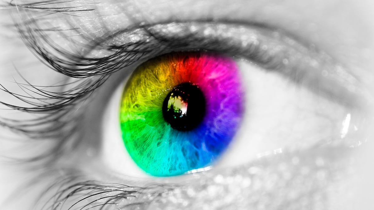
Humans are what are known as trichromats, meaning we have three types of cone cells in our retinas.
These are all sensitive to different wavelengths of light – red, green, and blue – and when you combine millions of them together, it allows us to enjoy all the familiar colors of the rainbow.
Not all animals have the same setup, of course.
Dogs are dichromatic, keenly seeing yellows and blues but missing reds and greens. Birds, meanwhile, boast tetrachromatic vision, giving them access to a world of brand-new colors we can’t even imagine.
But according to the new study, we should count ourselves lucky that we can tell the difference between a Granny Smith and a Red Delicious at all.
All the cones in the human eye absorb light using the same basic molecule, known as 11-cis-retinal. The differences in which wavelengths they absorb, and which colors we see as a result, come down to tiny variations in the protein structure.
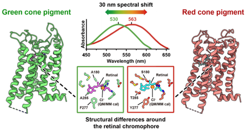
“The red- and green-sensitive pigments are particularly intriguing: They differ at only a few amino acid positions, yet their light absorption peaks are separated by about 30 nanometers, enabling red-green color discrimination,” the researchers write.
“By combining cryo-electron microscopy with vibrational spectroscopy and quantum chemical modeling, we aimed to clarify how cone pigments tune their spectral sensitivity.”
To investigate, the researchers turned to the crab-eating macaque (Macaca fascicularis), a species of primate that has very similar trichromatic vision to humans. They took cryo-electron microscope images of the red and green cone pigments in the macaques, then created digital models of them.
The team then used these digital versions to simulate what would happen with slightly different versions of the proteins, and how that affected light absorption.
In the end, the scientists discovered that almost the entire 30-nanometer difference between the red and green peaks could be attributed to variations in just three amino acids: A180S, F277Y, and A285T.
Intriguingly, the team found that these amino acids weren’t tuning which colors a cone was sensitive to by changing the physical shape of the proteins.
Instead, they seem to alter the electrostatic environment around them, which then changes which wavelengths are absorbed and by how much.

This close examination also revealed a new structure that may explain why your color vision doesn’t ‘wear out’ after a while in a bright room.
Cone cells are known to regenerate quickly after stimulation by light, and this could be thanks to a new ‘opening’ in the pigment structure that the team found. This opening isn’t present in rhodopsin, the pigment that the rod cells in your eye use to allow you to see in dim light.
Related: Scientists Say They Found a New Color Humans Have Never Seen Before
“These findings connect long-standing functional differences in color vision to concrete structural features,” the researchers write.
“Small sequence changes tune color by reshaping the electrostatic field acting on the retinal chromophore, whereas distinct membrane access pathways enable efficient chromophore exchange and rapid recovery.”
The research was published in the journal Science.
This article was fact-checked by Rachel Garner and edited by Peter Dockrill. While we pride ourselves on our process, we are only human. If you spot a mistake, please let us know.