
The brain keeps one of the strictest guest lists in the body.
Oxygen and nutrients may enter. Toxins, germs, and other substances traveling through the blood are turned away.
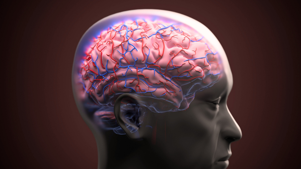
Controlling the door is the blood-brain barrier: a tightly sealed border around the brain’s blood vessels. But in Alzheimer’s disease, this protective border can begin to leak.
Scientists may now have caught one protein helping to hold that door open. Surprisingly, it normally belongs to the body’s repair crew.
A new study published in Nature Aging identifies fibronectin – a protein that helps injured tissue heal – as a key driver of blood-brain barrier damage in Alzheimer’s models linked to the APOE4 gene variant.
The researchers uncovered a possible chain of events: APOE4, inflammation, and Alzheimer’s-linked amyloid-beta prompt support cells in the brain to produce too much fibronectin. The protein piles up around blood vessels, disrupts the signals keeping them sealed, and allows the barrier to leak.
Crucially, the researchers reversed this process in experimental models.
“The most direct and most translatable finding is that reducing fibronectin itself rescues the barrier defects,” Columbia University neuroscientist Çağhan Kızıl told ScienceAlert.
This matters because blood-brain barrier breakdown can begin early in Alzheimer’s, sometimes before memory problems. Once the barrier weakens, unwanted substances can enter brain tissue and inflammation can increase.
Most Alzheimer’s research and several treatments focus on amyloid-beta and tau, the proteins forming plaques and tangles in the brain. These findings suggest another possible target: protecting the brain’s blood vessels and closing the leaks around them.
The trail began with APOE4, the strongest known genetic risk factor for late-onset Alzheimer’s.
Having APOE4 does not mean someone will develop the disease. But one copy increases the risk, and two raise it further. The variant has also been associated with early blood-brain barrier damage, though how it could create a physical breach was unclear.
Fibronectin acts like temporary scaffolding at an injury site, giving cells something to hold onto while they rebuild damaged tissue. In the brain, however, too much of this scaffolding may become part of the problem.
The team examined postmortem brain tissue and cerebrospinal fluid from people with Alzheimer’s, then tested its findings using human cells, three-dimensional blood-vessel models, APOE4 mice, and zebrafish.
Across these systems, fibronectin accumulated around brain blood vessels. Much of it came from astrocytes – star-shaped cells that surround vessels and help maintain the blood-brain barrier.
APOE4, inflammation, and amyloid-beta prompted astrocytes to produce more fibronectin.
The repair scaffold was beginning to crowd the doorway.
As fibronectin accumulated, it disrupted growth signals used by astrocytes and blood-vessel cells to communicate. Without those messages, the connections sealing the vessels weakened, creating gaps through which material from the blood could escape.
But fibronectin might simply have gathered around damaged vessels.
To find out, the researchers made support cells in zebrafish brains produce extra human fibronectin. The protein alone was enough to make the barrier leak.
Fibronectin was not merely present at the scene. It was helping cause the damage.
The researchers then attacked the problem at different points. Reducing fibronectin lowered leakage in zebrafish exposed to amyloid-beta. Blocking the cellular pathway through which fibronectin suppressed protective messages switched those signals back on.
In a separate experiment, the researchers blocked VEGF signaling, damaging the vessels’ tight junctions. They then added two downstream growth factors, HB-EGF and IGF-1, which restored tight-junction levels.
The experiments also revealed where researchers might intervene.
An earlier genetic finding makes fibronectin an especially interesting target.
“We previously identified a rare loss-of-function FN1 variant that lowers AD risk in APOE4 carriers by 71%, with no evidence of harm in carriers – nature has already run a version of this experiment,” Kızıl said.
That does not mean eliminating fibronectin. The body needs it for wound healing and blood-vessel maintenance.
“That reframes the goal: not eliminating fibronectin, which the body needs for wound healing and normal vascular maintenance, but developing a way to selectively curb its pathological buildup at the brain’s blood vessels,” Kızıl explained.

A future antibody or small-molecule drug might clear excess fibronectin from around those vessels. Another possibility is a gene-based treatment mimicking the protective variant’s effect in the brain.
However, this remains mechanistic, preclinical research. The interventions have not been shown to preserve memory or cognition in animals, let alone treat Alzheimer’s in people.
The human findings are correlational and need confirmation in larger groups. Before human trials, researchers must show that targeting fibronectin improves cognition in animals, establish its safety, and reach the brain without disrupting fibronectin’s useful roles elsewhere.
Related: New Unifying Theory May Explain How Alzheimer’s Emerges in The Brain
Still, the discovery introduces an intriguing suspect. In Alzheimer’s, part of the trouble may come from the repair crew leaving behind so much scaffolding that the door can no longer close.
The study was published in Nature Aging.
This article was fact-checked by Fiona MacDonald and edited by Fiona MacDonald. While we pride ourselves on our process, we are only human. If you spot a mistake, please let us know.