The hottest science in the prevention of heart disease awaits at ESC Preventive Cardiology 2024, a scientific congress of the European Society of Cardiology (ESC). The annual congress of the European Association of Preventive Cardiology (EAPC), a branch of the ESC, takes place 25 to 27 April at the Megaron – Athens International Conference Centre, Greece. Explore the scientific programme.
Don’t miss the late breaking science sessions for cutting-edge research in preventive cardiology, including unhealthy food and beverage trends in adolescents and the links between physical activity and smoking in children. Novel research will be presented in hundreds of scientific abstracts including data on stair climbing, insomnia, dairy products, and the potential connections between air pollution, mental health, and cardiovascular disease. Plus scientific sessions delving into burning issues about heart disease, sex, and much more…
Patients often have insecurities after a heart event and we will discuss important questions such as when sexual activity can be resumed after a heart attack. We know that exercise helps prevent cardiovascular disease, so is sexual activity enough ‘exercise’?”
Dr. Nicolle Kränkel, Congress Programme Committee Chair
Hear experts examine the links between the heart and brain in a session exploring common pathways between depression and heart disease, and how patients with cardiac conditions can stop worrying.
Dr. Kränkel said: “After a heart attack, patients are often scared and depressed. Depression and anxiety can also impact heart health. Additionally, awareness and cognition of one’s heart health play a large role in adhering to a healthy lifestyle. There is also crosstalk between the heart and other organs. That’s why this year’s congress theme is ‘Cardiovascular risk: The heart and beyond’ – exploring how we can harness these interactions to improve heart health and overall wellbeing.”
Other important questions that you should attend to hear the answers to:
Heart health and the young:
How do energy drinks affect the hearts of adolescents?
Is doping dangerous for the heart? Find out in a session dedicated to stimulants and their effects on the heart.
What is the impact of e-cigarettes on young hearts?
Lifestyle issues:
Weight loss update: different approaches to weight loss are needed from childhood to old age – hear how one size does not fit all. And it’s not only about losing fat: learn about personalising exercise in obese patients.
What’s new in smoking cessation, including digital tools?
Can heart healthy diets be affordable? And the latest evidence on demographic and socio-economic disparities in nutrition. Check out nutrition for a better heart.
And finally, could a vaccine prevent heart disease? Get up-to-the-minute scientific evidence on immunity and cardiovascular risk and what’s on the horizon.
A study conducted in mice found that intermittent fasting brought benefits beyond weight loss, suggesting the practice could help the body better process glucose and reduce age-related declines in intestinal function. Researchers will present their work this week at the American Physiology Summit, the flagship annual meeting of the American Physiological Society (APS), in Long Beach, California.
Our study suggests that intermittent fasting is a beneficial dietary practice to control weight gain, improve blood glucose levels and promote positive intestinal effects by reducing inflammation and oxidative stress while altering intestinal structure.”
Spencer Vroegop, the study’s first author and second-year student in the Arizona College of Osteopathic Medicine at Midwestern University
Intermittent fasting-;in which a person eats and then refrains from eating on a set schedule-;has gained attention in recent years as a strategy for weight management. The researchers sought to find out how it might affect health in older adults.
To do this, they used mice that were genetically altered to accelerate aging. Some of the mice had food available all the time while others had access to food only during alternating 24-hour cycles. After eight months, the mice that were fed every other day had gained less weight and also had structural changes in the small intestine associated with improved glucose control and reduced inflammation.
“Our data suggest that the weight loss induced by intermittent fasting is not likely only due to calorie restriction but also at least partially facilitated by a change in glucose metabolism,” Vroegop said. “This could imply that the weight loss induced via intermittent fasting is more likely to have longer effects than simple calorie restriction.”
The study focused specifically on the jejunum, a portion of the small intestine where most nutrient absorption occurs. “As mammals age, there are inherent damaging changes to the morphology of the small intestine that impact the ability to absorb nutrients and maintain its structure,” Vroegop said. “Our study suggests that an intermittent fasting diet may help prevent these age-related changes by returning the jejunum to a ‘younger’ version of itself.”
While the sample size was relatively small (32 mice in total), the researchers noted that the effects seemed more pronounced in female mice than in males, with females showing greater differences in the health and appearance of the small intestine and in the way sugars are transported. However, the effect on blood sugar levels was stronger in males than females. The team is working on follow-up studies to better understand the drivers behind these sex-specific differences.
Vroegop cautioned that it is difficult to extrapolate from mice to humans and the study should not be construed as providing medical advice. Because intermittent fasting is a relatively new area of study and there is wide variation in the fasting regimens used in different studies, there is not yet a scientific consensus on the risks and benefits or the optimal fasting strategy.
Two plant compounds with potential as GLP-1 agonist weight loss pills have been identified in an AI (artificial intelligence)-based study, the European Congress on Obesity (ECO 2024) (Venice 12-15 May), will hear.
Glucagon-like peptide-1 (GLP-1) receptor agonists such as semaglutide and tirzepatide are highly effective at helping people lose weight. By mimicking the action of a hormone called GLP-1 and binding to and activating the GLP-1 receptor in cells, they reduce appetite and feelings of hunger, slow the release of food from the stomach and increase feelings of fullness after eating.
There is, however, a need for alternatives, says Elena Murcia, of the Structural Bioinformatics and High-Performance Computing Research Group (BIO-HPC) & Eating Disorders Research Unit, Catholic University of Murcia (UCAM), Murcia, Spain.
Although the effectiveness of current GLP-1 agonists has been demonstrated, there are some side-effects associated with their use – gastrointestinal issues such as nausea, vomiting, and mental health changes like anxiety and irritability. Recent data has also confirmed that when patients stop treatment they regain lost weight.
In addition, most GLP-1 agonists are peptides – short chains of amino acids that can be degraded by stomach enzymes – and so they are currently more likely to be injected rather than taken orally.
Drugs that aren’t peptides may have fewer side-effects and be easier to administer, meaning they could be given as pills rather than injections. Other recent research has highlighted two promising non-peptide compounds, TTOAD2 and orforglipron.
These are synthetic and we were interested in finding natural alternatives.”
Elena Murcia, of the Structural Bioinformatics and High-Performance Computing Research Group (BIO-HPC) & Eating Disorders Research Unit, Catholic University of Murcia
Ms Murcia and colleagues used high-performance artificial intelligence (AI) techniques to identify non-peptide natural compounds that activate the GLP-1 receptor.
“We focused on plant extracts and other natural compounds because they may have fewer side-effects,” says Ms Murcia.
Virtual screening was used to sift through more than 10,000 compounds to identify those that bound to the GLP-1 receptor.
Next, further AI-based methods were used to look at how closely these bonds resembled those that occur between the GLP-1 hormone and its receptor. The 100 compounds that bound most similarly were then chosen for additional visual analysis, to determine whether they interacted with key residues – amino acids – on the receptor.
Finally, a Venn diagram (a mathematical graph using overlapping circles) was compiled to identify the compounds with the highest potential as GLP1-R agonists.
This resulted in a shortlist of 65 compounds, two of which, “Compound A” and “Compound B”, bound strongly to the key residues in a similar way to TTOAD2 and orforglipron.
Compound A and Compound B are derived from very common plants, extracts of which have been associated with beneficial effects on the human metabolism in the past. Further details of the plants and the compounds are being kept confidential until patents are granted. It is hoped both could be given in pill-form. The two compounds are now undergoing lab tests.
Ms Murcia says: “We are in the early stages of developing new GLP-1 agonists derived from natural sources. If our AI-based calculations confirmed in vitro and then in clinical trials, we will have other therapeutic options to manage obesity.
“Computer-based studies such as ours have key advantages, such as reductions in costs and time, rapid analysis of large data sets, flexibility in experimental design and the ability to identify and mitigate any ethical and safety risks before conducting experiments in the laboratory.
“These simulations also allow us to take advantage of AI resources to analyze complex problems and so provide a valuable initial perspective in the search for new drugs.”
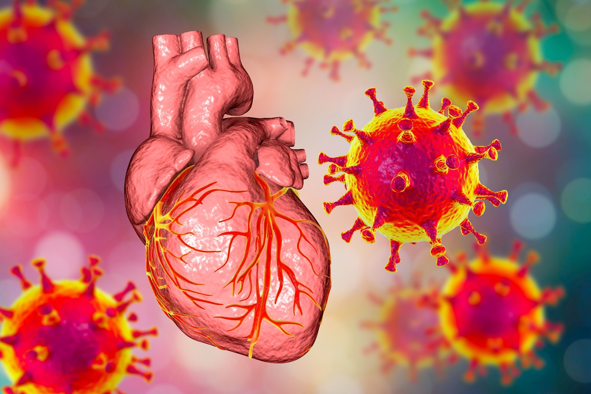
In a recent study published in the journal Circulation, researchers investigate the inflammatory response to acute respiratory distress syndrome (ARDS) within the heart.
The link between respiratory viral infections and CVD
Seasonal viral infections can range in severity from mild flu-like symptoms to potentially lethal ARDS. For example, despite being primarily a respiratory tract infection, coronavirus disease of 2019 (COVID-19) can lead to ARDS and other severe cardiovascular disease outcomes with high mortality rates.
Circulating immune cells may respond to COVID-19 by upregulating cytokine release, which can lead to myocardial injury. Cardiac macrophages, immune cells responsible for the myocardial inflammatory response, are increasingly being investigated for their role in ARDS. Recent evidence indicates that macrophage expansion, which can be accompanied by changes in the population size and relative abundances of various cardiac macrophages, is a characteristic feature of ARDS.
The main two types of cardiac macrophages include C-C chemokine receptor type 2 negative (CCR2–) and CCR2+ macrophages. Further research is needed to determine the viral-induced contributions of these macrophages to adverse cardiac outcomes.
These data would allow clinicians to make informed intervention decisions and elucidate whether these outcomes are COVID-19-induced or if observed inflammation is a systemic immune response to viral infection. Furthermore, this information could support the development of future therapies to prevent cardiovascular disease (CVD) following recovery from COVID-19.
About the study
In the present study, researchers investigate the role of viral- and non-viral-induced ARDS-associated immune signals in altering cardiac macrophage populations, thereby impacting CVD parameters, including systemic inflammation.
This study was conducted at Massachusetts General Hospital and involved 33 control samples obtained from patients who died between September and December 2019, prior to the onset of COVID-19, as well as 21 samples obtained between May and July 2020 from patients who died from COVID-19-associated complications. Samples consisted of autopsy tissue excised from the left ventricular or septal region.
Simultaneously, in vivo studies involved a daily intratracheal administration of an ARDS cocktail of immunostimulatory agents to mice, which included resiquimod, imiquimod, lipopolysaccharide (LPS), and angiotensin-converting enzyme 2 (ACE2) inhibitor MLN-4760. This model allowed the researchers to reproduce clinical ARDS features in mice without the severe acute respiratory syndrome coronavirus 2 (SARS-CoV-2).
Patient data included results obtained from electrocardiogram (ECG), echocardiography, lung computed tomography (CT) scan, blood gas analyses, body temperature evaluation, bronchoalveolar lavage fluid (BALF) characterization, blood pressure measurements, and flow cytometry. Both human and murine autopsy samples were processed using ribonucleic acid (RNA) isolation, real-time polymerase chain reaction (PCR) assay, and enzyme-linked immunosorbent assays (ELISAs) for protein and gene expression determinations.
Similar immune responses in non-viral- and SARS-CoV-2-associated ARDS
In the absence of viral infection, mice treated with the ARDS cocktail exhibited significant weight loss over the five-day cocktail treatment period. This was accompanied by hypothermia, a common feature of both ARDS and septic shock, as well as a mortality rate of over 40% by day five.
Mice with ARDS exhibited bilateral opacities and immune cell infiltrations within their lungs, as well as reduced blood oxygenation. Furthermore, increased D-dimer, neutrophil, and monocyte levels were observed, as well as reduced blood pressure and lower heart rates in ARDS mice. Other inflammatory pathways that were activated in ARDS mice included increased levels of interleukin 6 (IL-6), IL-1ß, tumor-necrosis factor α (TNF-α), and interferon y (IFN-y), all of which are also associated with SARS-CoV-2 infection.
In both non-infected ARDS and SARS-CoV-2-infected mice, an increased infiltration of interstitial macrophages and reduced levels of alveolar macrophages were observed. Although both mouse models exhibited increased levels of cardiac macrophages, this immune response was more pronounced in infected mice. Nevertheless, both models’ subsets of cardiac macrophages were altered to similar levels.
Upon comparison of control and COVID-19 patient myocardium samples, SARS-CoV-2 infection recruited a more significant number of CCR2+ CD68+ macrophages, thus indicating that a robust immune response is elicited after severe infection compared to other life-threatening diseases.
“Our findings indicate that systemic and myocardial inflammatory signals elicited by virally induced ARDS may contribute to the cardiovascular complications and high mortality rates of this condition. In addition, our study confirms previous reports that SARS-CoV-2 infection increases overall macrophage numbers in hearts.”
The cardiac benefits of TNF-α immune therapy
TNF-α neutralizing antibodies were also administered to mice to evaluate their effects on immune activation during ARDS. To this end, TNF-α immune therapy reduced weight loss, improved body temperature, increased blood oxygenation, and led to better survival rates. Histological analysis indicated that ARDS mice receiving anti-TNF-α therapy exhibited reduced macrophages, Cxcl2, IL-1ß, and IL-6 expression within the lungs.
TNF-α therapy also improved systolic dysfunction, cardiomyocyte apoptosis, and monocyte infiltration in ARDS mice. Total cardiac macrophage counts and reduced expression of IL-1ß, IL-6, and TNF-α within the myocardium were also observed, thus demonstrating the anti-inflammatory benefits associated with TNF-α immune therapy in the lungs and hearts of mice with ARDS.
Conclusions
The study findings demonstrate that SARS-CoV-2 infection leads to significant alterations in cardiac macrophage subset levels, particularly increased levels of CCR2+ macrophages, in both mice and humans. Even in the absence of SARS-CoV-2 or another virus, the immune response to ARDS-like injury is capable of inducing significant alterations in heart macrophage levels, which may increase the risk of cardiovascular complications and mortality associated with ARDS.
A new study in the peer-reviewed journal Diabetes Technology & Therapeutics (DTT) evaluated the use of tirzepatide in overweight/obese adults with type 1 diabetes.
Tirzepatide is approved for managing type 2 diabetes. It improves glucose control, facilitates weight loss, and improves cardiovascular disease outcomes.
Satish Garg, MD, from the University of Colorado Denver, and coauthors, compared a group of adults with type 1 diabetes who were prescribed tirzepatide (off-label) to a control group of adults with type 1 diabetes who were not using any weight-loss medication. The investigators reported significantly larger declines in body mass index (BMI) and weight in the treated group compared to controls. HbA1c decreased in the treated group as early as three months and was sustained through a one-year follow-up. Insulin dose decreased at 3 months in the treated group and throughout the study period.
“We conclude that tirzepatide facilitated an average 18.5% weight loss (>46 pounds) and improved glucose control in patients with T1D at one year,” stated the investigators.
“Most of the patients with diabetes, both type 1 diabetes (T1D) and T2D are either overweight or obese in the United States and Western Europe,” state Satish Garg, MD, and coauthors of an accompanying Editorial. The newer therapies for diabetes, which are known to not only improve glucose control but also cause significant weight loss and improve cardiovascular disease and diabetic kidney disease are currently not approved in the U.S. for use in type 1 diabetes. “Using GLP analogs in patients with T1D poses many challenges, but with close follow-up both patients and the healthcare provider may see many benefits such as significant weight loss and reduction of insulin dose, increased time-in-range on continuous glucose monitoring, and improve HbA1c levels,” state the authors. Long -term side-effects like gastroparesis, GERD, Cholelithiasis etc. from use of GLP analogs in patients with diabetes are not known. The authors recommend proper randomized control trials especially in patients with T1D.
Source:
Journal reference:
Garg, S. K., et al. (2024). Efficacy and Safety of Tirzepatide in Overweight and Obese Adult Patients with Type 1 Diabetes. Diabetes Technology & Therapeutics. doi.org/10.1089/dia.2024.0050.
Ozempic is often used to treat type 2 diabetes, but it might also lower the risk of cannabis use disorder
fcm82/Shutterstock
People prescribed Ozempic or Wegovy are less likely to develop a cannabis use disorder or relapse from the condition than those who take other diabetes or obesity medications, suggesting that the popular injections may treat cannabis addiction.
Ozempic and Wegovy are brand names for semaglutide, a drug that reduces appetite and regulates blood sugar levels. It is now routinely prescribed to treat type 2 diabetes and obesity, and some…
A recent Nature Metabolism study reports that circulating lactate levels are positively associated with weight loss in cancer cachexia patients. Mouse model experiments also revealed that adipose-specific G-protein-coupled receptor 81 (GPR81) is a key mediator of the catabolic effects of lactate.
Cachexia is a complicated metabolic syndrome that is associated with rapid body weight loss, including loss of fat and muscle mass.
Patients with cancer cachexia often develop anemia, fatigue, asthenia, and anorexia, which deteriorate their quality of life and reduce their tolerance to cancer therapies. As a result, cachexia accounts for around 20% of patients with cancer-related deaths.
To date, the precise mechanism responsible for the development of cancer cachexia is not well understood. Previous studies have shown that inflammatory cytokines, such as interleukin 6 (IL-6), tumor necrosis factor (TNF), interferon γ (IFN-γ), and transforming growth factor-β, induce the remodeling of adipose and muscle due to accelerated growth of cancer cells, all of which contribute to the pathogenesis of cancer cachexia.
Anti-inflammation treatments have not been associated with positive effects in alleviating cancer cachexia. Therefore, more research is needed to better understand the association between tumor manifestations and poor host metabolism.
About the study
The current study focuses on causally identifying the connecting factors between tumors and extensive catabolism in cancer cachexia. To determine serum lactate levels, samples collected from lung adenocarcinoma patients were used to calibrate the Biosen C-Line glucose lactate analyzer.
The systemic metabolic changes associated with cachexia were profiled using a mouse xenograft model of Lewis lung cancer (LLC) cells. Mice with tumor burden exhibited significant weight loss with reduced white adipose tissue (WAT).
Study findings
Metabolomics screening of a mouse model of cancer cachexia identified lactate as the top differential metabolite. The identity of this metabolite was corroborated by the peak in the mass spectrum, which was compared to the standard.
Lactate levels were strongly correlated with reduced body weight, particularly among patients with lung adenocarcinoma with cancer cachexia. Higher circulating and adipose interstitial lactate levels were observed before body weight loss. Additionally, the wasting phenotype lactate infusion results were similar to those induced by the tumor.
An osmotic minipump-mediated lactate infusion led to a persistent average increase of circulating lactate without a change in blood pH; however, d-lactate exhibited did not appear to influence weight loss. The sustained high lactate levels in many cancer patients were negatively associated with their prognosis.
Adipose GPR81 was identified as the primary mediator of lactate’s pro-catabolic effects. More specifically, GPR81 deficiency was found to block lactate infusion- and tumor-triggered cachectic manifestations, thus establishing lactate/GPR81 as the key connection between metabolic reprogramming in cancer cachexia and tumors.
The catabolic remodeling of WAT has also been identified as an early pathological event in cancer cachexia. In mouse models, depletion of key enzymes in lipolysis alleviated cachectic phenotypes, thereby confirming the crucial role of adipose tissue wasting in cancer cachexia.
A lactate-stimulated cachectic pathway activated the GPR81-Gαi/o-Gβγ-RhoA/ROCK1-p38 signaling cascade, not accompanied by the upregulation of parathyroid hormone-related protein (PTHrP). To trigger WAT browning and lipolysis, chronic elevation of blood lactate is sufficient.
Additionally, phosphoproteomics data showed the activation of extracellular signal-regulated kinase 1/2 (ERK1/2) in the GPR81−/− iWAT. This activation of ERK1/2 in GPR81-deficient mice could influence persistent adipogenesis, thereby muting lactate- and tumor-induced adipose wasting.
Conclusions
The current study identified host GPR81 as the key mediator of cancer cachexia, with lactate activating GPR81 to ultimately support tumor growth. This observation aligns with previous studies reporting the inhibition of GPR81 expression suppressing the growth of pancreatic and breast cancer cells. The experimental findings strongly suggest that the palliation of cachectic symptoms in GPR81−/− is mediated through GPR81 deficiency in the host.
Both in vitro and in vivo experiments associated with tumor growth revealed that the lack of GPR81 expression in LLC cells repressed cancer proliferation. Thus, lactate/GPR81 contributes to both cancer progression and cachexia, which deteriorates disease outcomes.
Mechanistically, lactate activates GPR81, which induces adipose metabolic remodeling through Gαi/o-Gβγ–RhoA/ROCK1–p38 signaling cascade. This leads to muscle dystrophy and systemic hypercatabolism.
Taken together, the study findings indicate that GPR81 could be targeted and blocked to alleviate metabolic impairments involved in cancer cachexia.
Journal reference:
Liu, X., Li, S., Cui, Q., et al. (2024) Activation of GPR81 by lactate drives tumor-induced cachexia. Nature Metabolism. doi:10.1038/s42255-024-01011-0
A recent study published in the journal Nature Metabolism showed that metformin treatment significantly increases blood levels of N-lactoyl phenylalanine (Lac-Phe), an appetite-suppressing metabolite.
Metformin, used for type 2 diabetes (T2D) treatment, reduces blood glucose and suppresses appetite. It is prescribed to more than 150 million individuals worldwide. However, the mechanisms of its therapeutic effects are not fully understood. It inhibits complex 1 of the electron transport chain at higher levels. Nevertheless, it is uncertain whether physiological levels of metformin are sufficient for complex 1 inhibition.
Lac-Phe is a metabolite produced by carnosine dipeptidase 2 and has been reported as an appetite suppressant in obese mice. Lac-Phe correlates with weight loss in humans with regular exercise. It also increases in mitochondrial disease and phenylketonuria. Nonetheless, whether Lac-Phe has a role in the appetite-suppressing activity of metformin has not been explored.
In the present study, researchers reported significant increases in Lac-Phe levels following metformin treatment. The study was conducted between August and December 2019 at Brigham and Women’s Hospital. Thirty-three volunteers who were 1) lean without T2D, 2) lean and prediabetic, 3) obese and prediabetic, 4) obese without T2D, or 5) obese with T2D were recruited.
Diabetes was primarily managed with metformin, albeit some participants also received insulin. The team collected sera from participants and performed untargeted metabolomic profiling. This showed notable increases in all N-lactoyl amino acids in the obese T2D group relative to obese participants without T2D. These elevations were not related to body mass index (BMI) but to T2D.
Moreover, Lac-Phe levels were 5.7 times higher in obese T2D subjects than in obese non-T2D volunteers. In addition, these increases were also significant compared to prediabetic individuals. Notably, Lac-Phe levels correlated with the concentrations of other N-lactoyl amino acids. Next, the team analyzed the metabolomic data from the TwinsUK cohort.
Lac-Phe levels in this cohort were elevated in individuals with T2D. Further, there was a robust correlation between metformin and Lac-Phe levels among T2D patients in the Brigham cohort. Notably, while metformin use was a criterion for inclusion, one volunteer lacked detectable metformin levels and had the lowest Lac-Phe levels; the volunteer discontinued metformin.
As such, the team speculated that metformin may elevate serum levels of Lac-Phe in individuals with T2D rather than T2D. This hypothesis was tested using the TwinsUK dataset; each participant had three samples collected during 1997-2012, a period when the role of metformin in T2D was growing considerably.
This enabled analyses of the Lac-Phe trajectories in participants whose metformin and T2D status changed with time. The researchers noted that metformin treatment significantly increased Lac-Phe levels in individuals newly diagnosed with T2D. By contrast, for newly diagnosed diabetic individuals without metformin therapy, there were no significant changes in Lac-Phe levels.
In participants who consistently had diabetes over successive sampling, metformin use caused substantial increases in Lac-Phe levels. On the other hand, those who did not receive metformin lacked significant changes in Lac-Phe levels. Further, the team analyzed two interventional studies from Denmark and Jordan to establish a causal link between metformin use and increases in Lac-Phe levels.
In the Danish study, significant increases in N-lactoyl amino acids were evident following a 12-week metformin intervention in non-T2D and T2D groups. The Jordanian study reported rapid increases in Lac-Phe levels over 36 hours after a single metformin dose, and the peak Lac-Phe concentration corresponded to the maximum metformin concentration.
Within the TwinsUK dataset, Lac-Phe levels correlated highly with the non-fasted state. As such, the researchers speculated whether metformin-related increases in Lac-Phe were influenced by feeding or fasting state. They observed a trend of higher Lac-Phe levels in the fed state. Moreover, while individuals not receiving metformin had lower Lac-Phe levels, Lac-Phe was significantly increased in fed subjects.
Conclusions
Taken together, the study highlighted that metformin elevates Lac-Phe levels. These increases were specific to metformin rather than T2D status and were evident in T2D subjects and healthy individuals. Further, Lac-Phe levels increase postprandially, paralleling other appetite suppressants’ patterns. Thus, pharmacological targeting of Lac-Phe could result in a more robust appetite-suppressing effect, leading to a new class of drugs for obesity.
Cancer treatment is often associated with undue weight gain, mostly due to fat deposition. The Mediterranean diet (MED diet) may help support such patients during this period. A new study published in the European Journal of Clinical Nutritionexplores the safety and benefits of this diet in adults with cancer, in addition to its feasibility in this population.
Almost 20 million people today have received a cancer diagnosis, making it the leading cause of illness and death globally. The treatment of cancer is also associated with multiple adverse effects that cause rapid aging, trigger chronic metabolic aberrations, and reduce the quality of life.
These side effects include early menopause, cognitive impairment, and cardiomyopathy, with persistent fatigue and weight loss. Such long-term ill effects could be mitigated by nutrition and exercise. Yet, there is little evidence to support the right nutritional pattern for such issues arising during or after cancer treatment.
About the Mediterranean diet
The MED diet has been long recognized as among the healthiest eating patterns. Compliance with this diet has been associated with reduced risk of many chronic illnesses, including type 2 diabetes and cardiovascular harm.
This dietary pattern is characterized by a high consumption of fish, vegetables, legumes, nuts, fruits, and extra virgin olive oil, a moderate intake of dairy and red wine, with little added sugar, processed foods, and red meat. The antioxidant and anti-inflammatory profile of this diet have been thought to mediate its beneficial effects on cardiac and metabolic health.
Previous evidence indicates that those on a MED diet have 22% and 13% lower odds of dying from prostate and breast cancer, respectively. This is very significant since the hormone treatment typically used in these cancers adversely affects the metabolic and body composition profile, increasing the risk of cardiometabolic disease.
This group of patients has been shown to benefit from nutritional manipulation with exercise by reducing body fat and body weight. Yet, there is little systematic evidence to support the recommendation of a MED diet for adults with cancer. This prompted the current study.
About the study
A systematic review was undertaken, including 15 articles covering the MED diet intervention among this group of patients. Of these, ten included breast cancer patients, all women, and one mostly female breast cancer patients. The remaining included prostate cancer, acute myeloid leukemia, and lung cancer.
In five of the studies, patients were being actively treated, while five were post-treatment studies. Three included patients who either had been or were being treated, with one starting within five years from the diagnosis.
In most studies, the aim was to lose weight, but a few were aimed at lessening fatigue, reducing inflammatory mediator levels, enhancing the diet, or maintaining a stable weight. Energy restrictions at varying levels were applied for those who were obese or overweight. Others used strategies such as reduced portion size or consuming more satiating foods.
What changes were observed?
Most participants adhered closely to the MED diet compared to controls, at 64% to 96% completion rates. In six studies, the body weight of the MED diet participants decreased compared to that of the control group. In seven studies, the body mass index (BMI) decreased in the intervention group vs controls.
A similar favorable change was reported in the body fat mass, though three also reported loss of lean mass.
Multiple metabolic biomarkers and inflammatory markers were favorably affected, including decreased glucose markers, reduced cardiovascular markers including triglycerides and total cholesterol, increased high-density lipoprotein (HDL, ‘good cholesterol’), higher albumin levels, and reduced interleukin-8 (IL-8).
The intervention was also associated with a higher quality of life and reduction in fatigue, as well as better emotional, physical, and cognitive health.
What are the conclusions?
The studies included in this review had widely varying study designs and MED diet content, making it difficult to provide a definitive recommendation to achieve these favorable outcomes. However, the safety and feasibility, as well as acceptability, of the MED diet as a nutritional intervention is established.
The positive role of nutritionists in this type of intervention, with accompanying advice on nutritional value, cooking demos, and recipes tailored to individual clients, appears from the significantly higher adherence seen in such studies.
Body weight reduction is particularly well supported by this diet with energy restriction, with overweight or obese subjects on hormone therapy or post-treatment for prostate or breast cancer losing up to 4 kg. Currently, evidence of the benefit of the MED diet is available only for women who have completed breast cancer treatment.
Further studies need to be done to confirm that the MED diet will help prevent and manage chronic disease in this population of cancer survivors who are at high risk for such conditions. Approaches that protect or increase muscle mass but are compatible with this dietary pattern need to be explored.
Confounding factors that may have mediated the improvement in cardiometabolic status and quality of life include frequent interactions with healthcare providers and other dietary components like green tea that have anti-inflammatory and antioxidant activity themselves. Careful documentation of the diet, adherence, and outcomes is necessary to rule out the role of such factors in preventing or managing chronic disease in future studies.
“Future longer term RCTs should focus on reducing the risk of, or managing, cardiovascular or metabolic disease after cancer treatment to improve the potential clinical implications of the MED-diet.”
A collaborative study between a bariatric and transplant surgery team has introduced new hope for patients suffering from end-stage renal disease (ESRD) who are also struggling with obesity. The study authors explored the outcomes of metabolic and bariatric surgery in ESRD patients and whether the surgery can improve their eligibility for kidney transplants. The findings are published in the Journal of the American College of Surgeons (JACS).
Obesity is a worsening problem in the United States, significantly impacting transplant eligibility. We established the CORT initiative – a collaborative for obesity research in transplantation – recognizing the urgent need to address this issue, especially in underserved populations who suffer the most from obesity-related diseases.”
Anil Paramesh, MD, MBA, FACS, corresponding study author, professor of surgery, urology, and pediatrics and director of the kidney and pancreas transplant programs at Tulane University School of Medicine
Patients with ESRD face many difficulties without a transplant; their only alternative is to prolong life through dialysis, a process that is not only costly and time-intensive but also significantly diminishes quality of life, Dr. Paramesh noted.
The study, conducted between January 2019 and June 2023, followed 183 ESRD patients referred for bariatric surgery, with 36 undergoing weight loss surgery and 10 subsequently receiving kidney transplants. Results showed a 27% reduction in average BMI at the time of transplant, alongside improvements in hypertension and diabetes management. This improvement in the management of comorbid conditions enhanced patients’ overall health and transplant viability.
With obesity being a major cause of transplant exclusion, this collaborative program represents a path forward for patients who previously would be ineligible, Dr. Paramesh said, and may help pave the way for increased patient education and access.
“We’ve seen that bariatric surgery is not just about weight loss; it significantly improves other serious conditions like diabetes, high blood pressure, and sleep apnea. This approach not only helps in reducing the patients’ weight to a level where they can safely receive a transplant, but also addresses the broader issue of health care disparities, particularly affecting Black and lower-income individuals,” said Dr. Paramesh.
However, the study also faced challenges, including a high drop-off rate of patients unwilling or unable to undergo surgery, and unique postoperative complications such as hypotension.
“Our findings indicate a pressing need to enhance patient education and support, making sure that potential candidates understand the benefits of weight loss surgery and its role in improving their eligibility for transplant,” said Dr. Paramesh.
The authors suggest further studies could examine the unique complications ESRD patients may have after weight loss surgery.
The study is published as an article in press on the JACS website.
Levy, S., et al. (2023). Collaborative Approach toward Transplant Candidacy for Obese End-Stage Renal Disease Patients. Journal of the American College of Surgeons. doi.org/10.1097/XCS.0000000000000962.