From day one of its very first menstrual cycle, an ovary works hard to develop follicles that produce and release the very best eggs for reproduction.
But that’s not all an ovary does.
“It has so many important endocrine functions beyond just reproduction,” says cardiovascular researcher Hattie Chung.
In humans, the ovaries produce hormones such as estrogen, progesterone, inhibin, and testosterone, all of which have far-reaching effects on the body.
Chung, who works at The Broad Institute of MIT and Harvard, is the senior author of a new study recently published in Nature Aging.
She and her team have found evidence that the long-understudied female reproductive organ undergoes major age-related changes long before menopause arrives – in mice, at least.
“At the menopause transition, humans typically still have about 1,000 eggs remaining,” says Chung.
“That suggests it’s not just the eggs themselves, but also the surrounding ecosystem that changes with age.”
While mouse studies obviously can’t tell us exactly what the equivalent human organs might do, as mammals, we share a similar evolutionary history, so they can offer hints.
By profiling 22 mouse ovaries harvested across a range of ages and stages of the reproductive cycle, the researchers began to see patterns in the way ovarian tissue changes across time.

frameborder=”0″ allow=”accelerometer; autoplay; clipboard-write; encrypted-media; gyroscope; picture-in-picture; web-share” referrerpolicy=”strict-origin-when-cross-origin” allowfullscreen>
Their study captured the way cells within each ovary interact with each other, spanning 358 oocytes (immature egg cells that mature into ova, the female reproductive cells), 668 follicles (the fluid-filled sacs containing the oocytes), and 236 corpora lutea (the temporary endocrine gland that forms from spent follicles after ovulation to produce progesterone).
Signs of aging emerged long before mouse ‘menopause’ officially arrived.
With age, ovarian tissue began to degenerate; cells that usually operate in sync to coordinate the development of follicles, ovulation, and tissue remodeling were falling out of time.
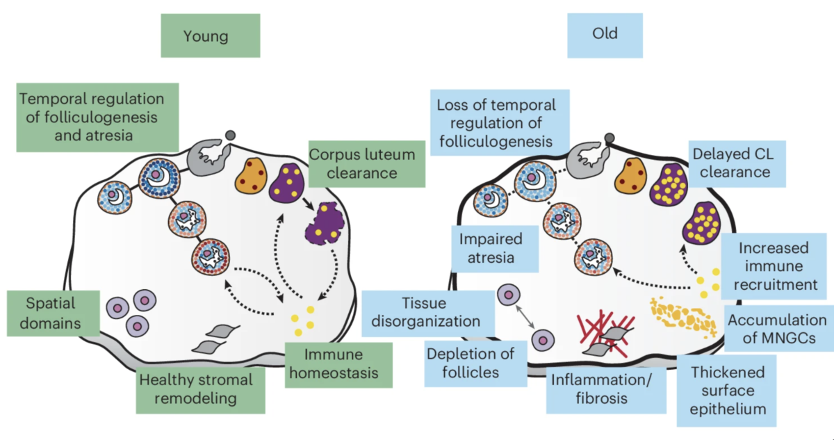
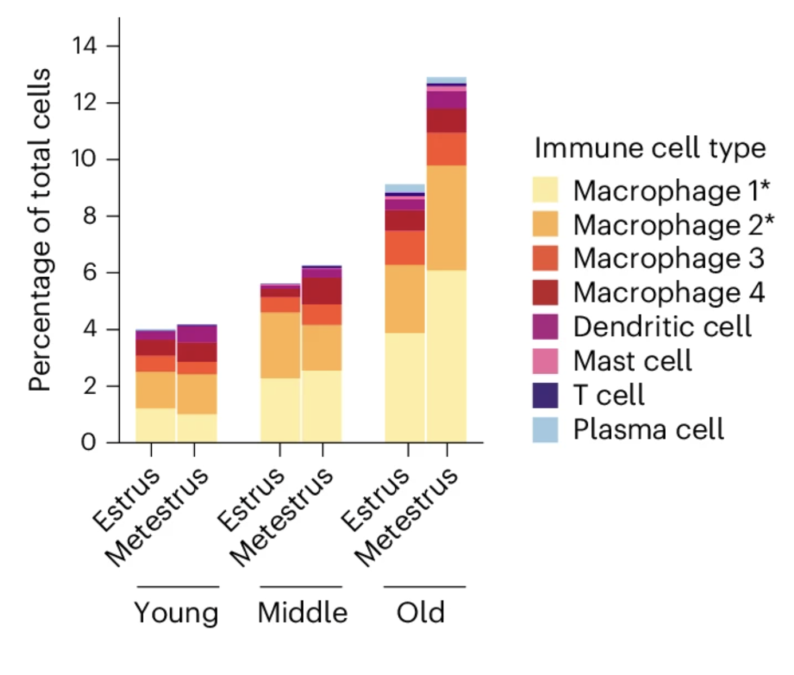
The researchers also noticed that the immune cell dynamics of aging ovaries shift, with increased signals of inflammation and a disorganization of tissues.
“These findings indicate that reproductive aging can be viewed as a progressive breakdown of tissue-level coordination, rather than solely the depletion of the follicle reserve,” the authors write.
Recent research suggests that ovaries continue to change even after menstruation ceases, both in mice and, likely, in humans.

A picture is emerging of the ovary in a constant state of flux throughout its life, when serving a reproductive role, and when it comes to other important hormonal and immune functions.
This could especially have implications for people whose ovaries are removed, a surgery known as oophorectomy.
People may undergo this procedure to treat cancer, endometriosis, or as a form of gender affirmation.
Knowing what ovaries are actually doing in the body – beyond just egg production – means that doctors can potentially get more precise in providing patient care post-oophorectomy, as well as through the normal course of ovarian aging.
“The next obvious step for us is to conduct these analyses in human tissues,” says Chung.
“We’re really keen on working with patient specimens to understand how these cellular interactions may be altered with aging and in pathologic conditions.”
Related: PCOS Is Officially Renamed, After Decades of Misinformation
Her team has already begun collaborating with Yale obstetrics and gynecology researchers to collect human ovarian samples that represent a range of age groups, in the hope of verifying these findings beyond the mouse model.
“Understanding how [the ovary’s] complex cellular ecosystem changes with age may ultimately provide insights into women’s health far beyond fertility alone,” Chung says.
The research was published in Nature Aging.
This article was fact-checked by Rachel Garner and edited by Rebecca Dyer. While we pride ourselves on our process, we are only human. If you spot a mistake, please let us know.


.jpg?w=527&resize=527,703&ssl=1 "White Supremacists Love Armie Hammer’s New Movie")