The top ten neurological conditions by disability-adjusted life year (DALY) globally are shown for 2021. Stroke has more than double the DALYs of any other condition, at 160 million. In 2021, stroke was the third leading cause of death globally, after coronary heart disease and COVID-19.
Uneven impact
Rankings DALY rates* for neurological conditions are shown for high-income regions and sub-Saharan Africa. Conditions with a high impact in African regions have some of the highest DALYs for children under five globally. Autism spectrum disorder has higher DALY rates in high-income regions, but a lack of diagnostic tools in lower-income countries is a contributing factor.
Source: Steinmetz, J. D. et al. Lancet Neurol.23, 344–381 (2021). Infographic by Bec Crew and Tanner Maxwell.
Time trend
The absolute number of global deaths attributed to neurological conditions has not changed much since 1990, but ageing and growing populations mean there are now more people living with these conditions. When age is standarized — all populations are given the same age structure, so the percentage of people under and over 40 years old is consistent, for example — death rates sharply decline.
Source: Steinmetz, J. D. et al. Lancet Neurol.23, 344–381 (2021). Infographic by Bec Crew and Tanner Maxwell.
Workforce spread
Europe overwhelmingly leads in the number of neurologists and neurosurgeons per capita. The World Health Organization reports a significant difference between income groups, with low-income countries reporting a median of 0.1 specialists per 100,000 population, compared with a median of 7.1 per 100,000 population in high-income countries. These workplace population numbers are self-reported.
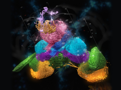
A fruit fly might not be the smartest organism, but scientists can still learn a lot from its brain. Researchers are hoping to do that now that they have a new map — the most complete for any organism so far — of the brain of a single fruit fly (Drosophila melanogaster). The wiring diagram, or ‘connectome’, includes nearly 140,000 neurons and captures more than 54.5 million synapses, which are the connections between nerve cells.
“This is a huge deal,” says Clay Reid, a neurobiologist at the Allen Institute for Brain Science in Seattle, Washington, who was not involved in the project but has worked with one of the team members who was. “It’s something that the world has been anxiously waiting for, for a long time.”
The map1 is described in a package of nine papers about the data published in Nature today. Its creators are part of a consortium known as FlyWire, co-led by neuroscientists Mala Murthy and Sebastian Seung at Princeton University in New Jersey.
A long road
Seung and Murthy say that they’ve been developing the FlyWire map for more than four years, using electron microscopy images of slices of the fly’s brain. The researchers and their colleagues stitched the data together to form a full map of the brain with the help of artificial-intelligence (AI) tools.
But these tools aren’t perfect, and the wiring diagram needed to be checked for errors. The scientists spent a great deal of time manually proofreading the data — so much time that they invited volunteers to help. In all, the consortium members and the volunteers made more than 3 million manual edits, according to co-author Gregory Jefferis, a neuroscientist at the University of Cambridge, UK. (He notes that much of this work took place in 2020, when fly researchers were at loose ends and working from home during the COVID-19 pandemic.)
An animation of the CT1 neuron in the fruit-fly brain. There are two of them; each one spans an entire eye and has more than 148,000 synapses. Credit: Amy Sterling, Murthy and Seung laboratories, Princeton University (ref. 1)
But the work wasn’t finished: the map still had to be annotated, a process in which the researchers and volunteers labelled each neuron as a particular cell type. Jefferis compares the task to assessing satellite images: AI software might be trained to recognize lakes or roads in such images, but humans would have to check the results and name the specific lakes or roads themselves. All told, the researchers identified 8,453 types of neuron — much more than anyone had expected. Of these, 4,581 were newly discovered, which will create new research directions, Seung says. “Every one of those cell types is a question,” he adds.
The team was surprised by some of the ways in which the various cells connect to one another, too. For instance, neurons that were thought to be involved in just one sensory wiring circuit, such as a visual pathway, tended to receive cues from multiple senses, including hearing and touch1. “It’s astounding how interconnected the brain is,” Murthy says.
Exploring the map
The FlyWire map data have been available for the past few years for researchers to explore. This has enabled scientists to learn more about the brain and about fruit flies — findings that are captured in some of the papers published in Nature today.
Cubic millimetre of brain mapped in spectacular detail
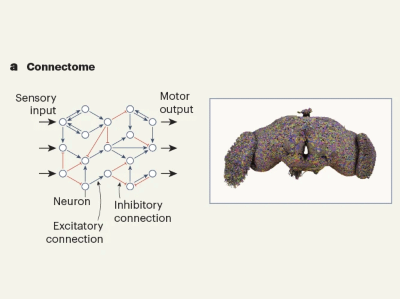
In one paper2, for example, researchers used the connectome to create a computer model of the entire fruit-fly brain, including all the connections between neurons. They tested it by activating neurons that they knew either sense sweet or bitter tastes. These neurons then launched a cascade of signals through the virtual fly’s brain, ultimately triggering motor neurons tied to the fly’s proboscis — the equivalent of the mammalian tongue. When the sweet circuit was activated, a signal for extending the proboscis was transmitted, as if the insect was preparing to feed; when the bitter circuit was activated, this signal was inhibited. To validate these findings, the team activated the same neurons in a real fruit fly. The researchers learnt that the simulation was more than 90% accurate at predicting which neurons would respond and therefore how the fly would behave.
In another study3, researchers describe two wiring circuits that signal a fly to stop walking. One of these contains two neurons that are responsible for halting ‘walk’ signals sent from the brain when the fly wants to stop and feed. The other circuit includes neurons in the nerve cord, which receives and processes signals from the brain. These cells create resistance in the fly’s leg joints, allowing the insect to stop while it grooms itself.
One limitation of the new connectome is that it was created from a single female fruit fly. Although fruit-fly brains are similar to each other, they are not identical. Until now, the most complete connectome for a fruit-fly brain was a map of a ‘hemibrain’ — a portion of a fly’s brain containing around 25,000 neurons. In one of the Nature papers out today4, Jefferis, Davi Bock, a neurobiologist at the University of Vermont in Burlington, and their colleagues compared the FlyWire brain with the hemibrain.
Some of the differences were striking. The FlyWire fly had almost twice as many neurons in a brain structure called the mushroom body, which is involved in smell, compared with the fly used in the hemibrain-mapping project. Bock thinks the discrepancy could be because the hemibrain fly might have starved while it was still growing, which harmed its brain development.
The FlyWire researchers say that much work remains to be done to fully understand the fruit-fly brain. For instance, the latest connectome shows only how neurons connect through chemical synapses, across which molecules called neurotransmitters send information. It doesn’t offer any information about electrical connectivity between neurons or about how neurons chemically communicate outside synapses. And Murthy hopes to eventually have a male fly connectome, too, which would allow researchers to study male-specific behaviours such as singing. “We’re not done, but it’s a big step,” Bock says.
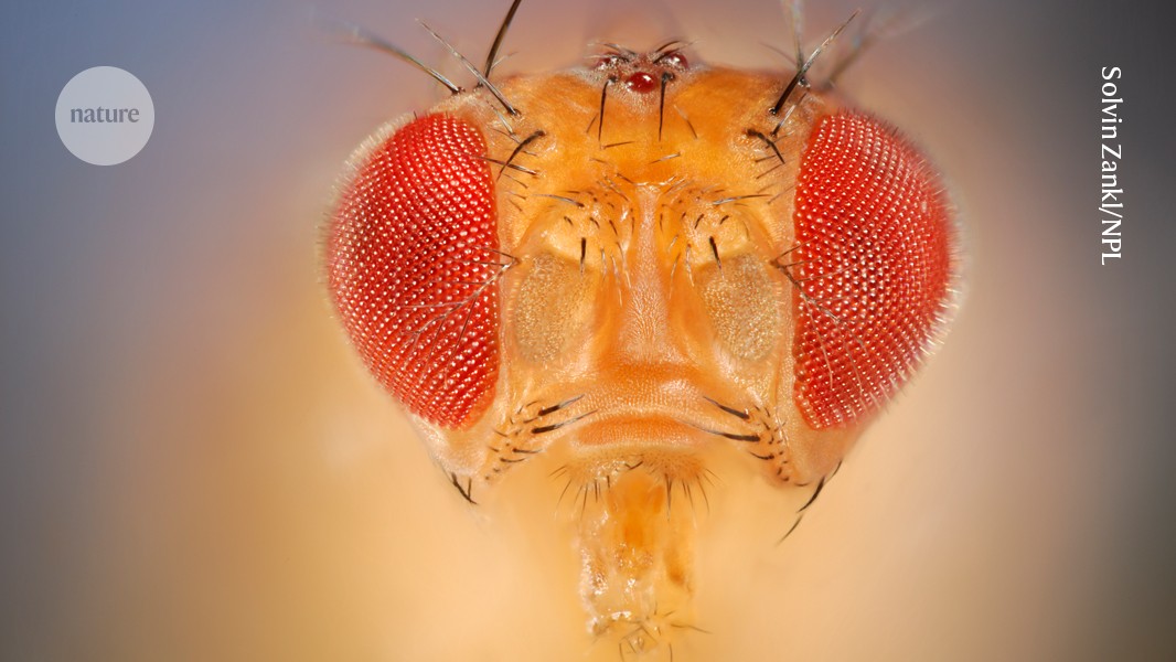
The fruit fly Drosophila melanogaster’s brain has been mapped in a project that discovered more than 4,500 new cell types.Credit: Solvin Zankl/Nature Picture Library
The brain of a fruit fly is so small that it can fit on the head of a pin. And yet, it is an inordinately complex object, encompassing nearly 140,000 neurons and 50 million synapses. It is no wonder, then, that it took 5 years, 50 laboratories and more than 200 people to map it — a monumental achievement that is the subject of a series of papers this week in Nature1–4.
Brain mapping is one of the most delicate and complex tasks in neuroscience, and even the smallest error can markedly affect the results. The achievement is all the more remarkable considering that around a dozen people working on the project were not full-time academic researchers, but volunteers from all walks of life, from around the world. They collaborated to validate results produced by artificial intelligence (AI). Hundreds of neuroscientists who study fruit flies also volunteered to support the project, and recruited their students. Together, they have been credited as authors. Amateur science is as old as science itself, but this project adds yet another way to involve more brain power in what is increasingly data-hungry research.
The FlyWire connectome: neuronal wiring diagram of a complete fly brain
The ‘FlyWire connectome’ map presented by Mala Murthy, a neuroscientist at Princeton University in New Jersey, and her colleagues1 is already helping to advance neuroscience. The project to create a ‘wiring diagram’ of connections between neurons discovered 4,581 cell types, many in understudied brain regions2. Other groups are using the map to study and make predictions about brain function. For example, researchers in the United States, the United Kingdom, South Korea and Puerto Rico have used it to investigate the neurons that control how flies feed and groom themselves3. The team simulated the relevant brain circuits and used the model to predict which neurons respond to food stimuli, such as sugar or water.
Another team, led by researchers at the University of California, Santa Barbara, used the map to predict how specialized neurons in certain parts of the fruit fly (Drosophilamelanogaster) brain receive and process visual information4. The researchers used imaging techniques to confirm their predictions in female flies that were between seven and nine days old, underscoring the connectome’s power to understand how the brain works.
One of the main tasks of the researcher volunteers was to help the academic neuroscientists label the map and double-check its accuracy. Initially, the project’s scientists used electron microscopy to take images of slices of the fly brain. They then trained an AI model on these images to create a wiring diagram of millions of synapses and thousands of neurons. These predictions needed to be verified against quality criteria and parameters.
Fact-checking millions of data points is time-consuming. Instead, project co-leader Sebastian Seung, a neuroscientist at Princeton University, and his team built an interactive game-like platform called EyeWire, in which volunteers were asked to assemble 3D models of neurons in a mouse retina and zebrafish hindbrain. The 100 best-performing players were then trained to identify various types of cell and then set to work on the real project, to proofread the AI-generated synapses and neurons — a task that otherwise would have needed another 33 neuroscientists working full time, for at least a year, to complete.
“We thought [this study] would be many, many years in the making,” says Murthy. “But as the technology developed, and as we opened up the data, and the [volunteer] community really stepped in and were excited to participate with us, it became a reality. And we completed it, in what I would consider record time.”
A complete wiring diagram of the fruit-fly brain
John Ngai, director of the Brain Research through Advancing Innovative Neurotechnologies (BRAIN) Initiative at the US National Institutes of Health in Bethesda, Maryland, which funded the studies, told Nature that such an approach could be used for larger connectome projects that the BRAIN Initiative is funding. This includes a programme called BRAIN CONNECTS, which involves training people in the research and technical skills required to generate wiring diagrams of mammalian brains.
Amateur scientists and volunteers are getting increasingly involved in other areas, too. Earlier this month, researchers at the United Nations Statistics Division in New York City and Open Data Watch, a non-profit organization based in Washington DC, described how citizen scientists are helping to fill in data gaps for the UN Sustainable Develop-ment Goals, especially for marginalized communities5.
Last year, a study6 in Nature described how the asteroid Dimorphos became temporarily brighter and redder when NASA’s Double Asteroid Redirection Test spacecraft deliberately hit it. The finding was confirmed by a small group of amateur astronomers who were able to share their observational data with professional scientists through an app.
Often, it’s the professionals who need to check the work of the amateurs. What’s different about the fly-brain work is that the volunteers are helping the full-time scientists to authenticate their findings — something that could have a much wider application in our data-rich world.
Among the top 25 countries for neuroscience output in the Nature Index, these ten have the highest proportion of neuroscience Share relative to their overall Share (neuroscience %). The United States, Germany, United Kingdom and Canada all rank within the top 10 overall for neuroscience; Norway and Portugal have the lowest overall ranks, at 22 and 23, respectively.
On the up
The Share of the fastest rising institutions in neuroscience for 2022–23 is shown over a five-year period. The University of Queensland in Australia is the only institution from outside China in the top five. The top-ranked institution in neuroscience overall, Harvard University in Cambridge, Massachusetts, was the sixth fastest riser, increasing its Share by 4.5% to reach 229.20 in 2023.
Institution outputs
Institutions with a special focus on neuroscience research are highlighted in this chart, which plots their neuroscience Share against their neuroscience %. Just over 10% of the top 200 institutions in neuroscience have more than 200 Share in the topic for the period 2019–23, and only 8.5% have more than 30% of their overall Share related to neuroscience.
The Chinese Academy of Sciences in Beijing has a relatively low proportion of its Nature Index output focused on neuroscience research, but it has the 6th highest Share in the topic, at 378.76. Harvard University’s Share in neuroscience (996.17) dwarfs that of all other institutions. With 19.6% of its total Share in the Index related to neuroscience, this is a clear priority area. Neuroscience-related outputs represented 89.7% of the total Share of the Allen Institute in Seattle, Washington, for 2019–23. The institution is ranked 144th in the topic overall, with a Share of 53.15.
Unravelling how the billions of interacting neurons in the human brain conjure consciousness is one of the greatest challenges in twenty-first-century science. Over the past decade, large, well-funded initiatives, including in the United States, Europe and China, have been launched to unlock the mysteries of cognitive function — mental processes such as memory, language, perception and problem-solving — by coming at it from all angles.
For the millions of people around the world who will develop an incurable or treatment-resistant brain disorder this year, the need to better understand cognitive function and dysfunction is pressing, says Christopher Rozell, a computational neuroengineer at Georgia Institute of Technology in Atlanta. Rozell co-leads a multidisciplinary team that is developing technology-based therapies for depression, the leading cause of ill health and disability worldwide. “Globally, more than 300 million people will have a major depressive episode this year — and that’s just one neurological disorder subtype,” he says.
Nature Index 2024 Neuroscience
Rozell is exploring a therapy for treatment-resistant depression based on deep-brain stimulation, in which implanted electrodes electrically stimulate specific brain areas to provide long-term symptom relief. The work is funded by the US National Institutes of Health’s (NIH) Brain Research Through Advancing Innovative Neurotechnologies (BRAIN) Initiative, a major project launched in 2013, which to date has invested more than US$4 billion across neuroscience research. The BRAIN Initiative’s strategy is to develop tools, and then use these advances to gain a deeper understanding of brain function. According to Rozell, the decade-long investment is beginning to pay off.
In depression treatment, for example, doctors have always had to make subjective clinical judgements and trial-and-error therapy adjustments when trying to manage the condition. But, in 2023, Rozell and his collaborators used new brain-implant and big-data processing technologies to identify changes in brain activity that can indicate a patient’s current clinical state, enabling doctors to adjust treatment in response1. At the end of the six-month trial, 90% of patients showed significant improvement and 70% were in remission or no longer depressed. BRAIN Initiative funding was key. “We work with clinicians and engineers in teams with a breadth of expertise that would have been very difficult to imagine under conventional funding programmes,” Rozell says. “Every week now, you see large, interdisciplinary teams making incredible advances that would not be happening if it were not for a programme like the BRAIN Initiative.”
Likewise, proponents of the Human Brain Project (HBP), one of the largest research endeavours ever funded by the European Union (EU), which spent €600 million (US$668 million) over ten years before its completion in 2023, point to several advances. New brain-implant technologies that could restore partial vision in certain forms of blindness and brain-like ‘neuromorphic’ computer chips for more sophisticated artificial intelligence (AI) are important outcomes.
But concerns remain that core questions in neuroscience have not been addressed by big projects. It’s not clear how cognitive function emerges from patterns of brain activity, for instance, let alone how these processes go awry caused by disease.
And although big-neuroscience funding has increased in China over the past few years, it has been cut significantly in the EU and the United States, threatening the trajectory of brain science advancement.
Uncharted territory
If understanding human brain function is the ‘moonshot’ of neuroscience, we’ll never make it without the right maps, says Rozell. Creating brain atlases, each focused on different structural features, has been a key aim. In late 2023, the BRAIN Initiative’s Cell Census Network (BICCN), a multi-centre effort led by the Allen Institute for Brain Science in Seattle, Washington, produced the most detailed map yet of the cells that make up the human brain. Using single-cell genome sequencing — a technique that allows all or part of an individual cell’s genome to be sequenced — the team identified more than 3,000 different cell types in the human brain, many previously undescribed.
BICCN researchers also produced the first complete cellular atlas of a mammalian brain, pinpointing the location and identity of each of the more than 32 million cells in a mouse brain2. When the team launched the project 10 years ago, it was unclear whether this was even feasible, says Allen Institute director, Hongkui Zeng, who led the work. But the rapid development and scaling-up of single-cell genomic technology has revolutionized the field.
“Previously, the brain was just an unknown number of faceless cells,” says Zeng. “Now, we have the molecular identities for specific cells in specific brain regions, and we can start to label each cell type and see what they do.”
Immune cells in a mouse brain, intertwined with tiny blood vessels, captured for a BRAIN-funded project.Credit: Josephine Liwang, Yongsoo Kim lab/Penn State College of Medicine, PA
BICCN’s open-access brain-cell atlases are an indispensable resource, says Sebastian Seung, a computer scientist and neuroscientist at Princeton University in New Jersey. “To go from mapping the brain as a bunch of regions, to mapping cell types, is a huge jump in precision,” he says. Brain-cell atlases are foundational data supporting Seung’s own research, which focuses on the wiring between brain cells, known as the connectome. Together with cell mapping, new tools in connectomics — including those developed in Seung’s lab with BRAIN funding, which use AI to automate brain-scan image processing — allow scientists to study the brain in ways they’ve never done before.
A different approach was used to build the Human Brain Atlas, the most detailed 3D anatomical map of a human brain yet assembled3. A team led by Katrin Amunts, a neuroscientist at the Jülich Research Centre, a large-scale national facility in the Helmholtz Association of German Research Centres, took a postmortem brain and analysed it, slice by slice, to build the atlas not from the cells up, but from a whole brain down. The Human Brain Atlas forms a core part of EBRAINS, an open-access digital platform that combines tools, services and data generated by the HBP, which has been used by more 10,000 people worldwide.
The platform’s ‘virtual brain’ tool is being used to create personalized patient brain models to guide clinical decision-making in epilepsy, multiple sclerosis, depression and Parkinson’s, and its brain atlases and data are being accessed by researchers in neuroimaging, neurology, AI and basic science. In January, the EBRAINS project won a further €38 million from the European Commission to fund its continued development.
There is an argument that although BRAIN and the HBP did not specifically focus on conceptual questions in neuroscience, the foundational resources that they have provided can help to fill major knowledge gaps that will benefit those working in both basic and applied neuroscience areas. Seung says this is why the BRAIN Initiative’s strategy of prioritizing neuroscience tool development was the right approach. “So much of the study of neuroscience has been limited by the scarcity of data,” he says. “The NIH would normally not necessarily fund technology development, but sometimes to get to important science, we need a technological revolution.”
New model
Still in its early phases, China’s big neuroscience project can benefit from lessons learned by its international counterparts. Conceived in 2013 — closely following the launch of BRAIN and the HBP — the China Brain Project (CBP) began in 2021 with ten-year funding of 12 billion yuan (US$1.66 billion) to advance brain-disease studies and basic neuroscience, as well as brain-inspired technologies and brain–computer interfaces. The project involves more than 500 laboratories across the country, and aims to build on China’s research strengths, including in connectomics and non-human primate animal models, a valuable, but contentious, aspect of neuroscience. “You cannot do invasive experiments in the human brain to understand what’s going on, so animal models are very important,” says Zeng.
The protocols and standards for non-human primate research in China are based on those set by the NIH, but the work is easier to conduct because animal-rights groups don’t protest against animal use in research like they do in the United States, says Muming Poo, scientific director of the Institute of Neuroscience at the Chinese Academy of Sciences in Shanghai, who has led the CBP organizing committee since 2020. “There is a great need in the community for using non-human primate disease models because mouse models for brain disease, especially psychiatric disease, are just not working,” says Poo. He notes the slow global pace of drug development for brain disease, which is mostly based on rodent models, and says non-human primates, as our closest living relatives, should offer better models of the human brain.
Poo’s group is developing a toolbox of genetic-engineering techniques to produce non-human primate models of disease that they hope can be used in drug testing. In late 2023, they reported the first live-born monkey chimaera4, created by taking stem cells from one macaque embryo and adding them to another. The work is a key step towards creating transgenic non-human primate models of human brain diseases, akin to way that transgenic rodent models of disease are currently made.
Another strength that the CBP hopes to build on is China’s vast population, from which researchers can draw on extensive patient cohorts. According to Jialin Zheng, dean of the Tongji University School of Medicine in Shanghai, autism spectrum disorder in children, depression in adults and Alzheimer’s disease in ageing populations are the priority conditions addressed by CBP research.
In parts of the CBP that are related to brain-inspired technology, such as AI and brain–computer interfaces, there is strong competition between institutions in China and abroad, says Poo. But in basic neuroscience and brain medicine, the CBP was specifically designed to complement work conducted by other countries. “We made a strong point of taking the directions that are deficient in the United States and Europe,” such as non-human primate models and large-cohort studies, says Poo. Some of the first internationally collaborative research conducted within the project are now close to publication, he adds. “I think it’s like the global-warming problem — brain disease is an urgent problem shared by all of society, and we should solve it together.”
In many ways, the approaches and priorities of the big-brain projects in the United States, Europe and China complement each other to make the most of international resources and talent. In the United States, for instance, the BRAIN Initiative pooled resources to push technology development, whereas the HBP’s strategy focused on coordinating multidisciplinary research, such as bringing neuroscientists together with computer scientists to develop new treatments. China’s strategy is to use its unique strengths to fill important gaps and expand on them through international collaboration.
There are challenges ahead if researchers want to build on the outputs of the three initiatives. For example, Zheng says it’s going to require coordination between governments to decide how genetic information and biological samples can safely be shared between countries. “Different countries have different regulation in terms of data. How can it be shared more openly? We are dealing with the same diseases, so, how can we work together to address these challenges?”
In addition to restrictions on data sharing, coordination between different data centres is a major issue, says Poo. “It has been difficult to set up a generally agreed, smooth way of data-sharing among many big projects, because each big project has its own data centre,” he says. “We are in international discussions about the data problem, but there is no solution yet.”
There are also concerns about whether long-standing questions around cognitive function can be answered by the kinds of projects being funded by big brain programmes. On the one hand, finding answers will require parallel studies of brain activity at the molecular, anatomical and physiological levels — something that large-scale initiatives are designed to facilitate, says Zeng.
Source: Nature Index
But knowing how to piece this information together to explain cognitive function will require new ideas and hypotheses at a foundational level that none of the big neuroscience projects has yet produced, says Yves Frégnac, emeritus research director in cognitive science at the University of Paris-Saclay in France. “New concepts are not evolving at the same pace as technologies,” he says. “Reading out signs of cognitive activity is very different from understanding the brain.”
For China, the CBP has brought a much-needed injection of cash to a field that has struggled to find funding in the past. Poo says the initiative, which so far seems to be on track to meet its decade-long funding promise, will not only advance neuroscience in highly applied areas, but also in fundamental research. “In other countries, there are avenues of support for basic research in brain science, through organizations such as the US NIH or National Science Foundation — but not in China,” he says.
As the CBP builds momentum, researchers in Europe are trying to regain their footing, a year after the end of the HBP. Raising just over half of the expected €1 billion in funding from the EU and its member states, the HBP feels to many scientists like an opportunity not quite fulfilled, despite the progress made. “This money was needed in the field of brain sciences,” says Frégnac, who wrote an opinion piece on how the initiative could have been done better5. “People talk about €1 billion, US$4 billion, but if you compare it to initiatives in physics, this is peanuts.” NASA’s James Webb Space Telescope, for example, cost $10 billion, and the $1.5 billion annual budget of the European particle-physics laboratory, CERN, dwarfs the HBP’s entire ten-year funding. “If we want to be serious about the brain, we need to put more money in,” says Frégnac, who adds that the possibility of a well-funded follow-up to the HBP looks remote.
The future of BRAIN Initiative-supported research is also unclear. In 2024, as the ten-year pot of funds set aside in 2016 entered its ramp-down phase, a budget cap across all federal spending constrained the US Congress from making up the shortfall. The result was a 40% cut to BRAIN Initiative funding, compared with 2023. Researchers such as Rozell, whose work on treating depression is directly threatened by the cuts, are worried. “We’ve made enormous progress, but this work is not finished — it is not an approved therapy,” says Rozell. With the global economic cost of mental disorders estimated at US$5 trillion, the need for investment is clear, he adds. “To have spent a decade of money, time and expertise to reach a place where we’re starting to see the returns, and then have the threat of these programmes being taken away, it’s enormously concerning.”
Three of the most prolific young researchers in neuroscience-related output in the Nature Index discuss the problems they’re trying to solve and what keeps them optimistic about their work.
CASEY PAQUOLA: Mind modeller
Casey Paquola is building a model that simulates brain changes from infancy to adulthood so researchers can track disease progression.Credit: Forschungszentrum Jülich/Sascha Kreklau
Roughly 75% of severe psychiatric disorders emerge before the age of 24, so “intervention in youth is likely to have the most significant impact in long-term psychiatric health”, says Casey Paquola, a computational neuroscientist at the Jülich Research Centre, one of the largest institutions in the Helmholtz Association of German Research Centres, Germany’s leading research organization.
Paquola and her colleagues are using large data sets of infant, child and adolescent brain scans to inform a new model that simulates changes from infancy to adulthood in the hope that it can predict the development of conditions such as schizophrenia and psychosis.
Nature Index 2024 Neuroscience
“What makes our approach unique, but also effective, is that we do this in a multiscale way,” says Paquola, referring to how the model accounts for changes at a cellular and DNA level, as well as larger shifts in the brain’s wiring and function. “That means we can cross-validate our theories for how cognition emerges.” Paquola received a €1.5-million (US$1.74-million) grant from the German Research Foundation, the country’s largest research funding organization, to support the work.
The model could inform future diagnosis and treatment protocols by allowing researchers to ‘reverse time’ and simulate the development of certain conditions backwards to find the cause. It could also help researchers to identify risk factors. In young children, for example, the brain’s cerebral cortex increases in thickness until adolescence, at which point it starts to thin back down again. Paquola and her colleagues observed unusually thick cortices in children with higher genetic risk of schizophrenia, which she says could be a symptom, or a cause, of the disease. “That’s the type of trajectory we’re interested in mapping,” she says.
Since moving to the town of Jülich in Germany from her native Australia, Paquola says she’s experienced strong support as an early career researcher. “It’s very hard for early career researchers to get a step up in Australia, I find. Whereas in Germany, they really do support younger researchers.”
For one thing, it is easier to acquire funding to support an entire research team for a multi-year project in Germany than it is in Australia, says Paquola. She adds that the local government in Jülich covers the primary costs of pre-school childcare for all residents, which is helping her plan her return to work after the birth of her son.
“It means I’ve been able to choose when to return to work based on what suits our family best,” she says. — Felicity Nelson
SOLOMIIA BOYKO: Alzheimer’s protein puzzler
Solomiia Boyko.Credit: Nancy Andrews
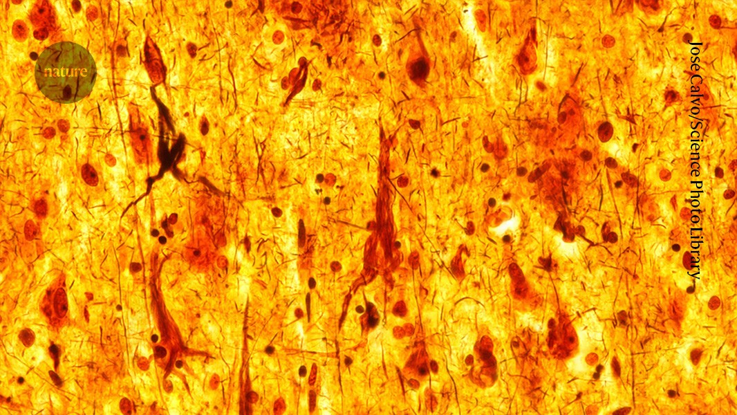
At Case Western Reserve University in Cleveland, Ohio, neuroscientist Solomiia Boyko is investigating how a type of brain protein called tau clumps inside the neurons of people with Alzheimer’s disease. Forming sticky, string-line accumulations called neurofibrillary tangles, this build-up blocks the synaptic lines of communication between neurons, which leads to the neuron cell death that drives dementia.
Past studies1 have shown that when droplets of tau protein are placed in a liquid, they will spontaneously gather around each other. Boyko wants to know if this dynamic holds true in living organisms or cells. “We know that [tau] droplets become aggregates,” says Boyko. “The missing part of the puzzle is whether this happens in the cell.”
The problem is that current tools and techniques are not advanced enough to show what happens to tau proteins within the swirling complexity of a living cell. So, Boyko and her colleagues created a liquid that chemically resembles a cell’s cytoplasm, the thick liquid that fills the inside of a cell, and watched what happened when they added tau droplets. Publishing their results in 2022, they describe behaviours similar to when oil comes into contact with water2. “They don’t mix,” says Boyko. “The tau form droplets and aggregate to each other.” What’s unclear is whether the process of tau droplets becoming tau aggregates have any physiological function in healthy brains, or if it is purely pathological.
As the global life expectancy improves, the number of people with Alzheimer’s, a disease that is strongly associated with ageing, will inevitably grow. From 1990 to 2019, the worldwide incidence of the disease ballooned by close to 150%. Researchers are struggling to keep pace — the Alzheimer’s drug-development pipeline for 2024 features fewer trials and fewer new drug candidates, compared with 2023.
There are methods in development that would allow in vivo observations of tau droplets, which Boyko is hopeful about. She says she is optimistic that the full biology of Alzheimer’s disease will eventually be decoded. “Alzheimer’s is such a big problem for the sufferers, the caregivers and the family. As a researcher, maybe I can be of some help,” she says. “I truly believe that the incremental changes of knowledge from research can make a difference.” — Benjamin Plackett
NICHOLAS BUSH: Neuron recorder
Nicholas Bush.Credit: Nicholas Edward Bush
As a first-year graduate student at Northwestern University in Evanston, Illinois, Nicholas Bush listened as researchers played the activity of a monkey’s sensory cortex — part of the brain that processes auditory, visual and other sensations — over a loudspeaker. When the monkey’s arm moved, he recalls hearing a ‘whoosh’ of electrical activity. At that moment, Bush knew he wanted to pursue a career in neuroscience. “To actually hear neurons in the brain of this monkey that was sitting right next to me, as it was actively moving, was just an ineffable moment,” he says.
Today, Bush is a postdoctoral fellow at the Seattle Children’s Research Institute in Washington, where he studies the circuits in the brainstem that control the ability to breathe. In a study3 published earlier this year, Bush and his colleagues showed how breathing disruptions associated with certain diseases can change the function of neurons in the brainstem that control breathing.
New implantable technologies can be invaluable for this kind of research, says Bush. He and his colleagues implanted silicon probes called Neuropixels, produced by Belgium-based non-profit research and development organization, Interuniversity Microelectronics Centre, into the skulls of mice to measure their neural activity at different points in the breathing cycle. When the mice were breathing normally, their neurons fired in a rhythmic cascade that reset each time they breathed out. Mice that were given morphine — an opioid drug that can cause breathing problems if misused — experienced a similar cycle of neuronal activity, but the wave of electrical activity from neuron to neuron was slower.
“Opioids are a huge concern right now,” says Bush, noting the millions of people around the world who are dealing with opioid-use disorder. “Knowing that [opioid use] is slowing down, or sort of restructuring, the dynamics of this system is particularly important.”
The team also measured how the neurons in the brainstem responded when mice were deprived of oxygen. When the mice gasped, their neurons stopped operating in a rhythmic pattern and instead fired all at once, facilitating a potentially life-saving increase in gas exchange. It’s hoped that such insights could inform research on sudden unexpected infant death syndrome in humans, which is thought to occur when infants experience a lack of oxygen but don’t wake up.
Technological advances such as Neuropixels are opening up many opportunities in neuroscience, says Bush. “The tools that are being developed are just astounding and are giving us new avenues of thinking about old problems,” he says. “It’s a super exciting time to be a neuroscientist.” — Felicity Nelson
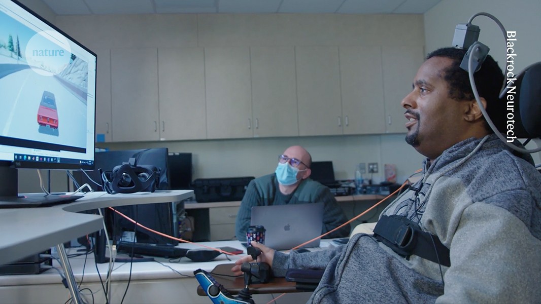
For seven years, British neuroscientist Luke Bashford trained as a postdoc in the United States, working on brain–computer interfaces (BCIs) — systems that directly link brain activity to external devices. By recording and decoding electrical signals from the brain to generate computer commands, BCIs allow people with limited movement to use their thoughts to control technologies such as smartphones, computers, wheelchairs and robotic arms. Unlike non-invasive BCIs — wearable devices such as caps or headbands that attach electrodes to the outside of the head — the implantable BCIs that Bashford works on require surgery to place the electrodes on or inside the brain to access more reliable and information-rich signals.
A lot of academic research — and most commercial investment — is focused on implantable BCIs, because their potential to provide high-performance assistive interfaces is stronger than their non-invasive counterparts. But implanting electrodes directly in the brain comes with obvious risks, so implantable BCI trials are tightly controlled by regulators. The most advanced implantable BCIs in development remain in early-stage clinical trials, and no such system has been approved for clinical use anywhere in the world.
Nature Index 2024 Neuroscience
The United States took an early lead on regulating implantable BCI, which has made it an attractive place for researchers such as Bashford to work. Since the first volunteer received an implantable BCI in California in 2004, most of the roughly 60 long-term recipients have been based in the United States. All of the world’s most established implantable BCI companies are based there, too. The US Food and Drug Agency (FDA), which oversees all US implantable BCI trials, is now very familiar with the technology, says Bashford. “You make a query, and they come back with a very nice framework of ‘Here’s what’s got to be done and how and why’.”
The dominance of the United States raises concerns about the potential for unequal access to implantable BCI technologies as they move from the laboratory to the clinic. When Bashford left his position at the University of California, Los Angeles, in 2023 to move to the University of Newcastle, in the United Kingdom, he realized how much work it would take to achieve his goal of running the country’s first clinical trials of implantable BCIs. When it comes to UK regulators, “there’s definitely an appetite for it”, he says. But the country’s inexperience with the technology makes getting approval for a clinical trial a lengthy process.
Bashford co-founded a National Consortium for Neurotechnology Regulation (NCNR) in February with a group of UK-based researchers and companies to help address the problem. By forging greater connections between academics, clinicians, industry, regulators and policymakers, the NCNR aims to set guidelines for human neurotechnology trials, which it hopes will ultimately accelerate patient access to such devices on the UK’s National Health Service.
Global private investment in BCIs and other neurotechnologies were worth an estimated US$7.3 billion in 2020 — a 22-fold increase from 2010. As research in this area becomes more widely distributed, national regulatory bodies are likely to play a key role in how trials progress and products develop, says Tim Denison, NCNR member and neurotechnology engineer at the University of Oxford, in the UK.
Global competition
Ruten, a company that makes implantable BCIs, has headquarters in both the United States and Japan. As a result, co-founder Kazutaka Takahashi has first-hand experience of the regulatory differences between the two countries. Japan, he says, lacks the expertise needed to evaluate new devices through the country’s Pharmaceuticals and Medical Devices Agency. “They’re still trying to come up with standards to be enforced in clinical trials,” he says. In the US, by contrast, the FDA has established protocols that it applies to initial feasibility trials of implantable BCIs. Ruten is working on implantable BCI-based therapies for paralysed people who have trouble swallowing. Almost certainly, any human trials of the device will be based in the United States, says Takahashi, following pathways set by the FDA.
Likewise, several of Europe’s top emerging neurotechnology companies are developing their implantable products in the United States, taking FDA pathways towards clinical approval and the market. Carolina Aguilar, chief executive of INBRAIN Neuroelectronics, says that for the company’s implantable epilepsy monitor, which requires similar implantation procedures to BCIs, going to the United States first is an obvious move. The device is designed to pinpoint where a patient’s epileptic activity originates and so needs to be implanted for only a month. In the United States, this qualifies it for non-implant status, which requires only animal testing to get FDA approval for clinical use, says Aguilar. In Europe, the device is classified as a chronic implant, which requires human testing for approval by the European Union’s Medical Device Regulation (MDR) agency.
The Utah Array, an implantable BCI developed by Blackrock Neurotech in Utah, can stimulate individual neurons or groups of neurons.Credit: Blackrock Neurotech
The relative ease with which researchers and companies can develop their products in the United States is a problem, says Takahashi, because early recipients of implantable BCIs should be more globally representative. He also worries about US health-care systems and insurers having an outsized influence on the industry, meaning only products that align with what they are willing to cover would make it to market. “If there’s only one country doing this, that’s bad,” says Takahashi.
US dominance in the area has a lot to do with the large investments that have come from government and venture-capital firms over the past 25 years, says Matt Angle, chief executive of Paradromics, a BCI company based in Austin, Texas. Today, the combination of an established regulatory landscape and the world’s most valuable medical-device market — worth an estimated US$180 billion last year — appeals to start-ups from all over the world. “The regulatory pathway for these kinds of devices is better defined and the wheels are better greased in the US than in Europe,” Angle says.
In addition to initiatives launched by the FDA in recent years, such as Early Feasibility Studies, which introduced exemptions for small exploratory studies in 2013, and its Breakthrough Devices Program, launched in 2016 to accelerate communication between developers and FDA officials, Angle also thinks a surge of new recruits at the agency has been a game-changer. “As recently as 2010, I would say the regulatory process was seen as an adversarial process, like a courtroom proceeding,” he says. “In 2024, it’s seen as a collaborative process. If you hadn’t had an influx of a new generation of people at the FDA, none of this would have worked.”
Vikash Gilja, chief scientific officer at Paradromics, adds that many newer recruits at the FDA who deal with neurotechnology were once researchers with direct experience in the field. “They can act as really impactful translators between the medical device innovators and the FDA,” says Gilja. He points to the Implantable BCI Collaborative Community, established by the FDA this year to bring together government regulators, companies, academics and patient advocates, as an important step in advancing implantable BCI-related policies.
Patient benefits
Whether the United States will remain the favoured route for international companies is uncertain. INBRAIN is seeking approval to run human trials of its epilepsy monitor in both the United States and the United Kingdom, the latter through its Medicines and Healthcare products Regulatory Agency (MHRA). Although the MHRA required a “huge amount of work” as part of its application process, the organization has been “super-supportive”, says Aguilar. She is also optimistic about how the EU’s MDR is updating its regulatory pathways and says INBRAIN intends to trial a speech-decoding BCI — an implantable device that records speech-related neural activity in patients — in Europe. “We’re talking to many investigators who want to make it happen from the European perspective,” says Aguilar. “Europe is waking up, because they have to — because they have seen the advantages of the FDA.”
Patient benefit is another major factor in how countries are choosing to regulate implantable BCIs. Denison says the globally accepted standards for keeping research participants safe means that no country’s approach is more dangerous than another’s. But regulators can differ in how they view the benefits of exploratory science to individual patients versus the potential clinical benefit for all future users. “Each country has a slightly different perspective on what they think is acceptable, in terms of the trade-offs,” he says.
Having moved from the United States to the United Kingdom, Bashford is experiencing this tension. In addition to not having the kinds of exploratory study programmes that the FDA runs, the UK’s apparent reluctance to have volunteers participate in early-stage medical-device research speaks to cultural differences between British and US regulators, says Bashford. In the United States, there is a broader view of patient benefit, where participating in research “can just improve someone’s outlook and give them a sense of purpose, where they might otherwise just be left in palliative care”, he says. Denison adds that compared with the FDA, the MHRA asks much earlier in the process how a device will help future users and how that can be assessed from the outset. “I like the MHRA approach because it really keeps me very focused on the translation stuff,” he says.
As one company draws closer to its goal of taking an implantable BCI to market, questions about patient benefit will need to be addressed. Synchron, a New York-based company founded on technology originally developed in Australia, has produced a device that allows recipients to control a smartphone using their thoughts. The company is in discussions with the FDA about what a large human trial must show in order to gain approval to go to market. “This is one of the biggest questions right now: how do we think about clinical endpoints in a pivotal study?” says Angle.
For example, should an implantable BCI be assessed on how efficiently signals are transferred from a user’s brain to a computer interface, or by how much it subjectively improves the user’s wellbeing? Or, perhaps more likely, will it be measured by how well the computer or other external device is controlled? The bar must be set just right, says Angle, to ensure that implantable BCIs can leave the lab and impart meaningful benefits to patients.
Neuroscience has undergone remarkable progress. Researchers can now study specific areas of the brain with unprecedented detail thanks to cutting-edge imaging and genetic tools. Advanced modelling techniques, driven by artificial intelligence, have facilitated whole-brain mapping to track cognitive development over a lifetime. But fundamental questions about how the brain’s core functions emerge from cellular and molecular processes remain unanswered, limiting treatment options for neurological conditions.
Nature Index 2024 Neuroscience
Countries are pooling their resources and expertise to up the ante. Large-scale neuroscience projects are making use of unique strengths, including China’s vast population data and the United States’ med-tech industry. But researchers are calling for more funding, pointing out that budgets in other areas, such as the European particle-physics laboratory, CERN, and NASA’s James Webb Space Telescope, dwarf those of the biggest neuroscience initiatives.
Raising more money is not the only challenge. Over the past decade, tens of billions have been spent on finding effective treatments for Alzheimer’s disease, with limited patient benefit. A greater understanding of how brain conditions relate to other organs and biological systems, and vice versa, is needed. Studies investigating long COVID, for instance, could have major implications for autoimmune diseases.
In the coming years, technological advances such as implantable brain–computer interfaces (BCIs) are expected to fundamentally change how neuroscience is researched, and how neurological disorders are treated and diagnosed. The United States, the leading country in Nature Index neuroscience output by some margin, is setting the pace for BCI regulation, and other nations will need to find their footing fast.
It is late at night. You are alone and wandering empty streets in search of your parked car when you hear footsteps creeping up from behind. Your heart pounds, your blood pressure skyrockets. Goose bumps appear on your arms, sweat on your palms. Your stomach knots and your muscles coil, ready to sprint or fight.
Now imagine the same scene, but without any of the body’s innate responses to an external threat. Would you still feel afraid?
Experiences like this reveal the tight integration between brain and body in the creation of mind—the collage of thoughts, perceptions, feelings, and personality unique to each of us. The capabilities of the brain alone are astonishing. The supreme organ gives most people a vivid sensory perception of the world. It can preserve memories, enable us to learn and speak, generate emotions and consciousness. But those who might attempt to preserve their mind by uploading its data into a computer miss a critical point: The body is essential to the mind.
How is this crucial brain-body connection orchestrated? The answer involves the very unusual vagus nerve. The longest nerve in the body, it wends its way from the brain throughout the head and trunk, issuing commands to our organs and receiving sensations from them. Much of the bewildering range of functions it regulates, such as mood, learning, sexual arousal, and fear, are automatic and operate without conscious control. These complex responses engage a constellation of cerebral circuits that link brain and body. The vagus nerve is, in one way of thinking, the conduit of the mind.
Nerves are typically named for the specific functions they perform. Optic nerves carry signals from the eyes to the brain for vision. Auditory nerves conduct acoustic information for hearing. The best that early anatomists could do with this nerve, however, was to call it the “vagus,” from the Latin for “wandering.” The wandering nerve was apparent to the first anatomists, notably Galen, the Greek polymath who lived until around the year 216. But centuries of study were required to grasp its complex anatomy and function. This effort is ongoing: Research on the vagus nerve is at the forefront of neuroscience today.
The most vigorous current research involves stimulating this nerve with electricity to enhance cognition and memory, and for a smorgasbord of therapies for neurological and psychological disorders, including migraine, tinnitus, obesity, pain, drug addiction, and more. But how could stimulating a single nerve potentially have such wide-ranging psychological and cognitive benefits? To understand this, we must understand the vagus nerve itself.
The vagus nerve originates from four clusters of neurons in the brain’s medulla, where the brainstem attaches to the spinal cord. Most nerves in our body branch directly from the spinal cord: They are threaded between the vertebrae in our backbone in a series of lateral bands to carry information into and out of the brain. But not the vagus. The vagus nerve is one of 13 nerves that leave the brain directly through special holes in the skull. From there it sprouts thickets of branches that reach almost everywhere in the head and trunk. The vagus also radiates from two major clusters of outpost neurons, called ganglia, stationed in critical spots in the body. For example, a large cluster of vagal neurons clings like a vine to the carotid artery in your neck. Its nerve fibers follow this network of blood vessels throughout your body to reach vital organs, from the heart and lungs to the gut.
Nature, Published online: 26 September 2024; doi:10.1038/d41586-024-02883-8
The protein tau is identified as the core component of neurofibrillary tangles — nearly 80 years after the structures were spotted in the brains of people with Alzheimer’s disease.