Tag: Protein

  • New trials show promise for immune checkpoint blockers in early-stage lung cancer

    New trials show promise for immune checkpoint blockers in early-stage lung cancer

    [ad_1]

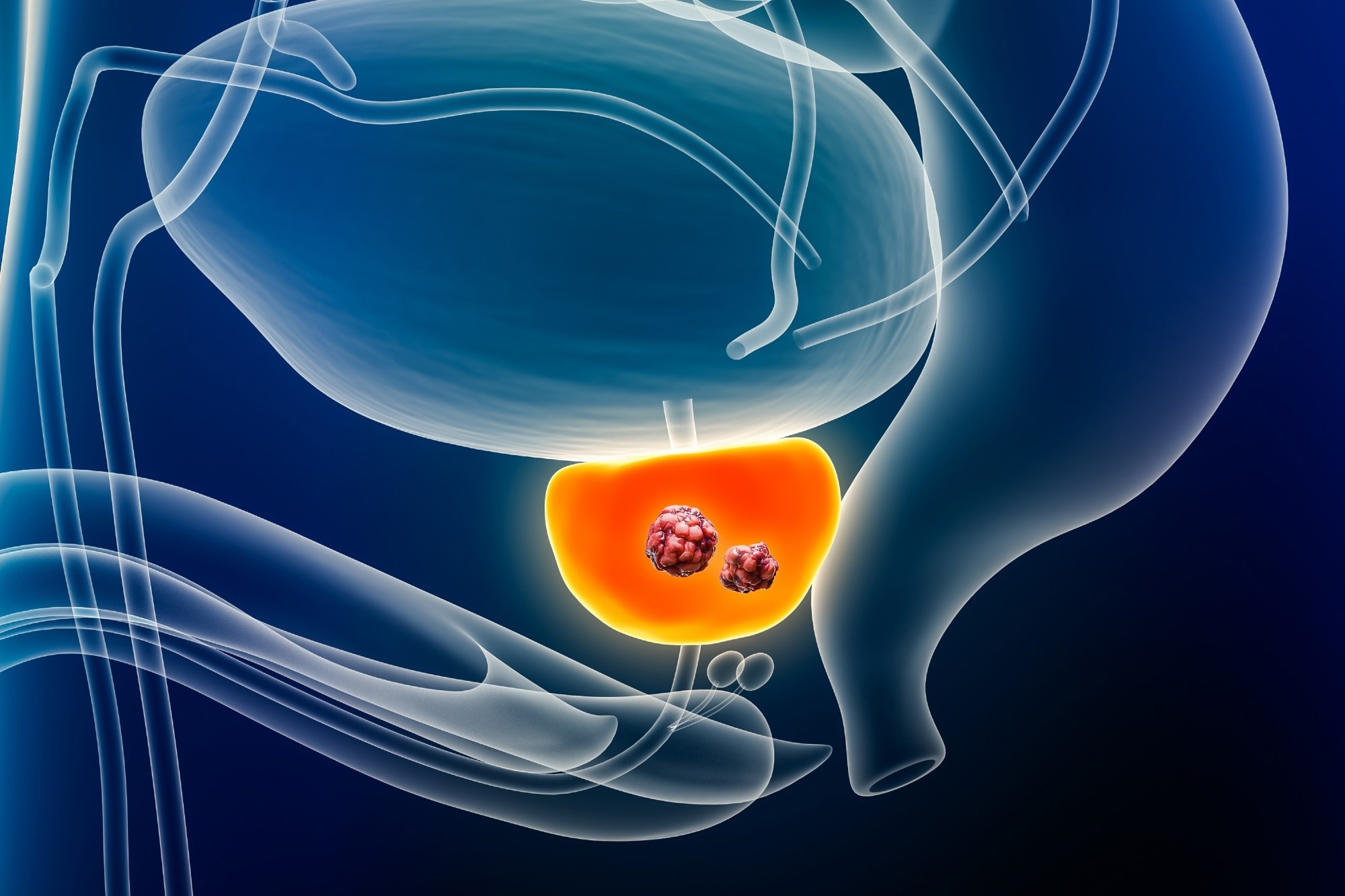
    In a recent phase I trial published in the journal Cell Reports Medicine, researchers from Belgium administered a dendritic cell (DC) vaccine targeting patient-specific neoantigens to patients with resected non-small cell lung cancer (NSCLC).

    They found that the vaccine was viable and led to limited toxicity and systemic T-cell responses, with 50% of patients experiencing disease recurrence during the study.

    Study: Neoantigen-targeted dendritic cell vaccination in lung cancer patients induces long-lived T cells exhibiting the full differentiation spectrum. Image Credit: PhotobyTawat/Shutterstock.comStudy: Neoantigen-targeted dendritic cell vaccination in lung cancer patients induces long-lived T cells exhibiting the full differentiation spectrum. Image Credit: PhotobyTawat/Shutterstock.com

    Background

    NSCLC, constituting over 80% of lung cancer cases, is primarily treated with surgical resection in early stages, yet recurrence rates remain high, with highly variable 5-year survival rates.

    Adjuvant chemotherapy offers modest survival benefits and often impacts the quality of life severely, emphasizing the need for more efficacious and tolerable adjuvant therapies. Recent phase 3 trials showed that immune checkpoint blockers (ICBs) benefit high-risk early-stage NSCLC, improving disease-free survival, but concerns exist over their toxicity.

    Neoantigen-targeting vaccines are extensively researched across different cancer stages, showing promising outcomes by inducing robust, high-affinity T-cell responses. Trials confirm their feasibility, safety, and potential clinical benefits, alone or combined with ICBs.

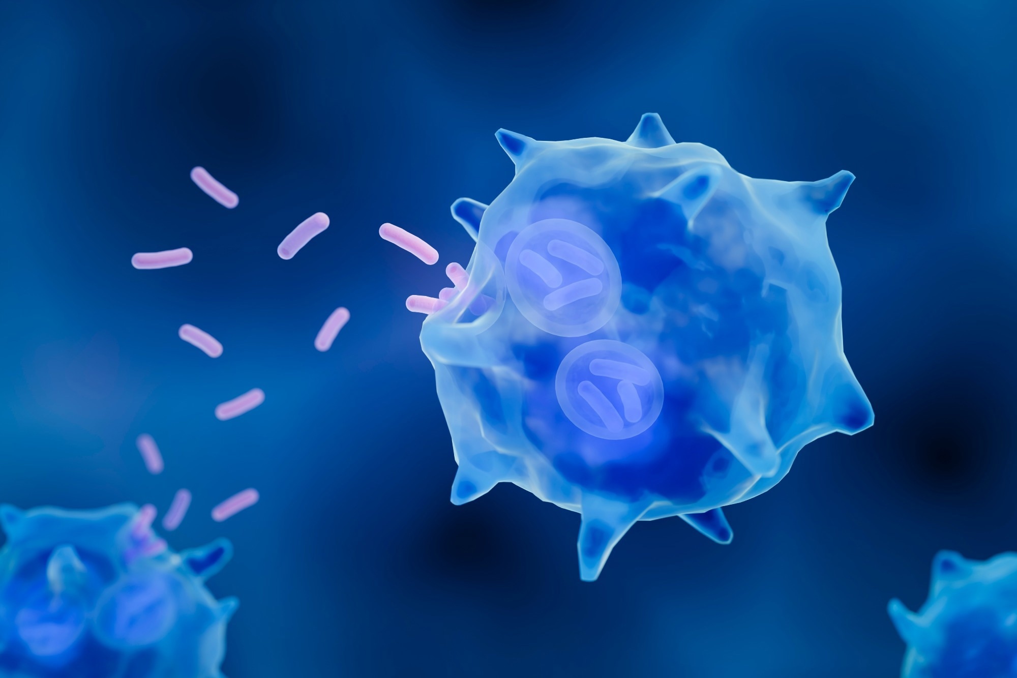
    DCs are highly efficient antigen-presenting cells, widely studied for cancer vaccination. However, their clinical efficacy is limited, partially due to their emphasis on tumor-associated antigens instead of mutanome-derived neoantigens.

    Therefore, in the present study, researchers investigated the safety and efficacy of an autologous DC vaccine targeting neoantigens for the treatment of resected NSCLC patients.

    About the study

    The study recruited ten resectable NSCLC patients without specific gene mutations in stages Ia3–IVb. Tumor material and peripheral blood samples were collected for whole-exome sequencing (WES) and ribonucleic acid (RNA) sequencing.

    A neoantigen identification pipeline was developed, prioritizing clonal variants with strong predicted binding to human leukocyte antigen (HLA) class I allotypes and RNA expression.

    Neoepitopes were selected based on their absence from the healthy human proteome and immunogenic potential. Mass spectrometry (MS)–based immuno-peptidomics, cell line engineering, and tumor-infiltrating lymphocyte (TIL) reactivity analyses were employed for neoantigen validation.

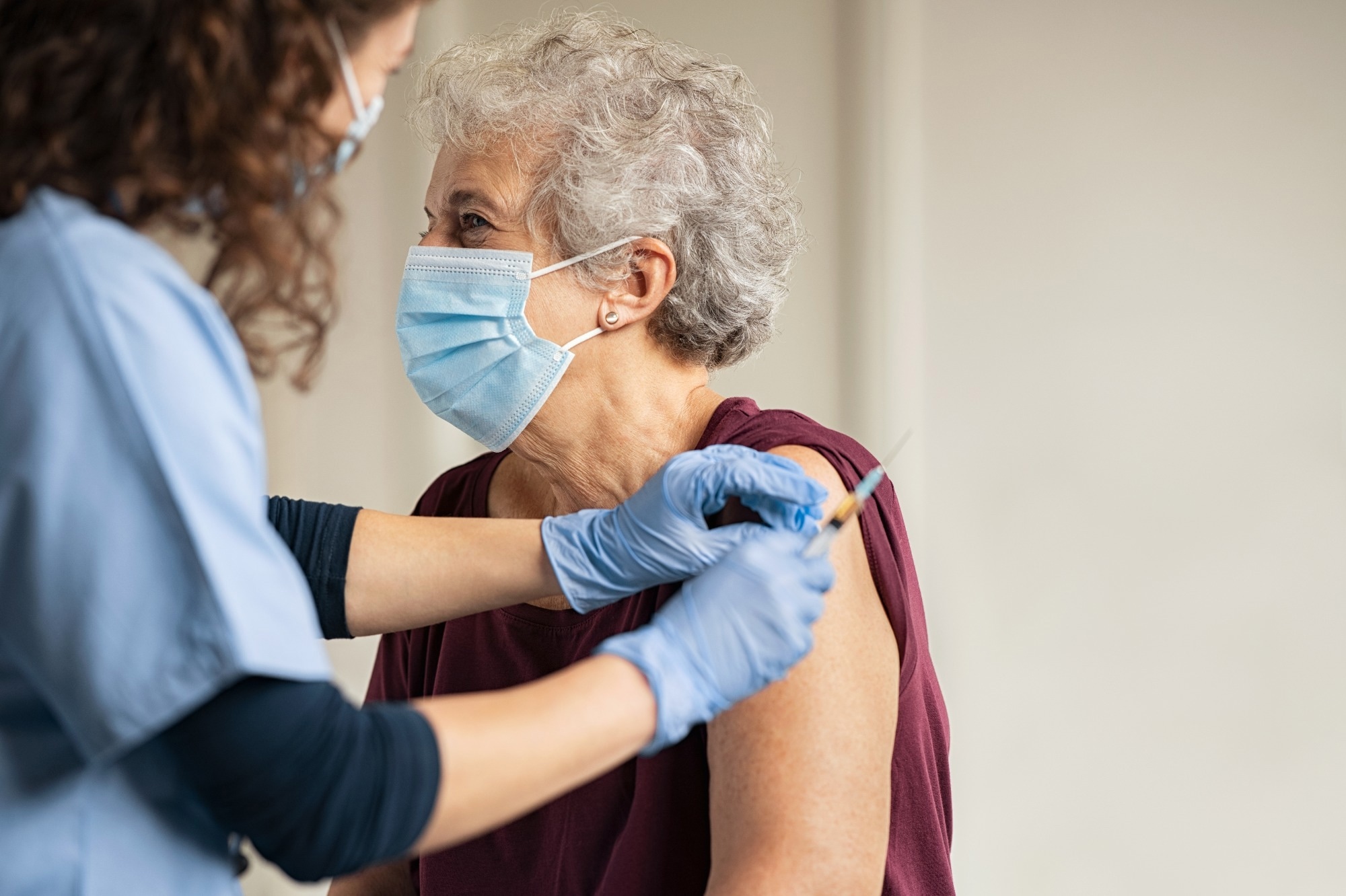
    Six patients received intravenous administrations of messenger RNA (mRNA)-loaded monocyte-derived DCs (Neo-mDCs) and were monitored for safety and clinical activity. The median time from surgery to the first vaccination was 198 days.

    Peripheral blood mononuclear cells (PBMCs) from patients receiving Neo-mDC treatment were analyzed for CD4+ and CD8+ T-cell responses against neoantigen 25-mers using interferon-gamma (IFN-γ) staining after in vitro stimulation.

    The correlation between T-cell responses and the detection of tandem protein in Neo-mDC batches was assessed. PBMCs were also analyzed to assess ex vivo T-cell frequency and differentiation phenotype.

    Individual clonotypes within neoantigen-specific T-cell populations were examined through single-cell analysis of the T-cell receptor (TCR) repertoire in tetramer-positive cells.

    Results and discussion

    Seven out of nine patients had five to six neoantigens selected for vaccination, with 0–56 potential neoepitopes identified per patient. Validation analyses confirmed that nine out of 33 selected neoantigens were naturally processed and presented on tumor HLA, supporting the reliability of the identification approach.

    Adverse events (AEs) were mild and self-limiting, with no grade 3–4 AEs reported. While three patients experienced disease recurrence, three patients remained free of disease recurrence during the follow-up period.

    Five out of six treated patients exhibited vaccine-induced T-cell responses, with 14 out of 33 neoantigens-inducing responses. Most responses were CD8+ T-cell-mediated and emerged after the first dose, persisting throughout treatment.

    Even at low doses, Neo-mDCs primed naive T-cells and/or expanded pre-existing T-cell responses.

    However, T-cell responses did not always correlate with detecting tandem protein in Neo-mDC batches. CD8+ T cell responses were highly specific for predicted neoepitopes, with no cross-reactivity observed toward wild-type epitopes. These results highlight the immunogenic potential of Neo-mDC vaccination in NSCLC patients.

    Post-vaccination, tetramer-positive T-cells expanded and were found to persist for ≥1.5 years. Analysis revealed naive, early, and late differentiated T-cell clusters with tetramer-positive cells across these states.

    These findings demonstrate the induction of diverse, long-lived neoantigen-specific T-cell responses by Neo-mDC vaccination in NSCLC patients.

    Further, the T-cells induced by vaccination included effector and long-lasting cells that could persist for several years. The T-cell populations generated by the vaccine were diverse and polyclonal, consisting of multiple clonotypes that exhibited a wide range of differentiation states.

    Overall, this first-in-human clinical trial provides valuable insights on using an autologous DC therapy delivering mRNA-encoded, patient-specific tumor neoantigens in NSCLC patients. However, it is limited by its small patient cohort and prolonged vaccine manufacturing time.

    Conclusion

    In conclusion, the study demonstrated neoantigen-targeted autologous DC vaccination’s safety, feasibility, and immunogenicity in surgically resected NSCLC patients.

    The findings highlight the potential of personalized immunotherapy strategies in combating NSCLC and possibly other malignancies to improve patient outcomes.

    [ad_2]

    Source link

  • New research pinpoints key pathways in prostate cancer’s vulnerability to ferroptosis

    New research pinpoints key pathways in prostate cancer’s vulnerability to ferroptosis

    [ad_1]

    In a recent review published in the journal Nature Reviews Urology, researchers examined the molecular mechanisms and metabolic processes that drive ferroptosis — a form of cell death that plays a significant role in prostate cancer. They also connected pathways involved in ferroptosis to the metabolic reprogramming that occurs in prostate cancer cells to highlight potential avenues of targeted therapeutic interventions.

    Perspective: Unlocking ferroptosis in prostate cancer — the road to novel therapies and imaging markers. Image Credit: MattL_Images / Shutterstock

    Perspective: Unlocking ferroptosis in prostate cancer — the road to novel therapies and imaging markers. Image Credit: MattL_Images / Shutterstock

    Background

    Although the five-year survival rate in cases of localized prostate cancer is very promising (greater than 99%), metastasis or the prostate cancer progressing to the castration-resistant form of prostate cancer reduces the five-year survival rate to between 30% and 40%. Furthermore, while treatment options such as radiotherapy, chemotherapy, immunotherapy, and second-generation androgen receptor-signaling inhibitors can be used to treat advanced prostate cancer, these therapies only increase the survival rate by two to three years. Understanding the underlying mechanisms of prostate cancer can help improve and initiate early treatment.

    Recent research has found that a pathway of regulated cell death, called ferroptosis, plays a significant role in the development of prostate cancer. Ferroptosis differs from the other forms of cell death, such as autophagy, apoptosis, and necrosis, in that it is iron-dependent and is driven by lipid peroxide build-up. Studies have found that the suppression of ferroptosis is linked to tumor pathogenesis, especially in prostate cancer.

    Ferroptosis

    Ferroptosis does not have the typical characteristics of apoptosis, such as the condensation of chromatin, apoptotic body formation, and cytoskeletal breakdown. Neither does it show the hallmarks of necrosis and autophagy, such as the swelling of organelles and the formation of the autophagosomes, respectively.

    During ferroptosis, the mitochondrial size and cristae of the cells reduce, and the membrane density increases. Additionally, the polyunsaturated fatty acids that are part of the phospholipid membrane form peroxides. The lipid peroxidation could be initiated due to oxidation from iron overload, the mitochondria, or the production of reactive oxygen species due to iron. The lipid peroxidation process results in widespread damage and oxidative injury, leading to cell death.

    Cells contain various intrinsic systems to circumvent lipid peroxidation and ferroptosis. The classical method involves the use of glutathione and glutathione peroxidase 4 to decrease the levels of lipid hydroperoxides, preserving the integrity of the phospholipid bilayer and preventing ferroptosis. Inhibition of ferroptosis can also occur through ferroptosis suppressor protein 1 or dihydroorotate dehydrogenase.

    Ferroptosis and cancer

    Given the association between ferroptosis and polyunsaturated fatty acids, cancer cells, which undergo substantial metabolic reprogramming and produce reactive oxygen species, are especially susceptible to ferroptosis. Cells in malignant tumors have higher energy and iron demands, which increases their susceptibility to ferroptosis. Prostate cancer cells depend on lipid metabolism for their high energy requirements, which causes the fatty acid metabolism in prostate cancer cells to be dysregulated.

    Furthermore, factors such as lipid metabolism gene upregulation, rewiring of the oxidative phosphorylation metabolism, and increased tricarboxylic acid flux have been observed in both early and late-stage prostate cancer cells. These processes could increase the intracellular reactive oxygen species burden, promote lipid peroxidation, and cause perturbations in iron homeostasis.

    The review discussed various mechanisms through which the susceptibility of cancer cells to ferroptosis could be exploited as potential treatment avenues for advanced cancers. Targeting the defense mechanisms that inhibit ferroptosis is a promising approach. Studies have suggested that targeting the glutathione peroxidase 4 inhibition mechanism could induce ferroptosis in cancer cells that do not respond to other treatment options.

    Research also indicated that dihydroorotate dehydrogenase was not the primary ferroptosis inhibitor in cancer cells, and therefore, targeting dihydroorotate dehydrogenase might not be as effective as deletion of ferroptosis suppressor protein 1.

    Furthermore, these studies highlighted the need to thoroughly understand the pitfalls and benefits of the various mechanisms to induce ferroptosis. Knockout studies in murine models revealed that glutathione peroxidase 4 was essential in various other processes and required for survival, while knocking out ferroptosis suppressor protein 1 resulted in no developmental changes, suggesting the latter to be a preferable method to induce ferroptosis.

    This comprehensive review provided a detailed discussion of the various metabolic processes that could be exploited to make cancer cells vulnerable to ferroptosis. These methods included modulating the balance between mono- and polyunsaturated fatty acids, de-novo lipogenesis, de-novo synthesis of polyunsaturated fatty acids, and β oxidation. The researchers also expanded on the role of iron, cystine, glutamate, and glutathione metabolism in ferroptosis.

    Conclusions

    To summarize, the review provided an in-depth view of the regulated cell death process of ferroptosis, the factors that make cancer cells susceptible to ferroptosis, and its importance in prostate cancer therapy. They discussed the pathways through which ferroptosis is suppressed in cancer cells and the metabolic mechanisms that must be targeted to induce ferroptosis in prostate cancer cells selectively.

    Journal reference:

    • Anh, H., Dominic, A., Lujan, F. E., Senthilkumar, S., Bhattacharya, P. K., Frigo, D. E., & Subramani, E. (2024). Unlocking ferroptosis in prostate cancer — the road to novel therapies and imaging markers. Nature Reviews Urology. DOI: 10.1038/s41585024008699, https://www.nature.com/articles/s41585-024-00869-9

    [ad_2]

    Source link

  • AI techniques massively accelerate the search for Parkinson’s disease treatments

    AI techniques massively accelerate the search for Parkinson’s disease treatments

    [ad_1]

    Researchers have used artificial intelligence techniques to massively accelerate the search for Parkinson’s disease treatments.

    The researchers, from the University of Cambridge, designed and used an AI-based strategy to identify compounds that block the clumping, or aggregation, of alpha-synuclein, the protein that characterises Parkinson’s.

    The team used machine learning techniques to quickly screen a chemical library containing millions of entries, and identified five highly potent compounds for further investigation.

    Parkinson’s affects more than six million people worldwide, with that number projected to triple by 2040. No disease-modifying treatments for the condition are currently available. The process of screening large chemical libraries for drug candidates – which needs to happen well before potential treatments can be tested on patients – is enormously time-consuming and expensive, and often unsuccessful.

    Using machine learning, the researchers were able to speed up the initial screening process by ten-fold, and reduce the cost by a thousand-fold, which could mean that potential treatments for Parkinson’s reach patients much faster. The results are reported in the journal Nature Chemical Biology.

    Parkinson’s is the fastest-growing neurological condition worldwide. In the UK, one in 37 people alive today will be diagnosed with Parkinson’s in their lifetime. In addition to motor symptoms, Parkinson’s can also affect the gastrointestinal system, nervous system, sleeping patterns, mood and cognition, and can contribute to a reduced quality of life and significant disability.

    Proteins are responsible for important cell processes, but when people have Parkinson’s, these proteins go rogue and cause the death of nerve cells. When proteins misfold, they can form abnormal clusters called Lewy bodies, which build up within brain cells stopping them from functioning properly.

    “One route to search for potential treatments for Parkinson’s requires the identification of small molecules that can inhibit the aggregation of alpha-synuclein, which is a protein closely associated with the disease,” said Professor Michele Vendruscolo from the Yusuf Hamied Department of Chemistry, who led the research. “But this is an extremely time-consuming process – just identifying a lead candidate for further testing can take months or even years.”

    While there are currently clinical trials for Parkinson’s currently underway, no disease-modifying drug has been approved, reflecting the inability to directly target the molecular species that cause the disease.

    This has been a major obstacle in Parkinson’s research, because of the lack of methods to identify the correct molecular targets and engage with them. This technological gap has severely hampered the development of effective treatments.

    The Cambridge team developed a machine learning method in which chemical libraries containing millions of compounds are screened to identify small molecules that bind to the amyloid aggregates and block their proliferation.

    A small number of top-ranking compounds were then tested experimentally to select the most potent inhibitors of aggregation. The information gained from these experimental assays was fed back into the machine learning model in an iterative manner, so that after few iterations, highly potent compounds were identified.

    Instead of screening experimentally, we screen computationally. By using the knowledge we gained from the initial screening with our machine learning model, we were able to train the model to identify the specific regions on these small molecules responsible for binding, then we can re-screen and find more potent molecules.”


    Professor Michele Vendruscolo, Co-Director of the Centre for Misfolding Diseases

    Using this method, the Cambridge team developed compounds to target pockets on the surfaces of the aggregates, which are responsible for the exponential proliferation of the aggregates themselves. These compounds are hundreds of times more potent, and far cheaper to develop, than previously reported ones.

    “Machine learning is having a real impact on the drug discovery process – it’s speeding up the whole process of identifying the most promising candidates,” said Vendruscolo. “For us this means we can start work on multiple drug discovery programmes – instead of just one. So much is possible due to the massive reduction in both time and cost – it’s an exciting time.”

    The research was conducted in the Chemistry of Health Laboratory in Cambridge, which was established with the support of the UK Research Partnership Investment Fund (UKRPIF) to promote the translation of academic research into clinical programmes.

    Source:

    Journal reference:

    Horne, R. I., et al. (2024). Discovery of potent inhibitors of α-synuclein aggregation using structure-based iterative learning. Nature Chemical Biology. doi.org/10.1038/s41589-024-01580-x.

    [ad_2]

    Source link

  • Exploring the versatile roles of tissue macrophages beyond immune defense

    Exploring the versatile roles of tissue macrophages beyond immune defense

    [ad_1]

    A recent study published in Science Immunology summarized the role of resident tissue macrophages (RTMs) in homeostasis and disease.

    Study: Resident tissue macrophages: Key coordinators of tissue homeostasis beyond immunity. Image Credit: ART-ur/Shutterstock.comStudy: Resident tissue macrophages: Key coordinators of tissue homeostasis beyond immunity. Image Credit: ART-ur/Shutterstock.com

    Background

    Macrophages are evolutionarily conserved phagocytes ubiquitously present in almost all organs and tissues. It is recognized that the umbrella term macrophage comprises highly heterogeneous cells with diverse functions and roles.

    RTMs are stable, long-lived subpopulations in different organs and tissues and have been linked to innate immunity and the pathogenesis of chronic inflammatory diseases. However, RTMs have broader functions beyond immunity.

    Recently, exploring RTM subsets to functional, developmental, and spatial levels has become feasible, helping identify mechanisms of tissue homeostasis.

    Notwithstanding these advances, substantial knowledge gaps remain. In the present review, researchers provided insights into conditions impacting RTM identity, division of labor among RTM subsets, and RTM dysfunction in disease.

    Tissue microenvironment impacts RTM development

    RTMs originate from embryonic progenitors or hematopoietic stem cell (HSC)-derived monocytes. The local microenvironment influences the trajectories of RTM differentiation upon seeding a tissue. In homeostasis, the local environmental cues shape RTM cell identity in a tissue-specific manner.

    Further, the phenotypic and functional convergence of HSC-derived monocytes towards a tissue-specific RTM program is driven by the local environment.

    However, inflammation or disease markedly impacts their differentiation. During such disturbance, the differentiation of HSC-derived monocytes skews toward pro-reparative, tumor-supportive, or pro-inflammatory phenotypes, differing from that of steady-state RTMs.

    These inflammation-associated macrophages (iMacs) are short-lived, and upon resolution (of the disturbance), the tissue transitions to a distinct state, i.e., inflammation aftermath.

    There may be permanent changes in the original homeostatic distribution and the composition of environmental factors. This was demonstrated in white adipose tissue, where HSC-derived RTMs acquired a more inflammatory phenotype following the resolution of chronic inflammation.

    This post-inflammation scar led to HSC-derived RTMs being unable to differentiate into their original cellular state.

    Coexistence of RTM subsets within tissues

    Historically, it has been believed that organs and tissues are populated by unique tissue-specific RTMs during homeostasis, such as Langerhans cells in the skin, alveolar macrophages (AMs) in the lungs, Kupffer cells in the liver, and microglia in the brain.

    However, a seminal work from 2010 showed that embryonic yolk sac progenitors, not monocytes, give rise to microglia.

    This was also instrumental in revealing the embryonic origin of other RTMs. Studies have demonstrated that two distinct conserved RTM subsets populate most tissues in the interstitial space.

    The authors term these RTM subsets as perivascular macrophages (PVMs). The PVMs precede with the name of the organ/tissue of residence.

    Of the conserved PVM subsets, T cell immunoglobulin and mucin domain containing 4 (TIM4+) PVMs emerge during embryogenesis in multiple organs and are characterized by low levels of major histocompatibility complex II (MHCII) and high levels of TIM4, folate receptor beta (FOLR2), lymphatic vessel endothelial hyaluronan receptor 1 (LYVE1), and cluster of differentiation 206 (CD206).

    By contrast, MHCII+ PVMs emerge from HSC-derived monocytes and are characterized by low/intermediate levels of FOLR2 and LYVE1 and high levels of CD206 and MHCII.

    While some studies have observed a C-C motif chemokine receptor 2 (CCR2+) PVM subset, they are likely to be recent organ immigrants. Although some organs have unique tissue-specific subsets of RTMs, almost all organs share these two conserved PVMs.

    Division of labor among RTMs

    Microglia are the only brain RTM subset in contact with neurons. Several fundamental microglial functions beyond immunity have been uncovered more recently. Animal studies have shown that microglia are essential in neuronal development and fitness.

    Microglia secrete growth factors critical for synapse formation. Additionally, they survey the brain microenvironment and modulate neuronal activity through synaptic engulfment and pruning.

    Recent studies have revealed the presence of PVMs in perivascular spaces of the central nervous system (CNS). Further, these PVMs regulate cerebrospinal fluid (CSF) dynamics, and TIM4+ PVMs in the brain facilitate proper extracellular matrix (ECM) dynamics.

    This idea was corroborated by the findings of abnormal ECM deposition and deterioration of CSF flow dynamics in aged mice, which are linked to a smaller ratio of brain TIM4+-to-MHCII+ PVMs.

    The distinct locations of lung PVMs indicate they have specialized roles. For instance, lung MHCII+ PVMs may regulate neuronal interaction with stromal cells, whereas lung TIM4+ PVMs contribute to lung homeostasis.

    Besides, lung TIM4+ PVMs may be involved in wound healing, while the MHCII+ counterparts may be involved in antigen presentation and immune activation.

    Heart MHCII+ and TIM4+ PVMs produce growth factors to support proper cellular functions and adjust to physiologic demands. Cardiac PVMs are in close contact with cardiomyocytes and participate in mutual electric conduction, supporting normal cardiac contractions.

    Gut muscularis MHCII+ and TIM4+ PVMs are close to blood vessels, myenteric plexus, and submucosal plexus. Gut MHCII+ PVMs are closely associated with neuronal bodies of the enteric nervous system.

    Mechanistically, gut muscularis PVMs secrete bone morphogenetic protein 2 (BMP2) to regulate enteric neurons expressing the BMP2 receptor. Besides, they regulate gastrointestinal motility independent of the enteric nervous system. Recent studies suggest that gut PVMs promote neuroprotection and limit neuronal cell death.

    RTM dysregulation and disease

    It is established that HSC-derived iMacs are linked to chronic inflammatory diseases. This chronicity is thought to be due to ongoing inflammation leading to tissue function loss.

    Nevertheless, how dysfunction or deviation of RTMs’ core homeostatic functions cause disease remains less studied. Usually, deviation is required for proper tissue repair.

    However, it is not clear how long-term and persistent deviation affects tissue physiology and disease severity. Pulmonary alveolar proteinosis is caused by AM dysfunction, characterized by protein and surfactant accumulation in the lung alveolar space, limiting proper gas exchange and increasing susceptibility to infections.

    This can occur due to mutations in the granulocyte-macrophage colony-stimulating factor (GM-CSF), autoantibodies against GM-CSF, or silica inhalation. Further, the absence or dysfunction of lung TIM4+ PVMs can result in increased fibrosis and loss of tissue function.

    Likewise, dysregulation of heart TIM4+ PVMs exacerbates fibrosis following cardiac infarction. Loss of RTM’s core homeostatic functions may impact cancer development.

    A recent study on breast cancer patients showed that those harboring tumors with increased breast TIM4+ PVMs had improved survival rates and T cell priming against the tumor.

    This suggested that enhancing the TIM4+ PVMs’ homeostatic functions while inhibiting the activity of HSC-derived tumor-related macrophages can be effective for treatment.

    Further, disruption of ECM remodeling in brain PVMs is associated with aging and Alzheimer’s disease (AD).

    Concluding remarks

    Taken together, ontogeny and local environmental cues shape the phenotype and heterogeneity of RTMs.

    There is a strong division of labor among RTM subpopulations. The study proposed a unifying nomenclature for the two conserved RTM subpopulations and explored the roles of several unique tissue-specific RTM subsets in homeostasis and disease.

    Nevertheless, further studies are required to delineate how RTM dysfunction leads to chronic inflammatory diseases fully.

    [ad_2]

    Source link

  • Vaccines targeting chronic diseases show promise in combatting age-related conditions

    Vaccines targeting chronic diseases show promise in combatting age-related conditions

    [ad_1]

    In a recent review published in Nature Aging, researchers explored vaccine-based therapeutics for age-related disorders.

    Study: Targeting aging and age-related diseases with vaccines. Image Credit: Ground Picture/Shutterstock.comStudy: Targeting aging and age-related diseases with vaccines. Image Credit: Ground Picture/Shutterstock.com

    Background

    Aging is a primary risk factor for chronic illnesses, marked by reduced physiological capabilities. Cell senescence, genomic instability, stem cell fatigue, and mitochondrial failure are characteristics.

    Aging also increases the risk of chronic illnesses such as Alzheimer’s disease, atherosclerosis, osteoarthritis, type 2 diabetes, chronic obstructive pulmonary disease (COPD), and cancer. Healthy habits like calorie control and regular physical exercise help prevent age-related disorders.

    However, small-molecule therapies have limits, and vaccines provide a potential technique to target specific antigens to generate immune responses.

    About the review

    In the present review, researchers present new developments in vaccines using senescent cells to target the etiological agents of aging and related diseases.

    Immunology of senolytic vaccines that target aging

    Vaccines stimulate the innate immunological system, making it rapidly respond to infection. This reaction activates adaptive immune cells, causing humoral antibodies to manufacture antibodies and cell-mediated immunity to attack infected cells. Vaccinations lead to pathogen-targeted memory cell development to accelerate response to reinfections.
    Vaccines targeting microbes, cell populations, or chemicals can prevent and treat disease development. Senolytic vaccines, which target senescent cells, have been demonstrated to reduce arterial plaque development.

    These vaccination techniques provide new pathways for managing age-associated disorders, with advantages such as fewer injections, increased patient adherence, cost-effectiveness, and improved targeting efficiency.

    Senolytic vaccinations target senescent cells by targeting chemicals on their surfaces, using peptide-based platforms to eradicate them through complement-dependent cytotoxicity (CDC) or natural killer (NK) cell-mediated and antibody-dependent cell-mediated cytotoxicity (ADCC).

    Membrane proteins overexpressed in senescent cells are used as vaccine antigens or immune cell targets to stimulate the development of specific antibodies.

    Tumor-associated antigens (TAAs) and tumor-specific antigens (TSAs) trigger a cluster of differentiation 4-expressing helper T cells, CD8+ cytotoxic T cells, and B cells to kill cancer cells.

    Cancer vaccines use cell-based, virus-based, peptide-based, and nucleic acid-based platforms to stimulate adaptive immunity.

    Research on vaccine-based strategies for age-related disorders

    Alzheimer’s disease (AD) vaccinations attempt to lower the brain’s amyloid-beta (Aβ) and tau protein levels by stimulating the adaptive immune system to produce antibodies. Peptides with neuroprotective properties are potentially possible therapies.

    Vaccines like ACI-24, UB-311, AV-1959D, ABvac40, ACI-35, AADvac1, and GV1001 target Aβ and tau proteins in innate immune cells. ACI-24 targets Aβ1-15 liposomes, UB-311 targets Aβ protein, AV-1959D targets deoxyribonucleic acid (DNA), ABvac40 targets Aβ protein, ACI-35 targets tau protein accumulation, and AADvac1 and GV1001 demonstrate long-term safety, tolerance, and immunogenicity.

    Type 2 diabetes vaccinations target molecules like dipeptidyl-peptidase 4 (DPP4) and interleukin-1 beta (IL-1β) to restore levels and prevent problems.

    The vaccines use peptide sequences to produce anti-DPP4 antibodies, increase glucose tolerance, restore beta cell mass, and reduce IL-1β production. They also couple prorenin epitopes with keyhole limpet hemocyanin (KLH), improving retinal blood circulation, decreasing microglia activation, and reducing gliosis and vascular leakage in diabetic rats.

    Vaccines for age-related vascular diseases such as hypertension, abdominal aortic aneurysm (AAA), and atherosclerosis target molecules that regulate blood pressure, cholesterol, and blood vessel function. PMD-2850, PMD-3117, CYT006-AngQb, AT1R, ATRQβ-001, and ADR-004 inhibit angiopoietin 1 (Ang I) and age-related accumulation in hypertensive rats.

    Vaccines containing KLH and Ang II decreased macrophage aggregation in the AAA wall, suppressed tumor necrosis factor (TNF) expression, and protected against elastic fiber injury.

    The nerve growth factor (NGF) protein is an osteoarthritis target. Researchers covalently linked recombinant NGF proteins to virus-like particles (VLPs) derived from a cucumber mosaic virus with tetanus toxoid epitopes.

    Vaccinated mice developed measurable antibodies against NGF and showed reversed pain behavior. ADAM metallopeptidase domain 12 (ADAM12) and GLI family zinc finger 1 (GLI1) levels are associated with fibrosis. Lentiviral vectors that encode these proteins decrease fibrotic activity in mice.

    Proprotein convertase subtilisin/kexin type 9 (PCSK9) can treat fibrosis. PCSK9Qβ-003, an epitope peptide, enhanced kidney fibrosis, and increased fatty acid beta-oxidation-associated variables among Ldlr+/− murine animals. The vaccine can treat kidney fibrosis, hypercholesterolemia, and atherosclerosis.

    Conclusions

    Based on the review findings, senescent cells play a significant role in age-related chronic disorders and demonstrate immunogenicity, making them candidates for targeted vaccine elimination.

    However, vaccinations can cause adverse effects such as injection site redness, discomfort, fever, and headaches. Continuous evaluation of vaccination safety is required to comprehend long-term adverse effects.  

    Vaccines targeting key mediators in age-related disorders have seldom proven efficacy in slowing disease development. Researchers must select antigens from key disease drivers or investigate epitopes generating robust immune responses to boost vaccination effectiveness.

    Combinatorial therapy incorporating vaccinations and immunomodulatory medications may be more effective.

    [ad_2]

    Source link

  • Genetically engineering skin bacteria to fight cancer

    Genetically engineering skin bacteria to fight cancer

    [ad_1]

    While studying a type of bacteria that lives on the healthy skin of every human being, researchers from Stanford Medicine and a colleague may have stumbled on a powerful new way to fight cancer.

    After genetically engineering the bacteria, called Staphylococcus epidermidis, to produce a tumor antigen (a protein unique to the tumor that’s capable of stimulating the immune system), they applied the live bacteria onto the fur of mice with cancer. The resulting immune response was strong enough to kill even an aggressive type of metastatic skin cancer, without causing inflammation.

    It seemed almost like magic. These mice had very aggressive tumors growing on their flank, and we gave them a gentle treatment where we simply took a swab of bacteria and rubbed it on the fur of their heads.”


    Michael Fischbach, PhD, associate professor of bioengineering

    Their research was published online April 13 in Science. Fischbach is the senior author, and Yiyin Erin Chen, MD, PhD, a former postdoctoral scholar at Stanford Medicine, now an assistant professor of biology at the Massachusetts Institute of Technology, is the lead author.

    Skin colonizers

    Millions of bacteria, fungi and viruses live on the surface of healthy skin. These friendly colonists play a crucial role in maintaining the skin barrier and preventing infection, but there are many unknowns about how the skin microbiota interacts with the host immune system. For instance, unique among colonizing bacteria, staph epidermidis triggers the production of potent immune cells called CD8 T cells — the “killer” cells responsible for battling severe infections or cancer.

    The researchers showed that by inserting a tumor antigen into staph epidermidis, they could trick the mouse’s immune system into producing CD8 T cells targeting the chosen antigen. These cells traveled throughout the mice and rapidly proliferated when they encountered a matching tumor, drastically slowing tumor growth or extinguishing the tumors altogether.

    “Watching those tumors disappear — especially at a site distant from where we applied the bacteria — was shocking,” Fischbach said. “It took us a while to believe it was happening.”

    The mystery of the T cells that do nothing

    Fischbach and his team didn’t start out trying to fight cancer. They wanted to answer a much more basic question: Why would a host organism waste energy making T cells designed to attack helpful colonizing bacteria? Especially as these T cells are “antigen-specific,” meaning each T cell has a homing receptor that matches a single fragment of the bacterium that activated it.

    Even stranger, the CD8 T cells induced by naturally occurring staph epidermidis don’t cause inflammation; in fact, they appear to do nothing at all. Most scientists thought colonist-induced T cells must be fundamentally different from regular T cells, Fischbach said, because instead of traveling throughout the body to hunt for their target, they seemed to stay right below the skin surface, somehow programmed to keep the peace between bacteria and host.

    To test whether these colonist-induced CD8 T cells could behave like regular killer T cells, the researchers engineered a strain of staph epidermidis to produce a different antigen — one that would generate T cells specific for a commonly studied tumor model in mice.

    They genetically grafted a small fragment of DNA encoding part of a protein called ovalbumin onto the surface of staph epidermidis. They chose ovalbumin because it’s been engineered into many commonly studied mouse tumor lines, including a type of aggressive melanoma, and therefore can act as a tumor antigen in multiple types of cancer. 

    The power of tumor-specific T cells

    Next, the scientists applied the genetically engineered bacteria to healthy mice. Because staph epidermidis is an efficient skin colonizer, they didn’t need to clean or shave the animals’ fur, but simply rubbed the bacteria on their heads. As expected, colonization didn’t cause any inflammation or infection.

    Source:

    Journal reference:

    Chen, Y. E., et al. (2024) Engineered skin bacteria induce antitumor T cell responses against melanoma. Science. doi.org/10.1126/science.abp9563.

    [ad_2]

    Source link

  • Researchers identify a novel mechanism connected to the early stages of Alzheimer’s disease

    Researchers identify a novel mechanism connected to the early stages of Alzheimer’s disease

    [ad_1]

    Alzheimer’s disease (AD) remains one of the most challenging and prevalent neurodegenerative disorders, affecting millions of individuals worldwide. In a new study published in Developmental Cell, researchers from the lab of Wim Annaert (VIB-KU Leuven) have identified a novel mechanism potentially connected to the early stages of AD. They demonstrated that a fragment of the amyloid precursor protein (APP), called APP-CTF, disrupts communication between cellular compartments crucial for calcium storage and waste disposal, which could be an early event preceding neuronal cell death. These findings, with potential implications for the development of new AD treatments, suggest that preventing APP-CTF accumulation needs to be taken into account to develop more effective treatments.

    Alzheimer’s disease is characterized by the progressive loss of cognitive function, memory impairment, and behavioral changes. One of the visible features in the brains of people with Alzheimer’s disease is the formation of amyloid plaques – clumps of β-amyloid (Aβ) peptides, which are degraded products of amyloid precursor protein (APP). These Aβ-fragments accumulate in neurons early in the disease, even before cognitive decline is observed.

    New research, however, suggests that there might even be earlier events happening in the AD brain before plaque formation and that the APP protein plays a role in these early stages. The mechanism behind this remained a mystery until now.

    In their latest study, the lab of Wim Annaert at the VIB-KU Leuven Center for Brain & Disease Research identified a mechanism explaining how APP may contribute to these early stages of AD. This discovery could lead to a new direction in AD research and treatment approaches.

    Disrupting cellular communication

    APP is found in the cell membranes of brain cells. The brain constantly produces new APP molecules while breaking down and removing old ones. This process involves enzymatic scissors, with gamma-secretase being the final one that generates the well-known and well-studied Aβ peptides in AD.

    For a long time, it was thought that blocking gamma-secretase would be the logical step to prevent the production of toxic Aβ fragments. However, this leads to the accumulation of their precursor, the APP-C-Terminal Fragments, or APP-CTFs. Now, the researchers have discovered that these fragments are also toxic to neurons. They appear to accumulate between the endoplasmic reticulum (ER), the compartment that is crucial for lipid synthesis and calcium storage, and the lysosomes, the so-called ‘waste bins’ of neurons, which are critical for degrading the cell’s waste products.

    “By doing so, APP-CTFs disrupt the delicate balance of calcium within lysosomes,” explains Dr. Marine Bretou, first author of the study. “This disruption triggers a cascade of events. The ER can no longer effectively refill lysosomes with calcium, leading to a buildup of cholesterol and a decline in their ability to break down cellular waste. This results in the collapse of the entire endolysosomal system, a crucial pathway for maintaining healthy neurons.”

    The new study further supports that the APP-CTFs resulting from suppressing gamma-secretase might actually be the culprit behind endolysosomal dysfunction, as observed in the very early stages of AD.

    A paradigm shift in understanding the early stages of AD pathogenesis

    This research significantly advances our comprehension of the potential causes of disease in the early stages of AD. A remarkable outcome of this study is that these early stages could be caused by another fragment of the same APP molecule rather than Aβ. This has significant implications for the current therapeutic approaches that aim to clear the AD brain from amyloid plaques, as they tend to ignore the toxic effects of other fragments. Other attempts focus on tau proteins or neuroinflammation, which are other hallmarks of AD progression that target later events. However, early intervention is likely the key to stopping or even preventing AD.

    “The failure of clinical trials using gamma-secretase inhibitors may be explained by the fact that we were focusing on only one culprit and at a too late stage in the disease,” explains Prof. Wim Annaert, lead author of the study. “Our research findings suggest that gamma-secretase modulators, which can help promote clearance of toxic APP-CTFs without blocking the enzyme completely, may be a more relevant target for early intervention in AD. The key might be finding the right balance between APP-CTF clearance and plaque prevention.”

    Looking ahead, the scientists are joining efforts with colleagues to develop these modulators based on these novel insights and will continue exploring cellular homeostasis in the early stages of AD.

    Source:

    Journal reference:

    Bretou, M., et al. (2024) Accumulation of APP C-terminal fragments causes endolysosomal dysfunction through the dysregulation of late endosome to lysosome-ER contact sites. Developmental Cell. doi.org/10.1016/j.devcel.2024.03.030.

    [ad_2]

    Source link

  • Study reveals how SARS-CoV-2 hijacks lung cells to drive COVID-19 severity

    Study reveals how SARS-CoV-2 hijacks lung cells to drive COVID-19 severity

    [ad_1]

    In a recent study published in the Journal of Experimental Medicine, researchers identified the cellular tropism and transcriptome consequences of severe acute respiratory syndrome coronavirus 2 (SARS-CoV-2) by infecting human lung tissue and using single-cell ribonucleic acid sequencing (scRNA-seq) to rebuild the transcriptional program in “infection pseudotime” for distinct lung cell types.

    Lower respiratory infections, such as coronavirus disease 2019 (COVID-19), are a leading cause of death worldwide, producing pneumonia and acute respiratory distress syndrome. Understanding their early phases is difficult. Researchers used classical histopathological approaches and single-cell multi-omic profiling to infer early phases in human pathogenesis from lung lavage, biopsy, or autopsy materials. These approaches reveal a thorough picture of COVID-19 pneumonia at unparalleled cellular and molecular resolution, implying infection models including alveolar epithelium, capillaries, macrophages, and myeloid cells.

    Study: Interstitial macrophages are a focus of viral takeover and inflammation in COVID-19 initiation in human lung. Image Credit: Dotted Yeti / ShutterstockStudy: Interstitial macrophages are a focus of viral takeover and inflammation in COVID-19 initiation in human lung. Image Credit: Dotted Yeti / Shutterstock

    About the study

    In the present study, researchers developed an experimental COVID-19 model to investigate early molecular processes and pathogenic mechanisms of SARS-CoV-2 infection at the cellular level in native tissues of the human lung.

    The researchers established SARS-CoV-2’s cellular tropism and its unique and dynamic impacts on host cellular gene expression in specific types of lung cells. Prominent targets were lung-resident macrophages, of which one SARS-CoV-2 takes over transcriptomes, inducing a targeted host interferon (IFN) antiviral program, and several chemokines and pro-fibrotic and pro-inflammatory and cytokines signaling to various structural and immunological cells of the lung.

    To determine the early stages of COVID-19 in human lungs, the researchers sliced lung tissue obtained from surgical specimens or organ donor individuals into thick sections and used them for tissue culture analysis. Subsequently, they exposed the tissues to the SARS-CoV-2 USA-WA1 2020 strain at 1.0 multiplicity of infection (MOI) for two hours before allowing the SARS-CoV-2 infection to continue for two to three days. They performed a plaque test on culture supernatants.

    The researchers separated the slices and examined them by scRNA-seq to evaluate host and viral genetic expression during the SARS-CoV-2 infection. They also examined the viral RNA molecules’ junctional structure and processing by analyzing the scRNA-seq dataset with the SICILIAN framework. They used molecular atlas markers to distinguish lung cell types in healthy lung slices and measure viral RNA levels in infected cells.

    The team performed multiplexed single-molecule fluorescence in situ hybridization (smFISH) to confirm lung cell tropism findings and show infected cells. They used single-cell gene expression patterns to identify cellular targets for inflammatory and pro-fibrotic signals elicited by the SARS-CoV-2 infection of a-IMs. They devised a technique for purifying macrophage populations from human lungs with a SARS-CoV-2 spike (S) protein-pseudotyped lentivirus (lenti-S-NLuc-tdT) to investigate lung macrophage entrance routes.

    The researchers productively infected human lung slices cultivated ex vivo with SARS-CoV-2, with production rising between 24 and 72 hours of culture. They heat-inactivated, ultraviolet (UV)-treated, or administered 10.0 µM remdesivir, an RNA-dependent RNA polymerase inhibitor used as a COVID-19 therapeutic, to prevent viral stock infection.

    Results

    The analysis showed that SARS-CoV-2 preferentially infects active interstitial macrophages (IMs), which can amass hundreds of SARS-CoV-2 RNA molecules, comprising >60% of the cell transcriptome and producing dense viral RNA bodies. Infected alveolar macrophages (AMs) exhibit no severe reactions, with spike (S) protein-dependent viral entrance into AMs utilizing angiotensin-converting enzyme 2 (ACE2) and the cluster of differentiation 169 (CD169) and IM entry via CD209.

    They found canonical sub-genomic junctions between the unusual sequence reads beyond their 39 terminal regions, indicating canonical-type SARS-CoV-2 messenger RNA (mRNA) production in the pulmonary cultures. They also found hundreds of new subgenomic junctions, showing a wide range of non-canonical and canonical sub-genomic SARS-CoV-2 RNAs produced during pulmonary infection.

    Model of initiation, transition, and pathogenesis of COVID-19 and the viral lifecycle in AMs and IMs. (a–d) Model of COVID-19 initiation in the human lung and transition from viral pneumonia to lethal COVID-19 ARDS. (a) SARS-CoV-2 virion dissemination and arrival in the alveoli. Luminal AM encounter virions shed from the upper respiratory tract that enter the lung. AMs can express low to moderate numbers of viral RNA molecules and can propagate the infection but “contain” the viral RNA from taking over the total transcriptome and show only a very limited host cell inflammatory response to viral infection. (b) Replication and epithelial injury. SARS-CoV-2 virions enter AT2 cells through ACE2, its canonical receptor, and “replicate” to high viral RNA levels, producing infectious virions and initiating viral pneumonia. (c) a-IM takeover and inflammation signaling. SARS-CoV-2 virions spread to the interstitial space through either transepithelial release of virions by AT2 cells or injury of the epithelial barrier, and enter a-IMs. Infected a-IMs can express very high levels of viral RNA that dominate (“take over”) the host transcriptome and can propagate the infection. Viral takeover triggers induction of the chemokines and cytokines shown, forming a focus of inflammatory and fibrotic signaling. (d) Endothelial breach and immune infiltration. The a-IM inflammatory cytokine IL6 targets structural cells of the alveolus causing epithelial and endothelial breakdown, and the inflammatory cytokines recruit the indicated immune cells from the interstitium or bloodstream, which flood and infiltrate the alveolus causing COVID-19 ARDS. Local inflammatory molecules are amplified by circulating immune cells, and reciprocally can spread through the bloodstream to cause systemic symptoms of cytokine storm. (e) Comparison of the SARS-CoV-2 viral lifecycle in AMs and IMs. Although both can produce infectious virions, note differences in viral entry receptors (AMs can use ACE2 and CD169/SIGLEC1, whereas IMs use CD209); viral RNA transcription of dsRNA intermediates (greater in AMs); replication of full-length genomic RNA (greater in IMs); viral takeover, formation of RNA bodies, and induction of a robust host cell inflammatory response (only in IMs), and cell destruction/death (only in IMs).Model of initiation, transition, and pathogenesis of COVID-19 and the viral lifecycle in AMs and IMs. (a–d) Model of COVID-19 initiation in the human lung and transition from viral pneumonia to lethal COVID-19 ARDS. (a) SARS-CoV-2 virion dissemination and arrival in the alveoli. Luminal AM encounter virions shed from the upper respiratory tract that enter the lung. AMs can express low to moderate numbers of viral RNA molecules and can propagate the infection but “contain” the viral RNA from taking over the total transcriptome and show only a very limited host cell inflammatory response to viral infection. (b) Replication and epithelial injury. SARS-CoV-2 virions enter AT2 cells through ACE2, its canonical receptor, and “replicate” to high viral RNA levels, producing infectious virions and initiating viral pneumonia. (c) a-IM takeover and inflammation signaling. SARS-CoV-2 virions spread to the interstitial space through either transepithelial release of virions by AT2 cells or injury of the epithelial barrier, and enter a-IMs. Infected a-IMs can express very high levels of viral RNA that dominate (“take over”) the host transcriptome and can propagate the infection. Viral takeover triggers induction of the chemokines and cytokines shown, forming a focus of inflammatory and fibrotic signaling. (d) Endothelial breach and immune infiltration. The a-IM inflammatory cytokine IL6 targets structural cells of the alveolus causing epithelial and endothelial breakdown, and the inflammatory cytokines recruit the indicated immune cells from the interstitium or bloodstream, which flood and infiltrate the alveolus causing COVID-19 ARDS. Local inflammatory molecules are amplified by circulating immune cells, and reciprocally can spread through the bloodstream to cause systemic symptoms of cytokine storm. (e) Comparison of the SARS-CoV-2 viral lifecycle in AMs and IMs. Although both can produce infectious virions, note differences in viral entry receptors (AMs can use ACE2 and CD169/SIGLEC1, whereas IMs use CD209); viral RNA transcription of dsRNA intermediates (greater in AMs); replication of full-length genomic RNA (greater in IMs); viral takeover, formation of RNA bodies, and induction of a robust host cell inflammatory response (only in IMs), and cell destruction/death (only in IMs).

    Heat, UV-C inactivation, or remdesivir therapy prevented the development of canonical and non-canonical connections. The team observed SARS-CoV-2 takeover of an activated IM subtype in 176,382 cells with high-quality transcriptomes obtained from infected lung slices of four donor lungs and in 112,359 cells from mock-infected slices (cultured without viral addition) and 95,389 uncultured control cells (directly from freshly cut lung slices). A differential gene expression study of a-IMs over infection pseudotime revealed host gene expression alterations corresponding to SARS-CoV-2 RNA levels.

    The study found that COVID-19 pneumonia infection and takeover cause an early antiviral cell response specific to activated interstitial macrophages, resulting in a powerful immunological and fibrotic signaling center. Inflammasome activation is uncommon and only detectable late in a-IM infection. Blocking antibodies against CD169 and CD209 prevented entrance into IMs and AMs. The study also highlighted IMs as the most vulnerable lung target, with initial emphasis on inflammation and fibrosis. Two unique molecular lineages of macrophage targets react differently to SARS-CoV-2, influencing etiology and treatments.

    [ad_2]

    Source link

  • Breakthrough study reveals melanoma’s resistance tactics to targeted therapy

    Breakthrough study reveals melanoma’s resistance tactics to targeted therapy

    [ad_1]

    Melanoma is the deadliest form of skin cancer. With global incidence rates rising, new, more effective treatments are necessary to alleviate the health burden of the disease. Important advances in recent years include doctors using genetic tests to look for specific mutations they can target for more personalised, effective treatment.

    Around 1 in 2 melanoma patients will have mutations in the BRAF gene. This gene normally makes a protein which helps control cell growth, but mutations can cause the cells to grow and divide uncontrollably instead, happening in many different types of cancer including melanoma.

    The discovery of BRAF mutations has led to development of targeted therapies to inhibit its function. One of the standard treatment options for melanoma over the last ten years has been to simultaneously target both BRAF mutations and MEK. These two genes are part of the MAPK signalling pathway, which, in cancer, is rewired for uncontrolled growth. Targeting two different critical points in the same domino chain helps slow or stop cancer growth.

    Despite great initial responses to the combined use of the first-generation inhibitors, around 50% of melanoma patients with BRAF mutations will relapse within one year. The cancer acquires resistance to the drugs, finding other ways to reactive the MAPK pathway through mechanisms which remain poorly understood.

    Melanoma drug resistance is a huge clinical problem because it occurs in almost all BRAF-mutated patients under BRAF/MEK inhibitor therapy and there are few or no therapeutic alternatives. There is an urgent need to understand the many different underlying mechanisms and find new strategies to deal with this constantly evolving arms race.”


    Dr. Francisco Aya Moreno, medically-trained oncologist and recent PhD graduate at the Centre for Genomic Regulation (CRG) in Barcelona

    A study published today in the journal Cell Reports has disentangled the mechanisms behind one of the ways cancer cells develop resistance to targeted therapy. The study found that, in response to treatment, melanomas can ‘break’ parts of their BRAF gene, also known as genomic deletions. This helps the tumour create alternative versions of the protein (altBRAFs) which lack regions targeted by BRAF inhibitors, reactivating the MAPK pathway and making the drugs less effective. The finding was consistent across various lab models and patient tumour samples.

    The findings are important because altBRAFs were thought to be made through alternative splicing, which is when cells use the same gene to synthesise different proteins. The discovery that genomic deletions, and not splicing, are the cause means a shift away from previous proposals for using drugs that target splicing as a therapeutic strategy.

    “For years, we’ve known that some patients produce altBRAFs and these help the cancer resist treatment, but we misunderstood the mechanism behind their creation. Knowing that genomic deletions are the cause opens new avenues for developing therapies that could more effectively help patients with BRAF mutations,” explains ICREA Research Professor Juan Valcarcel, co-author of the study and researcher at the Centre for Genomic Regulation.

    Surprisingly, the researchers found evidence of the same genomic deletions in melanomas which hadn’t been treated yet. In other words, melanomas can naturally develop mechanisms that mimic drug resistance, even without exposure to drugs. Identifying and targeting these early resistance mechanisms through profound genetic testing in a clinical setting before treatment begins could improve the efficacy of first-line therapies.

    Even more surprisingly, further analyses revealed that genomic deletions might be a more widespread mechanism of oncogenesis and resistance than previously thought. Though uncommon, researchers found evidence of altBRAFs in melanomas with a normal-functioning BRAF gene, as well as in other types of cancer including non-small cell lung cancer, breast cancer, kidney cancer and prostate cancer. The findings could increase the patient population benefiting from targeted treatments which are currently under clinical development.

    “There is an emerging class of drugs known as second generation RAF inibitors. Unlike BRAF inhibitors, these drugs have a broad spectrum, and so could potentially inhibit the function of altBRAFs. Clinical trials which are assessing their effectiveness should also expand to include melanoma patients with a normal functioning BRAF gene as well, and possibly to other cancer types which express altBRAFs,” explains Dr. Aya Moreno.

    Dr. Aya Moreno is part of the second cohort of the PhD4MD programme, a joint effort by Centre for Genomic Regulation (CRG), the Institute for Research in Biomedicine (IRB Barcelona), the August Pi i Sunyer Biomedical Research Institute (IDIBAPS) and the Vall d’Hebron Research Institute (VHIR) designed to leverage the medical insight of a doctor to drive research that benefits patients.

    “Having the opportunity to approach this research with both a clinician’s perspective and a scientist’s curiosity has been invaluable. It allowed us to uncover not just how melanomas resist treatment but also how this knowledge could lead to more effective therapies for patients. This fusion of clinical insight and scientific investigation is crucial for making real progress in our fight against cancer,” concludes Dr. Aya Moreno.

    The study was led by Dr. Aya Moreno and co-supervised by Professor Juan Valcarcel at the Centre for Genomic Regulation and Dr. Ana Arance at IDIBAPS. It was also carried out in collaboration with Nuria López Bigas’ research group at IRB Barcelona. Since completing his PhD at the CRG, Dr. Aya Moreno has returned to the Medical Oncology department in the Hospital Clinic in Barcelona.

    Source:

    Journal reference:

    Aya, F., et al. (2024). Genomic deletions explain the generation of alternative BRAF isoforms conferring resistance to MAPK inhibitors in melanoma. Cell Reports. doi.org/10.1016/j.celrep.2024.114048.

    [ad_2]

    Source link

  • Researchers unveil a novel strategy for identifying key proteins in organelle communication

    Researchers unveil a novel strategy for identifying key proteins in organelle communication

    [ad_1]

    A collaboration between POSTECH, Daegu Catholic University School of Medicine, and Seoul National University unveils a novel strategy for identifying key proteins in organelle communication. This approach advances our ability to pinpoint proteins essential for organelle interactions within specific spatial and temporal contexts.

    In cellular biology, unraveling the complexities of cellular function at the molecular level remains a paramount endeavor. Significant scientific focus has been placed on understanding the interactions at organelle contact sites, especially between mitochondria and the endoplasmic reticulum (ER). These sites are critical hubs for the exchange of essential biomolecules, such as lipids and calcium, which are vital for maintaining cellular homeostasis. Disruptions in this inter-organelle communication are implicated in the onset of various diseases, including neurodegenerative disorders, emphasizing the need to elucidate the mechanisms governing organelle interactions. However, the study of these dynamic complexes presents significant challenges due to the lack of available tools, complicating the quest to understand ER-mitochondria contact sites.

    Emerging from this need, a novel strategy called “OrthoID” has been developed through the collaborative efforts of scientists from POSTECH, Daegu Catholic University School of Medicine, and Seoul National University. Featured in Nature Communications, OrthoID addresses this challenge by refining our ability to identify proteins that act as mediators in these critical conversations.

    Traditional methods relied heavily on the streptavidin-biotin (SA-BT) binding pair system, derived from nature, for tagging and isolating these mediator proteins. However, this approach has its limitations, particularly in capturing the full spectrum of protein interactions between two different organelles. OrthoID overcomes these limitations by introducing an additional synthetic binding pair, cucurbit[7]uril-adamantane (CB[7]-Ad), to work alongside SA-BT. The combination of mutually orthogonal binding pair systems allowed a more precise identification and analysis of the mediator proteins that freely translocate between the ER and mitochondria, facilitating a deeper exploration of the proteins involved in the organelle contact sites and uncovering their roles in cellular functions and disease mechanisms.

    Through meticulous experiments, the researchers have demonstrated the efficacy of OrthoID in rapidly and accurately labeling proteins involved in the dynamic processes of organelle communication. By leveraging proximity labeling techniques (APEX2 and TurboID) with orthogonal binding pair systems, the method effectively labeled and isolated proteins facilitating the critical interactions between mitochondria and ER. This approach not only identifies known proteins involved in ER-mitochondria contacts but also uncovers new protein candidates, including LRC59, whose roles at the contact site were previously unknown. Moreover, they also successfully pinpointed the multiple protein sets undergoing structural and locational changes at the ER-mitochondria junction during critical cellular process such as mitophagy, where damaged mitochondria are targeted for degradation.

    “The flexibility and modularity of OrthoID are among its greates strengths.” states Prof. Kimoon Kim who led the research from POSTECH. This adaptability not only allows for the study of various organelle contact sites but also opens new avenues for exploring complex cellular communications, overcoming the technical limitations of existing methods.”

    OrthoID stands as a versatile and useful research tool, aimed to decode the complex language of cellular communication. It is expected to facilitate discoveries that will have profound implications for understanding cellular health, elucidating disease mechanisms, and fostering the development of new therapeutic strategies.”


    Prof. Kyeng Min Park from Daegu Catholic University School of Medicine

    The collaborative team included Prof. Kimoon Kim and Dr. Ara Lee from the Department of Chemistry, Dr. Gihyun Sung from the Division of Advanced Materials Science at Pohang University of Science and Technology (POSTECH), Prof. Kyeng Min Park from Daegu Catholic University School of Medicine, Professor Hyun-Woo Rhee from the Department of Chemistry and Professor Jong-Seo Kim from the School of Biological Sciences at Seoul National University.

    This work was supported by the National Research Foundation of Korea (NRF) and Institute for Basic Science (IBS).

    Source:

    Journal reference:

    Lee, A., et al. (2024). OrthoID: profiling dynamic proteomes through time and space using mutually orthogonal chemical tools. Nature Communications. doi.org/10.1038/s41467-024-46034-z.

    [ad_2]

    Source link