Tag: Protein

  • Gut-friendly psychobiotics could brighten moods and fight depression

    Gut-friendly psychobiotics could brighten moods and fight depression

    [ad_1]

    In a recent review published in the journal Nutrients, researchers investigated the psychobiotic treatment of depression by restoring microbial balance and regulating the microbiome-gut-brain (MGB) axis.

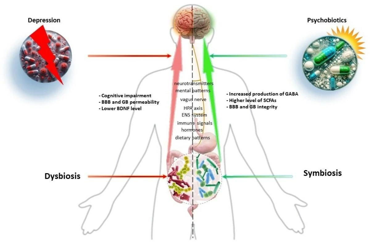
    Depression is a global health concern that causes pain, lost productivity, higher healthcare expenditures, and a high risk of suicide. Dysbiosis, a disruption in gut microbiome homeostasis, can affect the gut-brain axis (GBA), resulting in microbial alterations. Psychobiotics, which have favorable effects on the gut barrier, immunological responses, cortisol expression, and the hypothalamic-pituitary-adrenal (HPA) axis, might be used as a supportive treatment for depression, particularly in treatment-recalcitrant depression (TRD) cases.

    The Power of Psychobiotics in Depression: A Modern Approach through the Microbiota–Gut–Brain Axis: A literature Review. Image Credit: T. L. Furrer / ShutterstockThe Power of Psychobiotics in Depression: A Modern Approach through the Microbiota–Gut–Brain Axis: A literature Review. Image Credit: T. L. Furrer / Shutterstock

    About the review

    In the present review, researchers presented clinical evidence and elucidated the underlying mechanisms of psychobiotic therapies for depression via their effects on gut-brain communication.

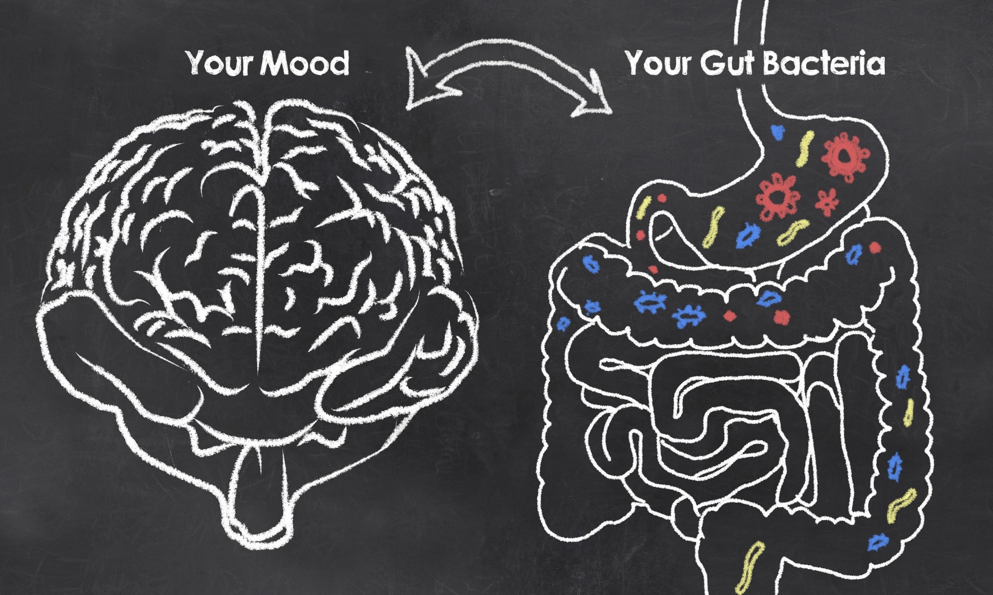
    Association between the gut-brain axis and depressive disorders

    Depression is a complex biological disorder influenced by a variety of molecular mechanisms, such as neurotransmitter reduction, a decrease in brain-derived neurotrophic factor (BDNF), an abnormally stressed HPA axis, an increase in pro-inflammatory gut microbial responses, and vagus nerve interaction between gut microbiota and brain. The GBA and intestinal microbes are inextricably linked, with MGB influencing neurobehavioral outcomes via endocrine, neuronal, and immunological mechanisms. Dysbiosis, or a disruption in the GBA axis, can alter the intestinal microbiome, influencing neuronal function, immunology, and gut inflammation.

    Chronic stress impairs intestinal homeostasis and changes gut microbial composition, increasing Faecalibaculum and Clostridium in individuals while decreasing Lactobacillus and Bifidobacterium. Recent animal models have demonstrated a relationship between the gut-brain axis and stress sensitivity and resilience. The intestinal microbiome influences inflammatory responses and brain states and is associated with psychiatric conditions such as major depressive disorder, bipolar disorder, psychosis, schizophrenia, anorexia nervosa, anxiety disorders, obsessive-compulsive disorder, post-traumatic stress disorder, and attention-deficit hyperactivity disorder (ADHD).

    Graphical Abstract

    Graphical Abstract

    Gut microbial metabolites involved in antidepressant actions

    The gut microbiome is a vital metabolite source, facilitating communications between the gut and the central nervous system. These metabolites consist of tryptophan, gamma-aminobutyric acid (GABA), serotonin, histamine, 5-hydroxytryptamine (5-HT), short-chain fatty acids (SCFAs), acetylcholine, and dopamine (DA). Microbial metabolites impact various mechanisms important for mental health, such as immunological and neuroendocrine system development, nutrition metabolism modulation, and xenobiotic transformation. They also help to maintain gut barrier function, strengthen the intestinal mucosa, and keep dangerous infections and poisons out of circulation. SCFAs are necessary for emotional states and cognition, impacting the host’s brain via G-protein-coupled receptors. They supply energy to colonocytes, protect the intestinal barrier, regulate inflammatory responses, and regulate hunger hormones. Increased SCFAs can reduce neuroinflammation and boost BDNF synthesis, boosting brain neuroplasticity.

    Impact of probiotic gut microbes on depression

    Psychobiotics are probiotic bacteria that boost mental health by improving the intestinal barrier and modifying the immune response in the gut-associated lymphoid tissue (GALT), which plays a role in inflammation development. The gut microbiota is crucial in the pathophysiology of depression since it regulates inflammatory processes. Bifidobacterium breve boosts BDNF levels, lowers interleukin-6 (IL-60) and TNF-alpha (TNF-α) levels, and enhances cognitive function.

    Lactic acid bacteria (LAB) reduce neuroinflammation, lower kynurenine levels, and promote tight junction (TJ) expression. Lactobacillus plantarum 299v boosts dopamine levels and helps with selective serotonin reuptake inhibitor (SSRI) therapy, resulting in better cognitive performance and lower kynurenine levels. Akkermansia muciniphila suppresses inflammatory cytokines in microglial cells, which lowers depressive-like behavior. Clostridium butyricum protects against neurological dysfunction, whereas Faecalibacterium prausnitzii lowers corticosterone and C-reactive protein (CRP) levels while boosting IL-10 levels and lowering cognitive impairment in Alzheimer’s disease rats.

    Clinical evidence highlighting the psychobiotic features of bacterial strains

    Postbiotics such as Bacillus coagulans MTCC 5856 and Bifidobacterium longum 1714 can help with irritable bowel syndrome (IBS) symptoms and depression. Probiotics such as Bifidobacterium longum 1714 and NCC3001 help to decrease stress and enhance memory. When coupled with antidepressants, these probiotics can effectively cure TRD. Probiotics such as Lactobacillus casei Shirota and Lactobacillus gasseri CP2305, at 2.5 × 10⁹ CFU/g, enhance general health and lower mood disorders. Multi-strain probiotic medication also boosts general health, alleviates anxiety symptoms, and reduces inflammation. Lactobacillus gasseri fermented black soybean beverage helps healthy individuals sleep better and feel less stressed. Probiotic milk drinks and fermented soybean seed paste improve cognitive performance in individuals with moderate cognitive impairment and Alzheimer’s disease.

    The review highlights probiotics’ involvement in lowering depressive symptoms and their importance in mental health. The gut microbiota is crucial for digestion, food absorption, and psychiatric concerns such as stress reduction and anxiety. With a shift in the emphasis in modern life from infectious disorders to more common mental illnesses such as depression, good dietary habits and optimal intestinal function are critical for mental well-being, with probiotics playing an important role.

    Journal reference:

    • Dziedzic, A.; Maciak, K.; Bliźniewska-Kowalska, K.; Gałecka, M.; Kobierecka, W.; Saluk, J. The Power of Psychobiotics in Depression: A Modern Approach through the Microbiota–Gut–Brain Axis: A literature Review. Nutrients 2024, 16, 1054. DOI: 10.3390/nu16071054, https://www.mdpi.com/2072-6643/16/7/1054

    [ad_2]

    Source link

  • Human neuron model identifies potential therapeutic targets for Alzheimer’s disease

    Human neuron model identifies potential therapeutic targets for Alzheimer’s disease

    [ad_1]

    Weill Cornell Medicine scientists have developed an innovative human neuron model that robustly simulates the spread of tau protein aggregates in the brain-;a process that drives cognitive decline in Alzheimer’s disease and frontotemporal dementia. This new model has led to the identification of novel therapeutic targets that could potentially block tau spread.

    The preclinical study, published April 5 in Cell, is a significant advancement in Alzheimer’s disease research.

    Currently no therapies can stop the spread of tau aggregates in the brains of patients with Alzheimer’s disease. Our human neuron model of tau spread overcomes the limitations of previous models and has unveiled potential targets for drug development that were previously unknown.”


    Dr. Li Gan, lead study author, director of the Helen and Robert Appel Alzheimer’s Disease Research Institute and the Burton P. and Judith B. Resnick Distinguished Professor in Neurodegenerative Diseases in the Feil Family Brain and Mind Research Institute at Weill Cornell Medicine

    Human pluripotent stem cells can develop into any cell of the body and can be coaxed to become neurons to model brain diseases in a lab dish. However, it had been nearly impossible to model tau propagation in these young neurons, as tau propagation requires decades in aging brains.

    Dr. Gan’s team used CRISPR technology to modify the genomes of human stem cells, prompting them to express forms of tau associated with diseased aging brains. “This model has been a game-changer, simulating tau spread in neurons within weeks-;a process that would typically take decades in the human brain,” Dr. Gan said.

    In their quest to halt tau propagation, Dr. Gan’s team employed CRISPRi screening to disable one thousand genes to ascertain their roles in tau spread. They discovered 500 genes that have a significant impact on tau abundance.

    “CRISPRi technology allowed us to use unbiased approaches to look for drug targets, not confined to what was previously reported by other scientists,” said one of the lead study authors Celeste Parra Bravo, a neuroscience doctoral candidate in the Weill Cornell Graduate School of Medical Sciences working in the Gan lab.

    One discovery includes the UFMylation cascade, a cellular process involving the attachment of a small protein named UFM1 to other proteins. This process’s connection to tau spread was previously unknown. Post-mortem studies of brains from patients with Alzheimer’s disease found that UFMylation is altered, and the team also found in preclinical models that inhibition of the enzyme required for UFMylation blocks tau propagation in neurons.

    “We are particularly encouraged by the confirmation that inhibiting UFMylation blocked tau spread in both human neurons and mouse models,” said paper co-author Dr. Shiaoching Gong, associate professor of research in neuroscience in the Appel Institute at Weill Cornell Medicine.

    Many Alzheimer’s disease treatments initially show promise in mouse models but do not succeed in clinical trials, Dr. Gan said. With the new human cell model, she is optimistic about the path ahead. “Our discoveries in human neurons open the door to developing new treatments that could truly make a difference for those suffering from this devastating disease.”

    Source:

    Journal reference:

    Bravo, C. P., et al. (2024) Human iPSC 4R tauopathy model uncovers modifiers of tau propagation. Cell. doi.org/10.1016/j.cell.2024.03.015.

    [ad_2]

    Source link

  • Protein intake during pregnancy affects offspring’s facial features

    Protein intake during pregnancy affects offspring’s facial features

    [ad_1]

    The protein content of the diet during pregnancy can affect the face of the offspring. This is shown in animal studies, and the underlying mechanism was also found in human genetic studies. The research is described in a study led by the University of Gothenburg.

    A child is expected to share facial features with their parents. However, the face is also influenced by factors beyond genetics, so-called environmental factors. Among those, lifestyle during pregnancy is an essential factor. For example, extensive alcohol consumption during pregnancy can lead to facial abnormalities in the child.

    The current study, published in Nature Communications, revealed a novel link between the child’s face and pregnancy lifestyle, specifically protein intake during pregnancy.

    A research team led by Andrei Chagin, Professor of Molecular Medicine, delved into the mechanisms that control the formation of the facial bone structure during the embryonic phase. The studies revealed that a particular signaling pathway in the cells seemed to play a crucial role in shaping the face.

    Noticeable differences in the face

    The signaling pathway, known as mTOR, controls several cell functions, including cell division and cell survival, and is known to be the target of an immunosuppressive medicinal product, rapamycin, which is administered during organ transplantation to avoid rejection.

    The mTOR signaling pathway acts as an intracellular nutritional sensor specifically tailored to amino acids, the building blocks of proteins. The researchers found that the mTOR pathway modulates the formation of facial skeletal structures in humans, mice, and zebrafish. This led researchers to investigate whether the protein content of the pregnancy diet affects mTOR and plays a role in the formation of the facial bone structure.

    Pregnant mice were given diets with high and low protein levels. As expected, the protein levels in the female’s diet were consistent with the activity level of mTOR in the developing face. In newborn offspring, the differences in the face were noticeable, albeit subtle.

    It is difficult to describe the exact effects, which can be caused by protein content in the diet during human pregnancy. But our data suggest that the mechanism is evolutionarily conserved and, from this perspective, likely serves to increase variability in the feeding apparatus, thus, allowing animals to adapt to various feeding sources in the wild. In mice, we see, for example, an enlarged nasal cavity in the offspring of mice fed a protein-enriched diet and a slightly elongated jaw in mice where the mother has eaten a low-protein diet.”

    Andrei Chagin, Professor of Molecular Medicine

    Good nutrition and clinical practices

    This international study was led by researchers at Sahlgrenska Academy at the University of Gothenburg in collaboration with colleagues at Karolinska Institutet, Belgium, Japan, China, Russia, the Czech Republic, and Austria.

    The aim has been to contribute to the future development of more effective clinical methods for the prevention and treatment of different types of facial congenital malformations and to increase knowledge of what a good pregnancy diet should contain.

    “The findings emphasize the importance of maintaining a well-balanced diet during pregnancy, with particular attention to protein intake. The insights open new avenues for understanding the intricate interplay between genetics, lifestyle, and the formation of our unique facial features,” concludes Andrei Chagin.

    Source:

    Journal reference:

    Xie, M., et al. (2024). The level of protein in the maternal murine diet modulates the facial appearance of the offspring via mTORC1 signaling. Nature Communications. doi.org/10.1038/s41467-024-46030-3.

    [ad_2]

    Source link

  • Lancet Commission predicts sharp increase in global prostate cancer cases

    Lancet Commission predicts sharp increase in global prostate cancer cases

    [ad_1]

    Cases of prostate cancer are projected to double from 1.4 million per year in 2020 to 2.9 million per year by 2040, with low- and middle-income countries (LMICs) predicted to see the highest increases in cases, according to The Lancet Commission on prostate cancer which will be launched by a presentation at the European Association of Urology Congress.

    The number of annual prostate cancer deaths worldwide is predicted to rise by 85% over the 20-year period, from 375,000 deaths in 2020 to almost 700,000 deaths by 2040. The true numbers will likely be much higher than the recorded figures due to under-diagnosis and missed opportunities for data collection in LMICs. 

    Most of these deaths are expected to be in LMICs, due to the rising number of cases and increasing mortality rates in these countries. Deaths from prostate cancer have declined in most high-income countries (HICs) since the mid-1990s.

    Prostate cancer is already a major cause of death and disability, accounting for 15% of all male cancers. It is the second most common cause of cancer deaths in UK men and the most common form of male cancer in more than half of the world’s countries.

    Aging populations and increasing life expectancy will lead to higher numbers of older men in coming years. As the main risk factors for prostate cancer – such as being aged 50 or older and having a family history of the disease – are unavoidable, it will not be possible to prevent the upcoming surge in cases through lifestyle changes or public health interventions.

    Professor Nick James, lead author of the Commission, Professor of Prostate and Bladder Cancer Research at The Institute of Cancer Research, London, and Consultant Clinical Oncologist at The Royal Marsden NHS Foundation Trust, said: “As more and more men around the world live to middle and old age, there will be an inevitable rise in the number of prostate cancer cases. We know this surge in cases is coming, so we need to start planning and take action now. Evidence-based interventions, such as improved early detection and education programs, will help to save lives and prevent ill health from prostate cancer in the years to come. This is especially true for low- and middle-income countries which will bear the overwhelming brunt of future cases.” 

    Global need for new and improved early detection programs

    In HICs, screening for prostate cancer often involves the PSA test, a blood test that measures levels of a protein called prostate-specific antigen (PSA). However, PSA tests often detect prostate cancer which may never cause symptoms and needs no treatment. The current approach to prostate cancer diagnosis in the UK and many other HICs relies on ‘informed choice’ PSA testing – when men aged 50 or over with no disease symptoms can request a PSA test from their doctor after a discussion of the risks and benefits. The Commission argues there is evidence to suggest this approach leads to over-testing in low-risk older men but does not increase detection of prostate cancer in younger men at higher risk. The authors also highlight huge variations in the likelihood of men being diagnosed with advanced prostate cancer with the ‘informed choice’ PSA testing strategy, for example The National Prostate Cancer Audit in the UK found that in 2022, 1 in 8 men (12.5%) with prostate cancer are diagnosed with advanced prostate cancer in London, whereas in Scotland more than 1 in 3 (35%) were diagnosed late.

    Instead, the authors recommend using MRI scans in combination with PSA testing to screen men at high risk of prostate cancer in HICs, such as those with a family history of the disease, those of African origin and those carrying the BRACA2 mutation. They argue that this approach would both reduce over-diagnosis and over-treatment, while detecting potentially lethal disease. MRI is effective in imaging cancers and can be used to provide information as to whether the disease is aggressive and likely to be life-threatening. However, biopsies are more effective at identifying aggressive cancers, so MRI alone should not be used to investigate men at high risk of disease.

    The effectiveness of population-level PSA testing has not been tested in LMICs and there is an urgent need for cancer screening trials in these countries. New approaches to enable earlier diagnosis in LMICs are vital, as most men in these countries present with metastatic cancer – an advanced form of disease where the cancer has spread to other parts of the body, often the bones. Men with late-stage prostate cancer are much less likely to survive for a long period of time than those who are diagnosed early.

    “With prostate cancer we cannot wait for people to feel ill and seek help – we must encourage testing in those who feel well but who have a high risk of the disease in order to catch lethal prostate cancer early. Pop-up clinics and mobile testing offer cost-effective solutions that combine health checks and education. In the UK we recently trialled a new innovative outreach programme called The Man Van which provided free health checks – including PSA tests – to high-risk men in London aged 45 and over. By bringing a van with quick and easy testing straight to men at work and in the community, and targeting those who have a higher risk of prostate cancer, we provided thousands of health checks which resulted in almost 100 cancer diagnoses in men who might otherwise have only seen a doctor once their cancer has progressed to a more advanced stage. The mix of education, outreach, testing and referral used in The Man Van trial may also be successful in LMICs and we hope to see similar initiatives rolled out globally to improve early detection of prostate cancer,” said Professor Nick James (also project lead for the ‘Man Van’).

    As well as being a major growing challenge, prostate cancer is also an indicator of a wider need to tailor future healthcare to cope with increases in several diseases, as the numbers of men reaching middle and old age increase worldwide. The Commission calls for trials of prostate cancer screening in LMICs to form part of holistic approaches with a broader focus on men’s health.

    Raising awareness of advanced prostate cancer and available therapies

    There is a need to raise awareness of the dangers and symptoms of metastatic prostate cancer among men and their families in LMICs. Public awareness of the key features of advanced prostate cancer – such as bone pain, caused by metastatic disease – is poor in many LMICs. Similarly, there is generally low public awareness that treatments can prolong survival and decrease suffering – including cheap, effective ones such as hormone therapy – are available in many LMICs. As with early diagnostic capacity, there is a need to scale-up availability and improve access to treatments for advanced disease in LMICs. 

    Improving education about the disease is critical, and the Commission authors suggest that programs should involve new technologies and channels such as smartphones, social media, and influencers. They highlight Project PINK BLUE, an organization that delivers a range of programs to raise awareness of breast, cervical, and prostate cancer in Nigeria, and provides free cancer screening. Many of Project PINK BLUE’s programs utilize digital technologies and involve well-known public figures and celebrities.

    Professor James N’Dow, Chair in Surgery, University of Aberdeen and Founder of Horizons Trust & Horizons Clinic, Gambia, said: “The issue in low- and middle-income countries is that late diagnosis of prostate cancer is the norm. Improved outreach programs are needed to better inform people of the key signs to look out for and what to do next. Implementing these in tandem with investments in cost-effective early diagnostic systems will be key to preventing deaths from prostate cancer as cases inevitably rise with a global aging population.” 

    He continues, “As well as the obvious direct effects on individual men’s health, rising numbers of cases and deaths from prostate cancer could have huge economic and social impacts on families in LMICs. Men in these countries are very often a family’s main breadwinner, so if they die or become seriously ill, this can lead to families facing major economic hardship. By preparing now for the upcoming surge in prostate cancer cases, with a particular emphasis on improved education and earlier diagnosis programs, many of these harms could be reduced substantially.” 

    Building capacity to diagnose and treat prostate cancer early in LMICs 

    Optimal management of prostate cancer requires the availability of specialist staff and infrastructure to support diagnosis, surgery and radiotherapy to treat localized prostate cancer, and radiotherapy and hormone therapy for metastatic disease.

    A major barrier to improved prostate cancer care in LMICs is a lack of trained staff and specialist facilities. These shortages are not limited to prostate cancer, and the 2015 Lancet Commission on Surgery found that 9 out of 10 people in LMICs cannot access basic surgical care. [4]

    Expanding early diagnostic capabilities in LMICs will increase the rates of detection of early-stage prostate cancer, further increasing demand for surgery and radiotherapy. Urgent measures are therefore needed to build surgical and radiotherapy capacity in these countries. The Commission authors state that establishing regional hubs could provide the infrastructure needed to increase specialist training and improve patient access to radiotherapy and surgery.

    For men with metastatic disease, earlier diagnosis and starting hormone therapy earlier will reduce deaths and prevent serious complications like painful spinal cord compression and urinary retention, which can lead to infection and kidney damage.

    More research is needed on ethnic inequities in care and survival

    The Commission authors highlight the need for more research to better understand prostate cancer in men who are not of White European origin, to enable improved detection and care in these groups. Research and knowledge of prostate cancer is heavily focussed on White European men, and most studies have been done in HICs. However, Black men, especially those of West African descent, have a higher risk of developing prostate cancer than White or Asian men, though the reasons for this are unclear. There is also a higher death rate from prostate cancer among Black men, but it is not known if this is driven by the differences in case rates or by other factors such as differing disease biology or societal factors such as deprivation or racism. More data is needed to identify the driving factors behind these trends. 

    The Commission authors call for mandatory recording of ethnicity in clinical trials, and that trials should reflect the ethnic mix of the populations being studied to ensure that the findings apply to all groups. The Commission authors also call for trials examining prostate cancer screening, early diagnosis, and treatment in LMICs. 

    Source:

    Journal reference:

    James, N. D., et al. (2024) The Lancet Commission on prostate cancer: planning for the surge in cases. The Lancet. doi.org/10.1016/S0140-6736(24)00651-2.

    [ad_2]

    Source link

  • Gene therapy and glycoside drugs offer new hope for polycystic kidney disease treatment

    Gene therapy and glycoside drugs offer new hope for polycystic kidney disease treatment

    [ad_1]

    Researchers have shown that dangerous cysts, which form over time in polycystic kidney disease (PKD), can be prevented by a single normal copy of a defective gene. This means the potential exists that scientists could one day tailor a gene therapy to treat the disease. They also discovered that a type of drug, known as a glycoside, can sidestep the effects of the defective gene in PKD. The discoveries could set the stage for new therapeutic approaches to treating PKD, which affects millions worldwide. The study, partially funded by the National Institutes of Health (NIH), is published in Cell Stem Cell.

    Scientists used gene editing and 3-D human cell models known as organoids to study the genetics of PKD, which is a life-threatening, inherited kidney disorder in which a gene defect causes microscopic tubes in the kidneys to expand like water balloons, forming cysts over decades. The cysts can crowd out healthy tissue, leading to kidney function problems and kidney failure. Most people with PKD are born with one healthy gene copy and one defective gene copy in their cells.

    Human PKD has been so difficult to study because cysts take years and decades to form. This new platform finally gives us a model to study the genetics of the disease and hopefully start to provide answers to the millions affected by this disease.”


    Benjamin Freedman, Ph.D., senior study author at the University of Washington, Seattle

    To better understand the genetic reasons cysts form in PKD, Freedman and his colleagues sought to determine if 3-D human mini-kidney organoids with one normal gene copy and one defective copy would form cysts. They grew organoids, which can mimic features of an organ’s structure and function, from induced pluripotent stem cells, which can become any kind of cell in the body.

    To generate organoids containing clinically relevant mutations, the researchers used a gene editing technique called base editing to create mutations in certain locations on the PKD1 and PKD2 genes in human stem cells. They focused on four types of mutations in these genes that are known to cause PKD by disrupting the production of polycystin protein. Disruptions in two types of the protein – polycystin-1 and polycystin-2 – are associated with the most severe forms of PKD.

    They then compared cells with two gene copy mutations in organoids to cells with only one gene copy mutation. In some cases, they also used gene editing to correct mutations in one of the two gene copies to see how this affected cyst formation. They found organoids with two defective gene copies always produced cysts and those that carried one good gene copy and one bad copy did not form cysts. 

    “We didn’t know if having a gene mutation in only one gene copy is enough to cause PKD, or if a second factor, such as another mutation or acute kidney injury was necessary,” Freedman said. “It’s unclear what such a trigger would look like, and until now, we haven’t had a good experimental model for human PKD.”

    According to Freedman, the cells with one healthy gene copy make only half the normal amount of polycystin-1 or polycystin-2, but that was sufficient to prevent cysts from developing. He added that the results suggest the need for a second trigger and that preventing that second hit might be able to prevent the disease.

    The organoid models also provided the first opportunity to study the effectiveness of a class of drugs known as eukaryotic ribosomal selective glycoside on PKD cyst formation.

    “These compounds will only work on single base pair mutations, which are commonly seen in PKD patients,” explained Freedman. “They wouldn’t be expected to work on any mouse models and didn’t work in our previous organoid models of PKD. We needed to create that type of mutation in an experimental model to test the drugs.”

    Freedman’s team found that the drugs could restore the ability of genes to make polycystin, increasing the levels of polycystin-1 to 50% and preventing cysts from forming. Even after cysts had formed, adding the drugs slowed their growth.

    Freedman suggested that a next step would be to test existing glycoside drugs in patients. Researchers also could explore the use of gene therapy as a treatment for PKD.

    The research was supported by NIH’s Nation Center for Advancing Translational Sciences, National Institute of Diabetes and Digestive and Kidney Diseases, and National Institute of General Medical Sciences through awards R01DK117914, UH3TR002158, UH3TR003288, U01DK127553, U01AI176460, U2CTR004867, UC2DK126006, P30DK089507, R21DK128638, and R35GM142902; an Eloxx Pharmaceuticals Award; the Lara Nowak-Macklin Research Fund; and a Washington Research Foundation fellowship.

    Source:

    Journal reference:

    Vishy, C. E., et al. (2024) Genetics of cystogenesis in base-edited human organoids reveal therapeutic strategies for polycystic kidney disease. Cell Stem Cell. doi.org/10.1016/j.stem.2024.03.005.

    [ad_2]

    Source link

  • SRF announces grant to support research on SynGAP-Related Disorder in adults

    SRF announces grant to support research on SynGAP-Related Disorder in adults

    [ad_1]

    The SynGAP Research Fund 501(c) today announced a grant to Dr. Danielle Andrade, Dr. Miles Thompson, Dr. Ryan Yuen, Dr. Rogier Kerssebook, and Dr. Anatoljevna Anna Kattentidt to support research on SynGAP-Related Disorder (SRD) in adults. SRD is a rare neurodevelopmental disorder that causes severe intractable epilepsy, and intellectual disability, and is one of the leading genetic causes of autism. 

    Dr. Andrade’s team recently published the only research on SRD in adults in Neurology Genetics, Rong 2023 which SRF was instrumental in encouraging and supporting via recruitment. See Dr. Andrade’s readout on this work at the 2022 SRF SYNGAP1 Conference here. The grant SRF is announcing today will build on that groundbreaking work. 

    Dr. Andrade, MD, the principal investigator for this work says “This research holds deep significance not just for the adults in this study but also for children affected by SYNGAP1. As children become adults, investigating the outcomes of SYNGAP1 is pivotal in empowering families and clinicians alike, equipping them to screen for, diagnose, and manage manifestations seen in adults. Also, findings of this study will be important to guide precision therapies as well as evaluate their efficacy.”

    Michael Graglia, Managing Director of SRF, and father to a child with SRD says “Dr. Andrade is the world’s leading expert in adults with DEEs, the SYNGAP1 community is very fortunate to have her attention focused on our disease. She and her colleagues are world-class.”

    Dr. Marta Dahiya, MD, SRF’s Head of Clinical and mother to an adult with SRD says, “While many patients today are not yet adults, they will all become adults and this research will inform and support their care and created the opportunity for adult patients to be included in therapeutic studies. Every family should be thrilled about this work.”

    It is exciting to see this transatlantic cooperation where the large number of adults with SYNGAP1 Related Disorders in the Netherlands are able to be studied and included in this essential work.”


    Katrien Deckers, head of SRF-Europe 

    How to sign up

    Families with a loved one with SYNGAP1 Related Disorder age 16 or older can join the accessible online study by emailing [email protected], more information is available in the study flyer

    About the project

    The primary goal is to comprehensively understand the adult phenotypes and natural history of SRD patients, as well as the differences between those with and without epilepsy, in order to promote optimal care among adult patients. 

    They will investigate the presence of seizures and their severity, movement abnormalities, adaptive skills, behavior, gastrointestinal symptoms, sleep difficulties, and comorbidities among patients at our Adult Genetic Epilepsy (AGE) clinic in Canada, at the adult SYNGAP1 clinic in the Netherlands and externally through patient organizations such as the SynGAP Research Fund.

    They will also determine the genotype/phenotype correlations between SYNGAP1 patients with and without epilepsy. This will be achieved via WGS sequencing to search for rare variants with large effect size that influences the phenotype, and the contribution of common variants to individual features of the phenotype, through calculating the polygenic risk score (PRS) for different comorbidities. This research will provide valuable insight into the unique experiences and clinical outcomes of adult patients with SRF while promoting dialogue on the importance of precise and timely diagnosis and treatment of adult patients.

    About the team

    Dr. Danielle Andrade, MD, MSc, FRCPC, is the Medical Director of the Epilepsy Program at University Health Network and founder and Director of Krembil Neuroscience Institute Adult Genetic Epilepsy (AGE) Program. Dr. Miles Thompson, Ph.D., GCGCVI Scientific Associate, is with Krembil Research Institute, Toronto Western Hospital, and University Health Network. Dr. Ryan Yuen, Ph.D. is the Senior Scientist, Genetics & Genome Biology Program, at The Hospital for Sick Children – Toronto, Canada. Dr. Rogier Kerssebook, MD, Ph.D. is a Consultant in Clinical Genetics, at Erasmus Medical Centre, Rotterdam, The Netherlands. Dr. Anatoljevna Anna Kattentidt, MD, is a Clinical Geneticist and Physician, at Erasmus Medical Center, Rotterdam, The Netherlands. 

    University Health Network, Toronto, Canada – https://www.uhn.ca

    The Hospital for Sick Children, Toronto, Canada – https://www.sickkids.ca 

    Erasmus Medical Centre, Rotterdam, the Netherlands – https://www.erasmusmc.nl/nl-nl/ 

    About SYNGAP1-related intellectual disability (SRD)

    SYNGAP1-related intellectual disability (US ICD-10 F78.A1 / Global ICD-11 LD90.Y) is a rare genetic disorder caused by variants on the SYNGAP1 gene that reduce SynGAP protein levels. This protein acts as a regulator in the synapses (where neurons communicate with each other). When SynGAP protein levels are too low, we see an increase in excitability in the synapses making it difficult for neurons to communicate effectively. This leads to many neurological issues seen in SynGAP patients.

    Symptoms of SYNGAP1 include intellectual disability; epilepsy; hypotonia (low muscle tone); gross and fine motor skill delays; autism spectrum disorder; gastro-intestinal issues; sleep and behavior disorders and visual abnormalities. 

    [ad_2]

    Source link

  • Researchers unlock the potential of whey-derived proteins for cancer prevention

    Researchers unlock the potential of whey-derived proteins for cancer prevention

    [ad_1]

    In a recent review article published in Food and Humanity, researchers summarized the current evidence regarding the significance of whey protein for cancer prevention and treatment.

    Their conclusions highlighted the emerging role of whey protein supplements as a cost-effective, practicable, and viable strategy for cancer treatment and prevention.

    Study: Emerging potential of whey proteins in prevention of cancer. Image Credit: Dan_photography/Shutterstock.comStudy: Emerging potential of whey proteins in prevention of cancer. Image Credit: Dan_photography/Shutterstock.com

    Background

    Cancer is a leading cause of mortality globally, and its prevalence has increased significantly, prompting research to guide the development of effective strategies for treatment and prevention.

    Whey protein, known for its nutritional value and popularity in fitness, has recently garnered attention for its potential anticancer properties.

    Studies suggest whey protein contains bioactive compounds, such as lactoferrin, which demonstrate anticancer effects by inhibiting cancer cell growth and boosting the immune system.

    Whey protein also appears to modulate signaling pathways involved in cancer development, potentially slowing its progression. Additionally, it may enhance the efficacy and reduce the side effects of conventional cancer treatments like chemotherapy and radiation therapy.

    While more research is needed to understand the mechanisms underlying the anticancer effects of whey protein, initial findings offer promising avenues for cancer prevention and treatment.

    Whey protein and its benefits

    Whey protein, formed during milk processing as a by-product, can be sweet or acid. Sweet whey is used widely in supplements, with about 50% of the nutrients found in milk constituting approximately 20% of the protein content of milk.

    Whey contains various nutrients, including bioactive peptides, minerals, B-complex vitamins, and growth factors. These bioactive components, such as lactoperoxidase, beta-lactoglobulin, and lactoferrin, demonstrate diverse bioactivities and functionalities.

    Whey protein is highly regarded for its role in providing essential amino acids and promoting quick absorption, making it ideal for people with cancer and individuals seeking protein-rich diets.

    Commercially, whey protein is available in different forms like whey protein isolates (WPI), whey protein hydrolysates (WPH), and whey protein concentrates (WPC), each with varying protein concentrations.

    Whey protein concentrates generally contain between 25% and 89% protein, while isolates contain between 90% and 95%.

    Whey protein offers numerous health benefits, including weight loss support, muscle preservation, digestive health promotion, hypertension regulation, and anti-carcinogenic effects.

    It has probiotic properties and is a precursor for bioactive compounds like lactulose and lactobionic acid, and exhibits a low glycemic index and cariogenicity compared to other protein sources.

    The therapeutic properties of whey protein are attributed to its antioxidant activity, glutathione enhancement, apoptosis induction, iron-binding capacity, cell proliferation regulation, and potential in treating cancer cachexia-anorexia syndrome.

    It stimulates glutathione synthesis, promotes apoptosis in cancer cells, and regulates cell growth and division through insulin-like growth factor 1 pathways.

    Further research into whey protein and its bioactive components holds promise for enhancing human health and well-being.

    In vivo and clinical cancer studies

    Animal studies demonstrate that whey protein shows promise against oxidative stress-induced tissue injuries and cancers. Its potential anticancer and antioxidant properties may be associated with its ability to increase glutathione levels.

    WPC exhibits advantages over soy, casein, and other proteins in reducing colorectal cancer incidence via glutathione elevation.

    Whey protein diets have also shown promise in managing mucositis for individuals undergoing chemotherapy while improving nutritional outcomes.

    Subfractions of whey protein, particularly bovine lactoferrin and alpha-lactalbumin, exhibit antitumor effects inhibiting tumor development. Researchers are exploring novel nanocarriers incorporating components of whey protein to prevent tumors without side effects.

    Some clinical trials with human participants have shown positive outcomes, which are consistent with the evidence from in vitro studies of whey protein’s antioxidant and anti-cancer.

    Regarding nutritional and performance parameters, interventions that combined supplementation with dietary assistance and exercise improved nutritional parameters and handgrip strength; WPI supplementation also showed promise for protein status strengthening, boosting immunity during chemotherapy, and raising glutathione levels.

    Studies also indicate both positive and complex effects of whey protein concentrate and lactoferrin supplementation on the health of cancer patients.

    While these results are promising, robust multicentric trials must be conducted across various forms of cancer to confirm the pervasive efficacy of whey protein supplementation as an adjuvant therapy.

    Conclusions

    The narrative review discussed the role of whey protein in cancer prevention and treatment based on both animal and clinical studies, highlighting the potential benefits of whey protein, including its antioxidant and anticancer properties, its ability to increase glutathione levels, and its effectiveness in managing mucositis during chemotherapy.

    Various subfractions of whey protein, such as alpha-lactalbumin and lactoferrin, show promising antitumor effects. Additionally, novel approaches like utilizing nanocarriers incorporating whey protein components are being explored for tumor prevention.

    Clinical trials suggest positive outcomes of whey protein supplementation, including improved nutritional and performance parameters, raised glutathione levels and strengthened immunity in cancer patients.

    However, robust multicentric trials across different cancer types are needed to confirm the widespread efficacy of whey protein supplementation as an adjuvant therapy.

    [ad_2]

    Source link

  • Lurie Children’s Hospital administers first gene therapy for Duchenne muscular dystrophy in Illinois

    Lurie Children’s Hospital administers first gene therapy for Duchenne muscular dystrophy in Illinois

    [ad_1]

    On March 27, 2024, Ann & Robert H. Lurie Children’s Hospital of Chicago treated its first patient with ELEVIDYS (delandistrogene moxeparvovec-rokl), the first gene therapy for Duchenne muscular dystrophy – a rare, genetic disease characterized by progressive muscle damage and weakness. Lurie Children’s is the first in Illinois to administer this treatment after ELEVIDYS received U.S. Food and Drug Administration (FDA) approval in June 2023.

    Developed by Sarepta Therapeutics, ELEVIDYS is approved for the treatment of Duchenne muscular dystrophy (DMD) in ambulatory patients aged 4 through 5 years with a confirmed mutation in the DMD gene.

    Our team at Lurie Children’s has had encouraging experience with this gene therapy for Duchenne through our active participation in clinical trials. Over the past two years, we have treated three boys with DMD with ELEVIDYS as part of a larger clinical trial, and it’s gratifying to see that their muscle strength and function stabilized. Without gene therapy, we would expect to see ongoing deterioration in muscle function in these boys. This therapy is not a cure, and unfortunately it cannot reverse previous muscle damage, but we anticipate that we can slow down the disease enough for science to step in and offer new treatments. This is the beginning of a very exciting journey.”


    Nancy Kuntz, MD, Director of Muscular Dystrophy Association Care Center at Lurie Children’s and Professor of Pediatrics and Neurology at Northwestern University Feinberg School of Medicine

    Duchenne occurs in approximately one in every 3,500-5,000 newborn males worldwide. It is caused by mutations in the dystrophin gene that lead to a lack of dystrophin protein, which acts as a shock absorber when muscles move. The first subtle signs of DMD may appear during infancy. Muscle weakness becomes increasingly noticeable between the ages of 3 and 5 years with the diagnosis being typically made around those ages. Most children living with Duchenne use a wheelchair by age 13 years. The leading causes of death in individuals with Duchenne are respiratory or cardiac failure, which typically occurs when patients are in their mid-20s/30s.

    Mason Flessner, now an energetic 6-year-old, was one of the clinical trial participants at Lurie Children’s who received ELEVIDYS about eight months ago. He now is able to run faster, climb stairs more easily and even jump – something he couldn’t do previously. His little brother, 3-year-old Dawson, who also has Duchenne, is waiting until he is old enough to qualify for gene therapy.

    “ELEVIDYS has been life-changing for Mason, and it has given us hope and optimism that Duchenne is no longer a fatal diagnosis,” said Dan Flessner, Mason’s father. “Thanks to research, gene therapy now gives us a pathway to a cure. With so much progress already, it’s not a pipedream anymore.”

    ELEVIDYS is administered as a one-time intravenous infusion. The gene therapy addresses the root genetic cause of Duchenne by delivering a gene that codes for a shortened form of dystrophin to muscle cells, known as ELEVIDYS-dystrophin. Because dystrophin gene is the largest known human gene, scientists engineered a shortened version of the gene that could fit inside current gene therapy delivery technologies and still retain key functional information. The therapy’s accelerated approval is based on an increase in ELEVIDYS-dystrophin protein expression in skeletal muscle cells.

    “Across the country, since the FDA approval, ELEVIDYS has only been administered a few times and we’re very excited to be the first site in Illinois to administer it,” said Abigail Schwaede, MD, one of the neuromuscular physicians on Mason’s care team at Lurie Children’s and Assistant Professor of Pediatrics at Northwestern University Feinberg School of Medicine. “This treatment has enormous potential to improve the quality of life and long-term outcomes for boys with Duchenne muscular dystrophy.”

    [ad_2]

    Source link

  • Ultrasound alone improves cognitive function in neurodegenerative disorders, UQ study finds

    Ultrasound alone improves cognitive function in neurodegenerative disorders, UQ study finds

    [ad_1]

    University of Queensland researchers have found targeting amyloid plaque in the brain is not essential for ultrasound to deliver cognitive improvement in neurodegenerative disorders.

    Dr Gerhard Leinenga and Professor Jürgen Götz from UQ’s Queensland Brain Institute (QBI) said the finding challenges the conventional notion in Alzheimer’s disease research that targeting and clearing amyloid plaque is essential to improve cognition.

    Amyloid plaques are clumps of protein that can build up in the brain and block communication between brain cells, leading to memory loss and other symptoms of Alzheimer’s disease.


    Previous studies have focused on opening the blood-brain barrier with microbubbles, which activate the cell type in the brain called microglia which clears the amyloid plaque.


    But we used scanning ultrasound alone on mouse models and observed significant memory enhancement.”


    Dr. Gerhard Leinenga, from UQ’s Queensland Brain Institute 

    Dr Leinenga said the finding shows ultrasound without microbubbles can induce long-lasting cognitive changes in the brain, correlating with memory improvement.

    “Ultrasound on its own has direct effects on the neurons, with increased plasticity and improved brain networks,” he said.

    “We think the ultrasound is increasing the plasticity or the resilience of the brain to the plaques, even though it’s not specifically clearing them.”

    Professor Götz said the study also revealed the effectiveness of ultrasound therapy varied depending on the frequency used.

    “We tested two types of ultrasound waves, emitted at two different frequencies,” he said.

    “We found the higher frequency showed superior results, compared to frequencies currently being explored in clinical trials for Alzheimer’s disease patients.”

    The researchers hope to incorporate the findings into Professor Götz’s pioneering safety trial using non-invasive ultrasound to treat Alzheimer’s disease.

    “By understanding the mechanisms underlying ultrasound therapy, we can tailor treatment strategies to maximize cognitive improvement in patients,” Dr Leinenga said.

    “This approach represents a significant step towards personalized, effective therapies for neurodegenerative disorders.”

    The research paper has been published in Molecular Psychiatry.

    Source:

    Journal reference:

    Leinenga, G., et al. (2024). Scanning ultrasound-mediated memory and functional improvements do not require amyloid-β reduction. Molecular Psychiatry. doi.org/10.1038/s41380-024-02509-5.

    [ad_2]

    Source link

  • Rare variants in the YKT6 gene cause new neurological disorder, study finds

    Rare variants in the YKT6 gene cause new neurological disorder, study finds

    [ad_1]

    A recent collaborative study has discovered rare variants in the YKT6 gene as the cause of a new neurological disorder characterized by developmental delays along with severe progressive liver disease and a potential risk for liver cancer. The study, published in Genetics in Medicine, was led by Dr. Hugo Bellen, Distinguished Service Professor at Baylor College of Medicine and Principal Investigator at the Jan and Dan Duncan Neurological Research Institute (Duncan NRI) at Texas Children’s Hospital, and Dr. Wendy Chung, the Chief of the Department of Pediatrics at Boston Children’s Hospital. 

    “It is known that the YKT6 gene plays important roles in many intracellular vesicular trafficking events in the cells but this is the first time it has been linked to a genetically inherited disorder,” Dr. Bellen said. “This study, using patient samples and fruit flies, provides a solid experimental foundation for future studies to better understand this new disease and to develop therapies.”

    YKT6 gene variants disrupt brain development and sometimes, liver function

    In collaboration with Dr. Mythily Ganapathi at Columbia University Irving Medical Center, Drs. Paula Hertel and Davut Pehlivan at Texas Children’s Hospital and Dr. James Lupski at Baylor College of Medicine, and by using the GeneMatcher tool and Baylor Genetics clinical diagnostics laboratory, this team of researchers and clinicians found three unrelated individuals with missense (analogous to misspellings in a word) variants in both copies of the YKT6 gene. 

    All three individuals had early onset of disease (four to six months of age) with failure to thrive. Two of them had an identical missense variant because of which the tyrosine amino acid at position 185 was changed to cysteine (Tyr185Cys). On the other hand, the third child carried a variant that caused the same amino acid change but in a different location (Tyr64Cys) of the YKT6 protein. Interestingly, in addition to developmental delays and neurological defects which were observed in all three children, only the two individuals with the Tyr185Cys variant had liver dysfunction and a potential risk for developing liver cancer.

    Interestingly, both individuals with the Tyr185Cys variant belong to the Syrian/Saint Thomas Christians of Kerala, India, a group currently estimated to be comprised of about 5 million individuals worldwide. Our genetic lineage analysis suggests this variant likely originated from a common ancestor before the community split.”


    Dr. Mythily Ganapathi at Columbia University Irving Medical Center 

    YKT6 gene variants impair autophagy

    To assess how YKT6 variants result in the observed disease pathologies, the Bellen team studied the fruit fly version of this gene which is quite similar to its human counterpart.

    “We found that the fly version of this protein is expressed in the fat body and brain which are analogous to the human liver and central nervous system respectively,” Dr. Mengqi Ma, one of the first authors and a postdoctoral fellow in the Bellen lab, said. “Moreover, fly strains with loss of function mutations in this gene were lethal.”

    Further, they observed that Ykt6 mutant flies expressing the normal fly version of the Ykt6 gene flies had an average lifespan. However, transgenic flies expressing the fly versions of the disease variants were less effective in restoring lifespan and other symptoms. While Ykt6 mutant flies expressing Tyr65Cys (equal to human Tyr64Cys) had normal lifespan and locomotion, those expressing Tyr186Cys (equal to human Tyr185Cys) had severely reduced lifespan and locomotor defects. “Our results showed that the fly Ykt6 Tyr186Cys cause more severe defects than Tyr65Cys,” Dr. Ma added, “suggesting that the corresponding human YKT6 Tyr185Cys is a more severe variant than Tyr64Cys.”

    To understand why these variants behaved differently, they delved deeper into their biology.

    YKT6 belongs to the SNARE family of proteins that regulate the flow of protein traffic to various compartments within the cell. In mammalian cells, YKT6 mediates the fusion of two cellular organelles – the autophagosomes and lysosomes to form autolysosomes – within which ‘used’ cellular proteins, lipids, and other molecules are degraded and recycled back for future use. This process called autophagy is critical for the proper function and health of the cells.

    The team found that the loss of fly Ykt6 led to an abnormal accumulation of proteins involved in autophagosome formation and autophagic cargo receptor, indicating a block in the autophagy pathway. Further studies revealed that just like lethality and other defects, fly Tyr186Cys (equal to human Tyr185Cys) was less efficient in reverting the symptoms compared to a normal copy of the Ykt6 gene. Furthermore, they observed that while autophagy initiation was normal, the steps involved in the breakdown of cellular waste were impaired in the absence of Ykt6.

    “Based on our findings, we recommend the YKT6 gene as a candidate for carrier screening in the Syrian/Saint Thomas Christian community of Kerala,” Dr. Mythily Ganapathi said.

    “Our work suggests children diagnosed with YKT6 liver disease will also need to be screened for hepatocellular carcinoma,” Dr. Paula Hertel said.

    “In summary, we have discovered YKT6 variants as the cause of a novel developmental disorder affecting brain function and in certain cases, also liver function, providing us valuable insights into a new genetic disease. However, additional studies with more patients will be needed to precisely understand the pathogenesis and to identify potential therapeutic targets for this condition,” Dr. Bellen added.

    Source:

    Journal reference:

    Ma, M., et al. (2024). Homozygous missense variants in YKT6 result in loss of function and are associated with developmental delay, with or without severe infantile liver disease and risk for hepatocellular carcinoma. Genetics in Medicine. doi.org/10.1016/j.gim.2024.101125.

    [ad_2]

    Source link