Tag: Science

  • Ancient DNA data hold insights into past organisms and ecosystems — handle them with more care

    Ancient DNA data hold insights into past organisms and ecosystems — handle them with more care

    [ad_1]

    An archaeological excavation of human skeletal remains.

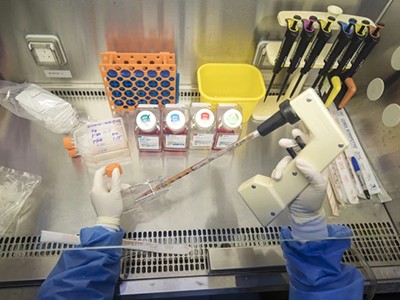
    DNA fragments extracted from archaeological human remains can be sequenced to identify the microorganisms that caused disease.Credit: Microgen/Getty

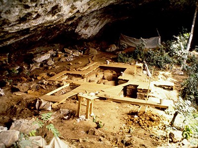
    Over the past ten years or so, investigations of degraded or ‘ancient’ DNA have skyrocketed. By extracting DNA fragments from diverse sources —from human teeth and faeces to soil samples and ice cores — researchers have uncovered the stories of all sorts of organisms and ecosystems stretching back for millennia.

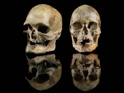
    Investigators have used ancient genomics to discover the previously unknown Denisovan hominin, an evolutionary cousin and contemporary of Neanderthals that left hardly any fossil record; to identify which microorganisms caused human disease thousands of years ago; and to establish the geographical origins of domesticated plants and animals, including maize (corn) and horses1. Ancient genomics has also been used to reconstruct the composition of Pleistocene ecosystems that existed up to two million years ago2; and even to identify the wearers of ice-age jewellery3.

    Most DNA sequence data are now archived in dedicated, publicly accessible databases, and the ancient DNA field has been heralded by some as a poster child for best practices in genetic data sharing4,5. However, as the pace of ancient DNA research has increased — largely thanks to the latest capabilities in DNA sequencing (see ‘A sampling surge’) — so, too, have problems with data archiving.

    A sampling surge. A stacked bar chart showing the cumulative number of ancient DNA samples with sequencing data in public archives rising between 2005 and 2024.

    Source: https://doi.org/10.5281/zenodo.14203618. Analysis by A. Bergström et al.

    Often, only some of the data obtained in any one study are uploaded to publicly available databases. Furthermore, the associated metadata — information on the age of the sample, where it was found, how the DNA was extracted and chemically treated, and so on — are frequently inaccurate or incomplete.

    Here, we set out the nature of these problems and outline steps to overcome them, so that this astonishing record of the genetic past can be digitally preserved and used again and again.

    Data loss

    Ancient genomic data have been obtained from more than 10,000 humans6, some 700 microbes and viruses7 and, by our estimate, more than 2,000 plant and non-human animal samples. At least 2,200 ancient host-associated and environmental microbiomes (communities of microorganisms) have been sequenced7 (see also go.nature.com/3bcaxtv).

    One major problem, however, is that not all sequences end up being archived.

    Earlier this year, one of us (A.B.) assessed what data and metadata had been uploaded into publicly accessible databases by the authors of 42 studies of ancient DNA. All studies involved the analysis of ancient DNA extracted from humans or non-human animals, and had been published in 2021, 2022 or 2023 in the journals Nature, Science and Cell. In about half of the papers, researchers archived only those sequences that they had managed to align to a reference genome, such as that for ancient human remains, leaving no record of the unaligned sequences (see ‘A snapshot of data-archiving troubles’). This represents a permanent loss of data for more than 3,000 ancient samples analysed in just these studies8.

    A snapshot of data-archiving troubles. A graphic showing 42 squares, 22 of which are coloured orange, representing the studies where the decision was made to not upload all the sequences generated. This resulted in a loss of data from more than 3,000 ancient samples.

    Source: Ref. 8

    It might seem that any sequence that does not align to the reference genome is irrelevant. But improvements in computational methods and more-complete reference genomes could enable researchers to align such sequences in the future. Also, even if some of the unaligned sequences are not from the species of interest, this does not mean that they have no scientific value. On the contrary, these sequences could be among the most interesting in the data set, especially if they originated from pathogenic microbes that infected the host.

    The study of ancient microbes has become a field in its own right, and is transforming our understanding of microbial evolution and the history of many infectious diseases. Until 2015, for instance, archaeologists and historians thought that plague (caused by the bacterium Yersinia pestis) emerged as a significant human disease only around 1,500 years ago. Analyses of non-aligned sequences from Neolithic and Bronze Age human remains have revealed, however, that outbreaks of the plague were occurring 5,000 years ago9. Researchers have likewise used studies of non-aligned sequences extracted from human remains to illuminate the evolutionary history of infectious agents such as smallpox, hepatitis B virus and parvovirus during the past 10,000 years10. Such work could help scientists to improve their understanding of current infectious-disease threats.

    Another problematic archiving practice is the uploading of digitally trimmed sequences. In their analyses, researchers sometimes remove the last few nucleotides from DNA fragments (usually the most degraded parts of the molecule) to increase the likelihood that the fragments will align to a reference genome. Just as with the exclusion of non-aligned sequences, uploading only these trimmed sequences to public databases limits future researchers’ abilities to replicate findings and to authenticate that the data carry the expected patterns of degradation. It leads to the permanent loss of potentially useful data from the scientific record.

    To further complicate efforts to reuse data, some researchers upload merged data that have been collected at different times and obtained using various laboratory protocols. We have also found that data sets obtained from different samples are sometimes incorrectly reported as coming from the same sample, and that data sets from a single sample are sometimes incorrectly registered to multiple samples8,11.

    Metadata mess

    Being able to reuse ancient DNA data easily and efficiently doesn’t just require researchers to make all their primary data available — it also requires them to annotate those data accurately and comprehensively.

    At a minimum, information is needed on the estimated age of the remains being studied, where in the world those remains were found, the type of tissue or material sampled, and key technical details, such as whether the ancient DNA was chemically treated to repair or remove the post-mortem damage that accumulates in such molecules1.

    Three woolly mammoths walk over snow covered hills, with mountains in the background.

    By analysing ancient DNA, researchers are uncovering the stories of extinct organisms, such as the woolly mammoth (Mammuthus primigenius; artist’s impression).Credit: Daniel Eskridge/Getty

    Public data archives, such as the European Nucleotide Archive (ENA), provide systems for reporting some of this information. But they are underused by the ancient DNA community. In A.B.’s survey of 42 studies, researchers submitting data to archives included information on the geographical origins of samples in only about 60% of studies, and on the age of the sample in only about 17% of studies8.

    Part of the problem is that the systems for recording metadata in standard archives are not designed with ancient DNA in mind. It is often unclear how researchers should record information on when an organism lived, for example, or how to indicate that only imprecise age and geographical information is available. Also, researchers often have different understandings about what information should be recorded. Does ‘geographical location’ mean the place of excavation or the museum from where the remains were sampled? For excavation sites, should the name of the nearest town be provided or the latitude and longitude? Does ‘collection date’ refer to when the organism lived, when the excavation took place or when the sampling at the museum occurred?

    Currently, managers of databases such as the ENA intend geographical location to refer to where specimens were sampled for DNA or RNA analysis, and collection date to refer to when they were sampled. This also applies to other resources in the International Nucleotide Sequence Database Collaboration (INSDC), an effort to coordinate databases containing DNA and RNA sequences. Most ancient DNA researchers assume, however, that these fields refer to where and when the sampled organism lived.

    These metadata-reporting problems lead to considerable confusion and inefficiencies. Researchers wanting to use published ancient DNA data often have to piece together a lot of the metadata themselves by digging through supplementary tables or by contacting the data producers11.

    Several initiatives are under way to recover metadata for published ancient genomic data and systematically package them into more user-friendly resources6,7,11,12. (This includes AncientMetagenomeDir, in which J.A.F.Y. and C.W. are involved.) Extensive volunteer work on such projects provides a bandage. But the fact that such post-publication metadata curation is needed speaks to the urgency of the problem. Furthermore, there is currently little coordination between the subfields that are trying to achieve more consistent standards.

    Cultural shift

    Data archiving in ancient genomics is not sufficiently prioritized. Too often, it seems to be delegated to inexperienced junior researchers or performed at the last minute to comply with journals’ publishing requirements.

    Portions of some genomic data sets might need to be withheld from public archiving13 — for instance, if descendant communities want to restrict data sharing or data reuse on ethical grounds14. When this is the case, it should be made explicit — for example, through the Biocultural Labels Initiative, which involves pairing sequence data with statements about community expectations around the appropriate use of biocultural collections and genomic data. But aside from these cases, comprehensive archiving of data and metadata should be standard practice.

    What is most needed now is a culture shift in the ancient DNA community.

    In developing better standards, researchers don’t have to start from scratch. The challenges around metadata reporting are not unique to ancient DNA research. In 2008, a group of biologists formed the Genomic Standards Consortium to promote the reporting of standardized metadata for the growing body of genomes and metagenomes being deposited in data archives15. The Minimum Information about any (x) Sequence (MIxS) framework developed by the consortium, comprising checklists with standardized metadata fields that researchers must fill out when submitting genomic data, have since been adopted by the INSDC databases16.

    Such checklists provide a model for ancient DNA researchers. Indeed, last year, a network of ancient DNA researchers (including J.A.F.Y. and C.W.) proposed exactly this — a Minimum Information about any Ancient Sequence (MInAS) scheme for ancient DNA.

    To make such checklists effective, the ancient DNA community needs to develop them in partnership with the INSDC, museum curators, archaeologists, radiocarbon-dating specialists and so on. This would enable researchers in all subfields to establish which metadata fields are common to everyone and what is needed for each subfield.

    Journal editors and research funders can help to ensure that all primary data are uploaded to public databases and annotated appropriately. Editors typically require authors to provide a ‘data accession identifier’ — a code obtained from a public database after an upload — to prove that they have complied with data-reporting standards. But reviewers and editors rarely check that the data have been archived correctly or completely. Journal guidelines should explicitly state that authors should submit to a public database all of the sequences generated in a study — not just those aligned to a reference genome — and that the archived data must be accompanied (in the same database) by at least a minimal set of metadata.

    Funders such as the European Research Council and the US National Science Foundation could likewise be more explicit about appropriate standards for data archiving. Similarly, the archaeologists and museum curators providing the biological materials used in ancient DNA research could provide researchers with specimens on the condition that any data obtained from them are archived appropriately.

    Data derived from existing cell lines or bacterial cultures can usually be regenerated. But with ancient remains, samples are always limited and often rare. Second tries are not always possible or desirable — say, if researchers have to destructively resample a bone or a tooth. All science should be reproducible. But for ancient genomics especially, everyone in the field stands to benefit if the data derived from these finite resources is handled with more care.

    [ad_2]

    Source link

  • Satellites are no silver bullet for methane monitoring

    Satellites are no silver bullet for methane monitoring

    [ad_1]

    Tracking methane emissions accurately is crucial for shaping environmental policies and regulations. This colourless and odourless gas, which is the main component of natural gas and a potent greenhouse gas, is emitted from a variety of sources, including oil drilling and farming. But finding and quantifying it is inherently challenging.

    A reliable system urgently needs to be put in place for methane monitoring. And there has been a lot of buzz lately around using satellites. In March, the MethaneSAT satellite was launched for this purpose. Some are heralding this technology as the next big thing in environmental monitoring.

    As someone who has spent decades working on satellite systems, I can appreciate the allure. Satellites offer the ability to cover vast expanses of land, capturing data from regions that are difficult to monitor by other means.

    But, before we get too carried away, it’s worth pausing to consider what satellites can — and, more importantly, cannot — do. Although satellites can provide crucial insights into methane releases, they are not a comprehensive solution. Their effectiveness is often hampered by limited spatial resolution, atmospheric interference and the challenge of accurately identifying specific emission sources.

    Satellites’ broad spatial coverage tends to come at the cost of precision. Take the Permian Basin — a prolific oil- and gas-producing area in the southwestern United States. Overlapping infrastructure, such as pipeline networks and storage facilities, combined with varying topography, fluctuating weather patterns and diverse land uses, make specific emission sources hard to pinpoint.

    Weather patterns can distort satellite readings, and offshore emissions are frequently missed. Given that oceans cover more than two-thirds of our planet, this is no small oversight.

    My experience managing large-scale satellite projects has taught me that remote-sensing data can sometimes raise more questions than they answer. This underscores the need for complementary monitoring methods.

    To verify findings and identify leaks, satellites must be paired with boots on the ground. Relying too heavily on satellite data without corroborating it risks painting an incomplete — and possibly inaccurate — picture. And modelled data should not replace on-the-ground observations.

    Such a multifaceted strategy can enhance the precision of methane monitoring, meaning that decisions are based on accurate and thorough data. More must be done to ensure that global players are investing in and deploying the most accurate methods, and are placing funding intentionally behind the technology that works best.

    That means taking a more realistic approach to missions such as MethaneSAT, which is a collaboration including the US Environmental Defense Fund, Harvard University in Cambridge, Massachusetts, and the New Zealand Space Agency. MethaneSAT represents a technological upgrade over previous satellites for monitoring methane. These include GHGSat, a series of satellites that monitor carbon dioxide and methane from industrial sources, and the European Space Agency’s Sentinel-5 Precursor, which is part of the Copernicus programme and equipped to detect various atmospheric gases. Nonetheless, several challenges can affect its data.

    Cloud and weather conditions can mask emissions and measurements cannot be performed at night. Emissions are hard to attribute to specific sources in densely populated areas, and data processing and interpretation challenges hinder detection in areas with dense forests or at high latitudes, where reduced sunlight reflection complicates measurements.

    MethaneSAT is unable to measure methane emissions over water bodies, although plans are under way to enhance its capabilities to monitor offshore methane emissions by observing sunlight glinting on the water’s surface. And for agriculture, there can be difficulties in distinguishing between emissions from livestock and those from wetlands.

    To enhance MethaneSAT’s accuracy, its data should be integrated with ground and aerial efforts. Ground teams and permanent monitoring stations can verify emissions, and drones and aircraft provide detailed coverage in challenging areas. Better algorithms and machine learning could fuse satellite, aerial and ground data for more precise emission attribution. Technological advances would allow night-time and offshore detection.

    Thus, the real work happens on the ground, where problems are actually solved. The US oil and natural-gas industry, for example, is working with the best minds to accelerate innovative technologies, including satellites, to detect and mitigate its methane emissions. It is also deploying response teams on the ground to quickly find and repair any leaks.

    Ultimately, my concern is that in our rush to embrace satellite monitoring, we end up missing the real picture. Methane detection is complex and no single technology can cover every angle. To make a difference, we need a balanced approach — one that values both the sweeping view from above and the granular, precise work done on the ground. Because, at the end of the day, methane monitoring is too important to leave to one tool alone. Let’s make sure we get this right.

    [ad_2]

    Source link

  • How CRISPR patent issues block Indian farmers from accessing biotech benefits

    How CRISPR patent issues block Indian farmers from accessing biotech benefits

    [ad_1]

    Increasingly, agricultural scientists in Asia and Africa are using the gene-editing tool CRISPR to develop disease-resistant, high-yield crop varieties. Many, especially those in government research laboratories, are underestimating the importance of the patent and licensing rules that surround tools such as CRISPR.

    The holders of patents on the CRISPR–Cas9 system have rights over discoveries made with it, because their invention makes those discoveries possible. Indian researchers have been able to use CRISPR–Cas9 legally since 2022, when the Indian Patent Office granted a local patent on the tool to the Dublin-based company ERS Genomics. ERS sets the rules of the tool’s use. As a result, scientists can use CRISPR for academic purposes, but cannot commercialize any resulting scientific breakthroughs.

    In agriculture, that’s a problem. I made a similar mistake when starting my career in 1997. As a biotechnologist at the Indian Council of Agricultural Research (ICAR), I was assigned as one of the principal investigators of the project to develop genetically modified, pest-resistant Bt cotton.

    Producing transgenic crops such as Bt cotton involves incorporating foreign DNA — in this case, genes from the bacterium Bacillus thuringiensis (hence ‘Bt’) — into a crop’s genome to induce desired traits. Indian scientists published a lot of research on transgenic crops in the 2000s, but the patent on the relevant Bt gene was held by the agrochemical company Monsanto (now owned by Bayer in Leverkusen, Germany), which did not allow commercial use of the research results. Ultimately, Indian farmers could not reap the benefits of state-subsidized seed varieties; those using Bt cotton now rely on expensive proprietary seeds.

    Having witnessed one failed project, I urge scientists in low- and middle-income countries to pay more attention to who owns CRISPR–Cas9 patents and under what terms they are willing to license this innovation.

    For instance, India’s ministry of agriculture has launched a 5-billion-rupee (US$60-million) initiative to expand genome-editing research to develop climate-resilient and bio-fortified seed varieties. Scientists in India who rely on patented toolkits to ramp up their research output will be wasting their time if those seeds cannot be sold.

    India already has fewer CRISPR patents than countries such as China and the United States. Acquiring commercial licences for full use of patent-protected CRISPR toolkits can be expensive. For example, in 2023, Vertex Pharmaceuticals in Boston, Massachusetts, secured approval to sell its CRISPR-based treatment for sickle-cell disease only after paying $50 million up front in fees to a licence-holder authorized by the Broad Institute, a genomic-research centre in Cambridge, Massachusetts, that holds the patents.

    Indian researchers could turn to freely available CRISPR toolkits offered by non-profit repositories such as AddGene in Watertown, Massachusetts. But these, too, can be used only for research, and not for commercial purposes. Another option is to negotiate licences for a patent pool, which would allow access to multiple patents at once.

    A ‘one nation, one licence’ policy for CRISPR toolkits could allow Indian researchers and institutions to access gene-editing technologies under a single government-negotiated agreement, reducing costs and simplifying access. It would be similar to the ‘one nation, one subscription’ initiative, launched last month, which provides researchers across India with universal access to scientific journals. ICAR’s public–private partnership programmes could be leveraged to raise the necessary funds.

    The best way forward, however, is to do the grunt work and develop home-grown CRISPR toolkits, as China seems to be doing. Until now, components of the kits, such as the protein Cas9, have been discovered mainly by studying microorganisms, which use molecular scissors to detect and destroy the DNA of invading viruses. India’s vast microbial biodiversity and its expertise in artificial intelligence, which can be used to analyse genetic data and predict viable proteins, could catalyse the discovery of molecular pathways by a different route.

    Already, the prohibitive cost of commercially available CRISPR-based gene therapy for sickle-cell disease — up to $3 million per recipient — has prompted researchers at India’s Institute of Genomics and Integrative Biology in Delhi to develop low-cost, locally made CRISPR tools to make such treatments more affordable. Agricultural scientists need to do something similar.

    Indian funding agencies should review proposed projects to genetically modify seeds, to ensure that they address intellectual-property concerns and have clear commercial applications. This would help to align funding with projects that can have real-world impact and economic value for farmers. Agricultural scientists in low-income countries also need adequate training in the business and economic principles of modern biotechnology research.

    Farmers in India want to grow climate-resilient rice or sweet pink pineapples, which could be developed with the aid of CRISPR. Under current standards, these crops face fewer regulatory hurdles for field trials than do transgenic varieties, making their adoption easier. To improve the economic well-being and food security of millions of people, agricultural scientists must devise ways to navigate the intellectual-property thicket and develop products that can be commercialized.

    Competing Interests

    The author declares no competing interests.

    [ad_2]

    Source link

  • Is the telescope pointing the right way?

    Is the telescope pointing the right way?

    [ad_1]

    Nature, Published online: 10 December 2024; doi:10.1038/d41586-024-03859-4

    This week’s excerpts from Nature’s archive feature reviews of a visit to the Lick Observatory in California and a children’s adaptation of a book on anthropology.

    [ad_2]

    Source link

  • Why the genetic-testing revolution left some people behind — and what to do about it

    Why the genetic-testing revolution left some people behind — and what to do about it

    [ad_1]

    Mary-Claire King sitting on a lab bench next to a microscope and a computer

    Evolutionary geneticist Mary-Claire King did not anticipate the impact of her discovery.Credit: Rina Castelnuovo/New York Times/Redux/eyevine

    When Mary-Claire King embarked on a painstaking 17-year-long hunt for a gene linked to breast cancer, she had no inkling that its discovery would be saving lives some three decades later.

    King, an evolutionary geneticist, was trying to solve the mystery of why breast cancer was common in some families. This was during the 1970s, decades before the first human genome was sequenced. In the absence of modern tools such as PCR tests, sequencing and mapping genes took a heroic effort. Cancer researchers at the time were mostly studying tumour-causing viruses, but several individuals having the disease across generations of the same family suggested that considerable danger could lurk in the human genome, too.

    “The worldwide impact of something like this just never crossed my mind,” says King, who is at the University of Washington in Seattle. “I was absolutely gobsmacked.” King named the gene BRCA1. Since then, it has become clear that mutations in BRCA1 are responsible for about 35% of hereditary breast cancers, and that genetic variants of it and a related gene called BRCA2 are also linked to ovarian, prostate and pancreatic cancers. Drugs have been developed that target cancers with these variants, and genetic tests are available to identify people who are at risk.

    But looking back on the impact of BRCA1’s discovery also highlights how far there still is to go. Too few people have access to genetic tests and, even when they do, they have few options to reduce their risk of cancer. Researchers must advocate for and study ways to improve access and to expand the cancer-prevention options available to people who carry BRCA1 and BRCA2 mutations.

    BRCA1 encodes a protein that is important for repairing damaged DNA. Although King identified the BRCA1 gene and pinpointed its location1 in 1990, the team that first sequenced it in 1994 included researchers at the precision-medicine firm Myriad Genetics in Salt Lake City, Utah2. Myriad promptly applied for patents on the gene and used this intellectual property to prevent competitors from developing tests for cancer-associated BRCA1 mutations. The high price tag of Myriad’s genetic tests kept them out of many people’s reach, until a landmark US Supreme Court decision in June 2013 found that such gene patents were invalid.

    Following the court’s decision, test prices in the United States plummeted from around US$3,800 to $250, as other providers surged into the field. Yet, testing remains limited, despite studies3 finding that expanding BRCA1 and BRCA2 testing to all women could be cost-effective, particularly for those screened between the ages of 20 and 35. There are several reasons for this, including limited health-care access and concerns about privacy. Lack of awareness among primary-care physicians about genetic testing and conflicting guidelines from professional organizations about who should be tested contribute, too. For now, however, even in places where testing is an option, it is often made available only to those at high risk of carrying a cancer-associated form of BRCA1, including people with a high rate of cancer in their family (see ‘Testing times’).

    Testing times: Chart showing that the percentage of patients with breast cancer who received genetic testing within one year of their diagnosis increased from around 37% in 2011 to about 68% in 2020.

    Source: Ref. 4

    Many who are eligible do not get tested for BRCA1 and BRCA2 mutations: one US study4 found that only about 35% of eligible individuals with ovarian cancer and 56% of eligible people with breast cancer had been tested. Other problems limit the tests’ practical benefits, too. Reports provided to physicians and people with cancer are often unnecessarily complicated, because they list not only mutations known to increase risk, but also any other unusual DNA sequences in the genes — even if their relevance is unknown. Many tests also provide data on genes unrelated to cancer, launching fresh medical odysseys for people already dealing with a cancer diagnosis. When King accompanied a friend diagnosed with breast cancer to a clinical appointment, the attending doctor waved off suggestions for BRCA1 testing. “The difficulty with genetic tests,” they said, “is that they simply beget more tests.”

    Simplifying tests and equipping medical staff with the knowledge to interpret the results could improve uptake. People who learn that they carry worrisome BRCA1 mutations need better options to either prevent cancer from developing or intercept it at an early stage. This is particularly crucial for reducing the risk of ovarian cancer and aiding its early detection. Whereas mammograms can detect some breast tumours early, there is no equivalent test for ovarian cancer, which is often diagnosed at late stages. At present, cancer detection and prevention are typically achieved by careful monitoring or, in some cases, surgery to remove the breasts and ovaries. “When I see a 25-year-old woman newly found to have a BRCA1 mutation, I’m mostly having the same conversations now that I did long ago for her options for risk reduction,” says Susan Domchek, a breast-cancer specialist at the University of Pennsylvania Perelman School of Medicine in Philadelphia. “We have a lot of work to do.”

    To improve on this, researchers must develop better means of detecting cancers early, and learn more about the biology of early tumours and why some will go on to become malignant whereas others do not. They must also investigate ways to treat people at earlier stages — an effort that will require learning more about early cancers’ biological hallmarks. By contrast, most treatments are first developed for and tested in people who have advanced disease.

    By filling the gaps on testing and giving people with harmful mutations better ways to reduce their risk, BRCA1 and BRCA2 testing could become a model for how genetic tests for other cancer risk factors should be implemented. Then, King’s crucial discovery will save even more lives.

    [ad_2]

    Source link

  • Researchers uncover the physical chemistry behind tick adhesion to skin

    Researchers uncover the physical chemistry behind tick adhesion to skin

    [ad_1]

    Researchers uncover the physical chemistry behind tick adhesion to skin
    Glycine-rich protein present in tick saliva is intrinsically disordered and shows a high propensity for LLPS. Credit: Nature Chemistry (2024). DOI: 10.1038/s41557-024-01686-8

    A team of physical chemists at Wageningen University and Research, Maastricht University and EnzyTag BV, all in the Netherlands, has uncovered the physical chemistry behind the ticks’ ability to adhere to the skin of its host. In their study, published in the journal Nature Chemistry, the group observed the evaporation of a drop of artificially synthesized amino acid similar to the kind produced in tick saliva to see if it would show phase separation.

    Prior research has shown that when a tick catches a ride on a passing host, it adheres, then pierces the skin and feeds. In this new study, the research team noted that the sticking mechanism has not previously been well studied. They began by collecting samples of the saliva protein produced by the ticks, which they noted formed into solid cones when extruded onto the skin of its host. That meant that it was a bio-adhesive, the only one known to stick to a living substrate.

    They found that the tick saliva had glycine-rich proteins, which was due to the tick upping its production just prior to latching onto a host. Prior research has shown that such proteins can prevent protein folding, which accounts for the degree of hardness of the cone that forms.

    In studying the tick saliva and its proteins, the team found evidence of a possible liquid-to-liquid phase separation. To confirm it, they created a synthetic version of one of the main amino acids found in the saliva and placed a drop on a flat surface and watched it evaporate.

    Prior research has shown that other liquid-to-liquid phase separations, such as those that occur in coffee, result in the creation of rings as they dry. After a few minutes, the research team found the rings they were expecting—they also noted fluorescence at the ring boundary and the creation of a rim. Finally, they observed tiny droplets of the synthesized protein floating in the rim.

    Taken together, the behavior of the drop showed that liquid-to-liquid phase separation. The addition of salt helped to strengthen the bonds in the fluid, resulting in harder cones. To confirm that the natural tick saliva exhibited phase separation, they captured enough ticks to extract a quantity of saliva sufficient to repeat the earlier work using real saliva and found the same results.

    More information:
    Ketan A. Ganar et al, Phase separation and ageing of glycine-rich protein from tick adhesive, Nature Chemistry (2024). DOI: 10.1038/s41557-024-01686-8

    © 2024 Science X Network

    Citation:
    Researchers uncover the physical chemistry behind tick adhesion to skin (2024, December 9)
    retrieved 9 December 2024
    from https://phys.org/news/2024-12-uncover-physical-chemistry-adhesion-skin.html

    This document is subject to copyright. Apart from any fair dealing for the purpose of private study or research, no
    part may be reproduced without the written permission. The content is provided for information purposes only.


    [ad_2]

    Source link

  • Organic LED material achieves faster organic phosphorescence for better display tech

    Organic LED material achieves faster organic phosphorescence for better display tech

    [ad_1]

    Faster organic phosphorescence for better display tech
    Photophysical properties of DDT and DDT/MoS2. Credit: Nature Communications (2024). DOI: 10.1038/s41467-024-51501-8

    Screens for TVs, smartphones or other displays could be made with a new kind of organic LED material developed by an international team, co-led by University of Michigan engineers. The material maintains sharp color and contrast while replacing the heavy metal with a new hybrid material.

    Curiously, the material also seemed to break a quantum rule.

    OLED devices currently on the market include heavy metal components like iridium and platinum, which improve the efficiency, brightness and color range of the screen. But they come with drawbacks—significantly higher cost, a shorter device lifetime and increased health and environmental hazards.

    In OLEDs, light emission through the more energy-efficient phosphorescence is preferred over fluorescence, but phosphorescence happens more slowly, taking milliseconds or longer without the heavy metal component. Speeding up phosphorescence to happen in microseconds is necessary to keep up with modern displays, which operate at 120 frames per second, without producing a lingering “ghost” image. This is a key role of the heavy metals.

    “We found a way to make a phosphorescent organic molecule that can emit light on the microsecond scale, without including heavy metals in the molecular framework,” said Jinsang Kim, U-M professor of materials science and engineering and co-corresponding author of the study published in Nature Communications.

    Dong Hyuk Park, professor of chemical and biomedical science and engineering at Inha University, and Sunkook Kim, professor of advanced materials science and engineering at Sungkyunkwan University, both in the Republic of Korea, are also co-corresponding authors.


    Nature Communications (2024). DOI: 10.1038/s41467-024-51501-8

    The speed difference between fluorescence and phosphorescence is driven by what happens after electrons from the electrical current running through the OLED material slide into the high energy level within the molecule’s available electron orbitals, known as an excited state—sort of like jumping onto a rung of a ladder. In fluorescence, they can immediately release the energy as light, jumping back down to the ground state. But in phosphorescence, they have to make a conversion first.

    The conversion has to do with the electron’s spin. Each electron has a partner in its ground state, and a quantum mechanical rule—Pauli Exclusion Principle—demands that they spin in opposite directions. But when an electron slides into that higher rung, it can end up spinning in either direction because each electron is now alone in its orbital. It only remains opposite its partner a quarter of the time, and this is the case that results in fluorescence.

    Phosphorescence is three times more efficient because it harnesses the other 75% of excited electrons too, but it requires the electron to flip its spin before it can come back down. In conventional phosphorescent materials, the large atomic nucleus of the heavy metal generates a magnetic field that forces the same spin direction excited electron to turn quickly, resulting in faster light emission as it returns to its ground state.

    The new material positions a 2D layer of molybdenum and sulfur near a similarly thin layer of the organic light emitting material, achieving the same effect by physical proximity without any chemical bonding. This hybrid construction sped up light emission by 1,000 times, achieving speeds fast enough for modern displays.

    Light emission happens entirely within the organic material without having the weak metal-organic ligand bonding, helping the material last longer. Phosphorescent OLEDs that rely on heavy metals also use the metals to help produce the color, and the weaker chemical bonds between the metal and organic material can break apart when two excited electrons come into contact, dimming out the pixel.

    Discover the latest in science, tech, and space with over 100,000 subscribers who rely on Phys.org for daily insights.
    Sign up for our free newsletter and get updates on breakthroughs,
    innovations, and research that matter—daily or weekly.

    Pixel burnout is a particular problem for high energy blue light that has yet to be solved, but the research team hopes their new design approach can help work towards stable, blue phosphorescent pixels. Current OLEDs use phosphorescent red and green pixels and fluorescent blue pixels, avoiding blue pixel burnout at the expense of lower energy efficiency.


    Credit: Nature Communications (2024). DOI: 10.1038/s41467-024-51501-8

    Beyond the potential applications, analysis of this molecular hybrid system measured something once thought to be impossible—paired electrons sharing an orbital seemed to have a combined spin under dark conditions, suggesting a forbidden ‘triplet’ state when instead their spins should cancel one another out.

    “We don’t yet fully understand what causes this triplet character in the ground state because this violates the Pauli Exclusion Principle. That is very impossible, but looking at the measurement data, yes, that seems to be the case,” Kim said. “That’s why we have a lot of questions about what really makes that happen.”

    The research team will explore how the material achieves triplet character ground states while also pursuing potential spintronics device applications.

    Collaborators from the University of California, Berkeley and Dongguk University contributed to the study. Jinsang Kim is also a director of academic programs for macromolecular science and engineering and a professor of chemistry.

    More information:
    Jinho Choi et al, Microsecond triplet emission from organic chromophore-transition metal dichalcogenide hybrids via through-space spin orbit proximity effect, Nature Communications (2024). DOI: 10.1038/s41467-024-51501-8

    Provided by
    University of Michigan


    Citation:
    Organic LED material achieves faster organic phosphorescence for better display tech (2024, December 9)
    retrieved 9 December 2024
    from https://phys.org/news/2024-12-material-faster-phosphorescence-display-tech.html

    This document is subject to copyright. Apart from any fair dealing for the purpose of private study or research, no
    part may be reproduced without the written permission. The content is provided for information purposes only.


    [ad_2]

    Source link

  • Scientists demonstrate catalyst activation and degradation during oxygen evolution reaction in hydrous iridium oxides

    Scientists demonstrate catalyst activation and degradation during oxygen evolution reaction in hydrous iridium oxides

    [ad_1]

    Catalyst activation and degradation during the oxygen evolution reaction in hydrous iridium oxides
    DFT supported analysis of the in situ Ir L3-edge XAS of am-hydr-IrOx at applied potentials. Credit: Energy & Environmental Science (2024). DOI: 10.1039/D4EE02839B

    Water electrolysis is a cornerstone of global sustainable and renewable energy systems, facilitating the production of hydrogen fuel. This clean and versatile energy carrier can be utilized in various applications, such as chemical CO2 conversion, and electricity generation. Utilizing renewable energy sources such as solar and wind to power the electrolysis process may help reduce carbon emissions and promote the transition to a low-carbon economy.

    The development of efficient and stable anode materials for the Oxygen Evolution Reaction (OER) is essential for advancing Proton Exchange Membrane (PEM) water electrolysis technology. OER is a key electrochemical reaction that generates oxygen gas (O₂) from water (H₂O) or hydroxide ions (OH⁻) during water splitting.

    This seemingly simple reaction is crucial in energy conversion technologies like water electrolysis as it is hard to efficiently realize and a concurrent process to the wanted hydrogen production. Iridium (Ir)-based materials, particularly amorphous hydrous iridium oxide (am-hydr-IrOx), are at the forefront of this research due to their high activity. However, their application is limited by high dissolution rates of the precious iridium.

    A collaborative effort led by scientists from the Department of Interface Design at the Helmholtz-Zentrum Berlin für Materialien und Energie GmbH and the Theory Department at the Fritz-Haber-Institut der Max-Planck-Gesellschaft provided now fundamental insights into the intertwined mechanisms of OER and Ir dissolution in amorphous, hydrous iridium oxides (am-hydr-IrOx). Traditionally, the understanding of these processes has been limited by reliance on crystalline iridium oxide models. The paper is published in the journal Energy & Environmental Science.

    In this joint effort, Hydrous Iridium Oxide Thin Films (HIROFs) was explored as a model system, which revealed a unique iridium suboxide species associated with high OER activity. In situ X-ray photoelectron and X-ray absorption spectroscopy at BESSY II and ALBA synchrotrons and Density Functional Theory (DFT) was employed to investigate the local electronic and geometric structures of these materials under operating conditions, leading to the introduction of a novel surface H-terminated nanosheet model.

    This model better represents the short-range structure of am-hydr-IrOx, revealing elongated Ir-O bond lengths compared to traditional crystalline models.

    Moreover, Ir dissolution was identified as a spontaneous, thermodynamically driven process, already occurring at potentials lower than OER activation, while the prevalent mechanistic picture assumes degradation to be driven by rare events during OER. This discovery required the development of a new mechanistic framework to describe Ir dissolution through the formation of Ir defects.

    The study also offered insights into the relationship between activity and stability of am-hydr-IrOx by systematically analyzing the DFT-calculated OER activity across different Ir and O chemical environments.

    Overall, the current research results challenge conventional perceptions of iridium dissolution and OER mechanisms, offering an alternative dual-mechanistic framework. By examining a highly active and porous catalyst with a singular hydroxylated Ir suboxide species, the study develops a nanosheet atomistic model that surpasses conventional crystal-based models.

    This research not only challenges traditional understanding but also offers a new atomistic perspective on the delicate relationship between OER activity and durability of precious metal oxide catalysts. The findings are expected to be broadly applicable, potentially guiding the development of more efficient and stable anode materials for advancing PEM.

    More information:
    Marianne van der Merwe et al, Unravelling the mechanistic complexity of the oxygen evolution reaction and Ir dissolution in highly dimensional amorphous hydrous iridium oxides, Energy & Environmental Science (2024). DOI: 10.1039/D4EE02839B

    Provided by
    Max Planck Society


    Citation:
    Scientists demonstrate catalyst activation and degradation during oxygen evolution reaction in hydrous iridium oxides (2024, December 9)
    retrieved 9 December 2024
    from https://phys.org/news/2024-12-scientists-catalyst-degradation-oxygen-evolution.html

    This document is subject to copyright. Apart from any fair dealing for the purpose of private study or research, no
    part may be reproduced without the written permission. The content is provided for information purposes only.


    [ad_2]

    Source link

  • Efficient filtering method uses metal-organic framework compounds to remove PFAS chemicals from drinking water

    Efficient filtering method uses metal-organic framework compounds to remove PFAS chemicals from drinking water

    [ad_1]

    Removing hazardous PFAS chemicals from drinking water
    Sorbent structures and characterization. Credit: Advanced Materials (2024). DOI: 10.1002/adma.202413120

    The chemicals known as PFAS are considered a severe threat to human health. Among other things, they can cause liver damage, cancer, and hormonal disorders. Researchers at the Technical University of Munich (TUM) have now developed a new, efficient method of filtering these substances out of drinking water. They rely on so-called metal-organic framework compounds, which work much better than the materials commonly used to date. Even extremely low concentrations of per- and polyfluoroalkyl substances (PFAS) in the water can still be captured.

    Per- and polyfluoroalkyl substances (PFAS) are considered “forever chemicals”; they generally do not decompose on their own even after centuries and, therefore, pose a long-term threat to humans and animals. PFAS have been used in numerous products such as textiles, fire-fighting foams, and food packaging, and have thus been released into the environment. The substances can accumulate in the body via food and drinking water, and thus cause serious health issues.

    The team led by Nebojša Ilić from the TUM Chair of Urban Water Systems Engineering and Prof. Soumya Mukherjee, a former Alexander von Humboldt postdoctoral researcher at the TUM Chair of Inorganic and Organometallic Chemistry during the study period and now Assistant Professor of Materials Chemistry at the University of Limerick, identified water-stable metal-organic framework compounds made of zirconium carboxylate as particularly effective PFAS filters.

    The bespoke class of materials is characterized by adaptable pore sizes and surface chemistry. The materials are water-resistant and highly electrostatically charged. By specifically designing the structures and combining them with polymers, the filter capacity has been significantly improved compared to materials already in use, such as activated carbon and special resins.

    Prof. Jörg Drewes, Chair of Urban Water Systems Engineering, emphasizes the great social significance of the research results, “PFAS pose a constant threat to public health. For too long, the negative effects of the chemicals, which, among other things, ensure that rain jackets are waterproof and breathable, have been underestimated. The industry has now started to rethink this, but the legacy of PFAS will continue to affect us for several generations to come.”

    Researchers from the TUM School of Natural Sciences worked together with colleagues from the TUM School of Engineering and Design and simulation experts from the TUM School of Computation, Information, and Technology to develop and research the new filters.

    Prof. Roland Fischer, Chair of Inorganic and Organometallic Chemistry, emphasizes, “When solving such major challenges, experts from a wide range of disciplines have to work together. You simply can’t get anywhere on your own. I am delighted that this approach has again proved its worth here.”

    However, it will be some time before this new filter material is adopted at large scale in waterworks. The newly discovered principle would have to be implemented with sustainably available, inexpensive materials that are safe in every respect. This will require considerable further research and engineering solutions.

    More information:
    Nebojša Ilić et al, Trace Adsorptive Removal of PFAS from Water by Optimizing the UiO‐66 MOF Interface, Advanced Materials (2024). DOI: 10.1002/adma.202413120

    Provided by
    Technical University Munich


    Citation:
    Efficient filtering method uses metal-organic framework compounds to remove PFAS chemicals from drinking water (2024, December 9)
    retrieved 9 December 2024
    from https://phys.org/news/2024-12-efficient-filtering-method-metal-framework.html

    This document is subject to copyright. Apart from any fair dealing for the purpose of private study or research, no
    part may be reproduced without the written permission. The content is provided for information purposes only.


    [ad_2]

    Source link

  • Muscle Implants Could Allow Mind-Controlled Prosthetics—No Brain Surgery Required

    Muscle Implants Could Allow Mind-Controlled Prosthetics—No Brain Surgery Required

    [ad_1]

    Alex Smith was 11 years old when he lost his right arm in 2003. A drunk driver operating a boat collided with his family’s vessel on Lake Austin, sending him overboard. He hit a propeller, and his arm was severed in the water.

    A year later, he got a myoelectric arm, a type of prosthetic powered by the electrical signals in his residual limb’s muscles. But Smith hardly used it because it was “very, very slow” and had a limited range of movements. He could open and close the hand, but not do much else. He tried other robotic arms over the years, but they had similar problems.

    “They’re just not super functional,” he says. “There’s a massive delay between executing a function and then having the prosthetic actually do it. In my day-to-day life, it just became faster to figure out other ways to do things.”

    Recently, he’s been trying out a new system by Austin-based startup Phantom Neuro that has the potential to provide more lifelike control of prosthetic limbs. The company is building a thin, flexible muscle implant to allow amputees a wider, more natural range of movement just by thinking about the gestures they want to make.

    “Not many people use robotic limbs, and that’s largely due to how horrible the control system is,” says Connor Glass, CEO and cofounder of Phantom Neuro.

    In data shared exclusively with WIRED, 10 participants in a study conducted by Phantom used a wearable version of the company’s sensors to control a robotic arm already on the market, achieving an average accuracy of 93.8 percent across 11 hand and wrist gestures. Smith was one of the participants, while the other nine were able-bodied volunteers, which is common in early studies of prosthetics. The success of this study paves the way for testing Phantom’s implantable sensors in the future.

    Current myoelectric prosthetics, like the ones Smith has tried, read electrical impulses from surface electrodes that sit on the amputated stump. Most robotic prostheses have two electrodes, or recording channels. When a person flexes their hand, their arm muscles contract. Those muscle contractions still occur in an upper limb amputee when they flex. The electrodes pick up electrical signals from those contractions, interpret them, and initiate movements in the prosthetic. But surface electrodes don’t always capture stable signals because they can slip and move around, which decreases their accuracy in a real-world environment.

    [ad_2]

    Source link